Loading…
arrow_back View All Dates
Sunday, July 12
 

8:15am ADT

Registration
Sunday July 12, 2026 8:15am - 5:00pm ADT

Sunday July 12, 2026 8:15am - 5:00pm ADT
TBA

9:00am ADT

Announcements
Sunday July 12, 2026 9:00am - 9:10am ADT

Sunday July 12, 2026 9:00am - 9:10am ADT
Ballroom B1

9:10am ADT

Keynote 2: Mac Shine, "The Neurobiological Basis of Consciousness"
Sunday July 12, 2026 9:10am - 10:10am ADT
Understanding the neural basis of consciousness requires mechanistic accounts that span multiple scales of brain organisation. Yet most existing theoretical frameworks operate at the macroscale, offering systems-level predictions without prescribing the cellular and circuit-level mechanisms that implement them. Here I argue that the multiscale architecture of the thalamocortical system offers a principled solution to this problem. Drawing on theoretical and computational work from our group, I show how the core/matrix organisation of the thalamus, in combination with the nonlinear dendritic integration properties of L5B pyramidal neurons, generates the conditions necessary for both the global state of consciousness and the specific contents of experience. A biophysical microcircuit model, extended to a corticothalamic neural mass framework, reproduces key empirical phenomena across multiple perturbation regimes - including anaesthesia, optogenetic manipulation, and pharmacological intervention - and makes predictions at the macroscale that are consistent with leading theoretical accounts.


Sunday July 12, 2026 9:10am - 10:10am ADT
Ballroom B1

10:10am ADT

Coffee Break
Sunday July 12, 2026 10:10am - 10:40am ADT

Sunday July 12, 2026 10:10am - 10:40am ADT
Ballroom Salon

10:40am ADT

FO1: The Synapse-Pairing Tradeoff: How Clustering, Bursts, and Dendritic Location Enable Robust Plasticity In-Vivo
Sunday July 12, 2026 10:40am - 11:10am ADT
Dhuruva Priyan Gowri Mariyappan*1,2,3, Nghi V Nguyen2,3,4, Giuseppe Chindemi5, András Ecker6, Sabrina Tazerart2,4, James Isbister7, Darshan Mandge7, Diana E. Mitchell2,4, Michael W Reimann7, Roberto Araya4,2, Eilif B Muller2,3,4
Department of Computer Science and Operations Research, Faculty of Arts and Science, Université de Montréal, Montréal, Canada
Centre de Recherche Azrieli du CHU Sainte-Justine, Montréal, Canada
Mila Quebec AI Institute, Montréal, Canada
Department of Neurosciences, Faculty of Medicine, Université de Montréal, Montréal, Canada
ETH AI Center, Zurich, Switzerland
Cytocast Hungary Kft, Budapest, Hungary
Open Brain Institute, Lausanne, Switzerland
*Email: [email protected]


Introduction
Cortical representations are thought to arise from stable network motifs formed by neuronal assemblies, with synaptic plasticity between pyramidal cells (PCs) playing a central role in their formation, competition, and maintenance. While rules governing such synaptic changes have been well characterized in slice conditions, their implications for learning in awake behaving animals remain an active area of research. Here we use biophysically detailed simulations with calibrated ion channels, background synaptic activity, and calcium-based plasticity rules to investigate mechanisms enabling reliable plasticity in-vivo. We find that spatially clustered activation and burst firing offer robust pathways for LTP under physiological conditions.

Methods
We used biophysically detailed simulations of a large-scale in-silico cortical microcircuit of rat somatosensory cortex with a calcium-based plasticity model capturing LTP and Long-Term Depression (LTD) as a function of integrated postsynaptic calcium. We parameterized voltage-gated Na⁺, K⁺, Ca²⁺, and Bk channels throughout the dendritic tree based on experimental data. To reproduce the high-conductance state of awake cortex, we incorporated stochastic background activity using Dendritic Extra-Excitatory Synapses (DEES) at 1.1 synapses/μm. We investigated clustered plasticity in L2/3 PC and L5-TTPC basal and apical dendrites under both in-vitro and in-vivo-like extracellular calcium concentrations.

Results
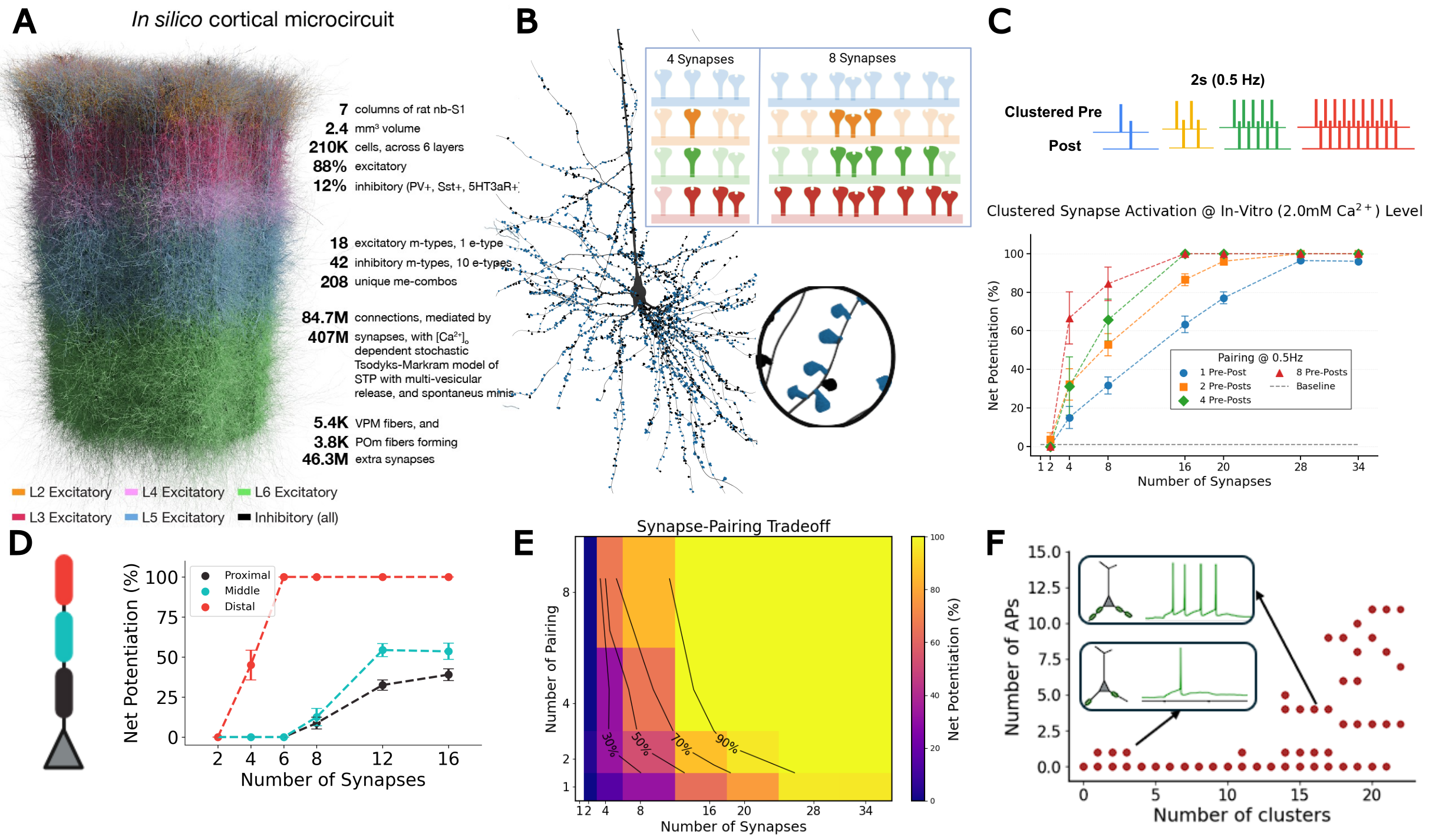
Synchronous activation of ≥11 clustered synapses generates dendritic plateau potentials (≥100 ms) that induce LTP in distal basal dendrites (Fig. 1). We identify a synapse-pairing tradeoff, where dendrites effectively trade the number of co-activated synapses for pairing repetitions: 16-synapse clusters achieve one-shot learning, while 8-synapse clusters require 3+ pairings. This tradeoff exhibits spatial gradients: distal dendrites achieve LTP independent of backpropagating action potentials, while proximal clusters require spike-timing coincidence. When multiple basal clusters coactivate, summated depolarizations trigger somatic bursts; both presynaptic and postsynaptic bursts drive robust LTP with all-or-none threshold dynamics.

Discussion

These findings establish multiple plasticity mechanisms within a single neuron—spatial clustering, location-dependent learning modes, and dual burst pathways—providing testable predictions for how cortical circuits implement flexible, hierarchical learning. Distal dendrites enable unsupervised learning via cluster-based LTP independent of bAPs, while proximal regions implement supervised learning requiring spike-timing coincidence. Apical dendrites receiving top-down signals can generate bursts or couple with somatic spikes via backpropagation-activated calcium (BAC) firing, a substrate for top-down plasticity modulation. These mechanisms reveal how dendrites trade synapse number for pairing repetitions under noisy physiological conditions.

Figure 1. A, In silico cortical microcircuit. B, L5-TTPC with magnified cluster showing plasticity for 4 vs 8 co-active synapses. C, Clustered pre-post pairing (0.5 Hz); net potentiation vs synapse number. D, Spatial learning gradient E, Synapse-pairing tradeoff heatmap. F, Basal cluster coactivation triggers somatic burst.

References
1. Chindemi, G., Abdellah, M., Amsalem, O., Benavides-Piccione, R., Delattre, V., Doron, M., Ecker, A., Jaquier, A. T., King, J., Kumbhar, P., Monney, C., Perin, R., Rössert, C., Tuncel, A. M., Van Geit, W., DeFelipe, J., Graupner, M., Segev, I., Markram, H., & Muller, E. B. (2022). A calcium-based plasticity model for predicting long-term potentiation and depression in the neocortex.
2. Ecker, A., Egas Santander, D., Abdellah, M., Alonso, J. B., Bolaños-Puchet, S., Chindemi, G., Gowri Mariyappan, D. P., Isbister, J. B., Ki
Sunday July 12, 2026 10:40am - 11:10am ADT
Ballroom B1

11:10am ADT

O1: Biologically plausible credit assignment via neuronal frequency multiplexing
Sunday July 12, 2026 11:10am - 11:30am ADT

Li Ji-An1,2,3Marcus K. Benna*3

Neurosciences Graduate Program, University of California San Diego, La Jolla, CA, USA
Department of Psychology, New York University, New York, NY, USA
Department of Neurobiology, University of California San Diego, La Jolla, CA, USA

*Email: [email protected]

Introduction
Backpropagation has been highly successful for training artificial neural networks. However, whether the biological brain implements any variant of backpropagation remains unknown. A key challenge concerns the capability of a single biological neuron to simultaneously encode and transmit feedforward predictions and feedback errors with minimal interference. Here, we propose a neuronal frequency multiplexing framework to address this challenge.

Methods
Each model neuron has multiple compartments that multiplex signals in the frequency domain. One dendrite acts as a low-pass filter, and extracts feedforward prediction signals from the low-frequency, direct-current components of the inputs. Another dendrite acts as a high-pass filter, and extracts feedback error signals from high-frequency, oscillatory components of the inputs. The soma integrates both signals, transmitting them to other neurons through its firing rate, which consists of a slowly varying prediction component and an oscillatory error component.

Results
We demonstrate that this frequency multiplexing algorithm using a simple, local learning rule closely approximates backpropagation in fully connected networks trained on the MNIST dataset and in convolutional networks trained on the CIFAR-10 dataset, achieving comparable performance and similar learning speed as a function of the number of training epochs.

Discussion
Our framework implements backpropagation-like training of functionally feedforward neural networks using continuously running, recurrently connected neuronal populations that simultaneously encode and propagate both prediction and error signals with minimal interference. This represents a new solution to the long-standing problem of biologically plausible credit assignment, suggesting a potential computational role for oscillatory signals in coordinating synaptic plasticity.

Speakers
MK

Marcus K Benna

UC San Diego
Sunday July 12, 2026 11:10am - 11:30am ADT
Ballroom B1

11:30am ADT

O2: A unifying account of rTMS and rTFUS neurostimulation effects based on calcium-dependent synaptic plasticity theory and an equivalent-energy principle
Sunday July 12, 2026 11:30am - 11:50am ADT
John D. Griffiths*1,2,3,4, Kevin Kadak1,3, Yupeng Tian1,5,6

1Krembil Centre for Neuroinformatics, Centre for Addiction and Mental Health, Toronto, Canada
2Department of Psychiatry, University of Toronto, Canada
3Institute of Medical Sciences, University of Toronto, Canada
4Institute of Biomedical Engineering, University of Toronto, Canada
5Dept. Mathematics, University of Toronto, Canada 
6Fields Institute for Mathematical Sciences, Toronto, Canada


*Email: [email protected]

Introduction
Repetitive transcranial magnetic stimulation (rTMS) and transcranial focused ultrasound stimulation (rTFUS) are noninvasive neuromodulation techniques with established and promising clinical applications, respectively.Though their primary mechanisms of action differ (electromagnetic vs. acoustic),both exert clinically relevant effects through stimulation-induced synaptic plasticity. Despite rich neurophysiological understanding of plasticity, a validated theoretical framework describing noninvasive neurostimulation-induced plasticity remains to be developed. We present a unified mean-field modelling framework for rTMS- and rTFUS-induced plasticity, grounded in calcium-dependent plasticity theory embedded in a corticothalamic circuit[1,2,3].

Methods
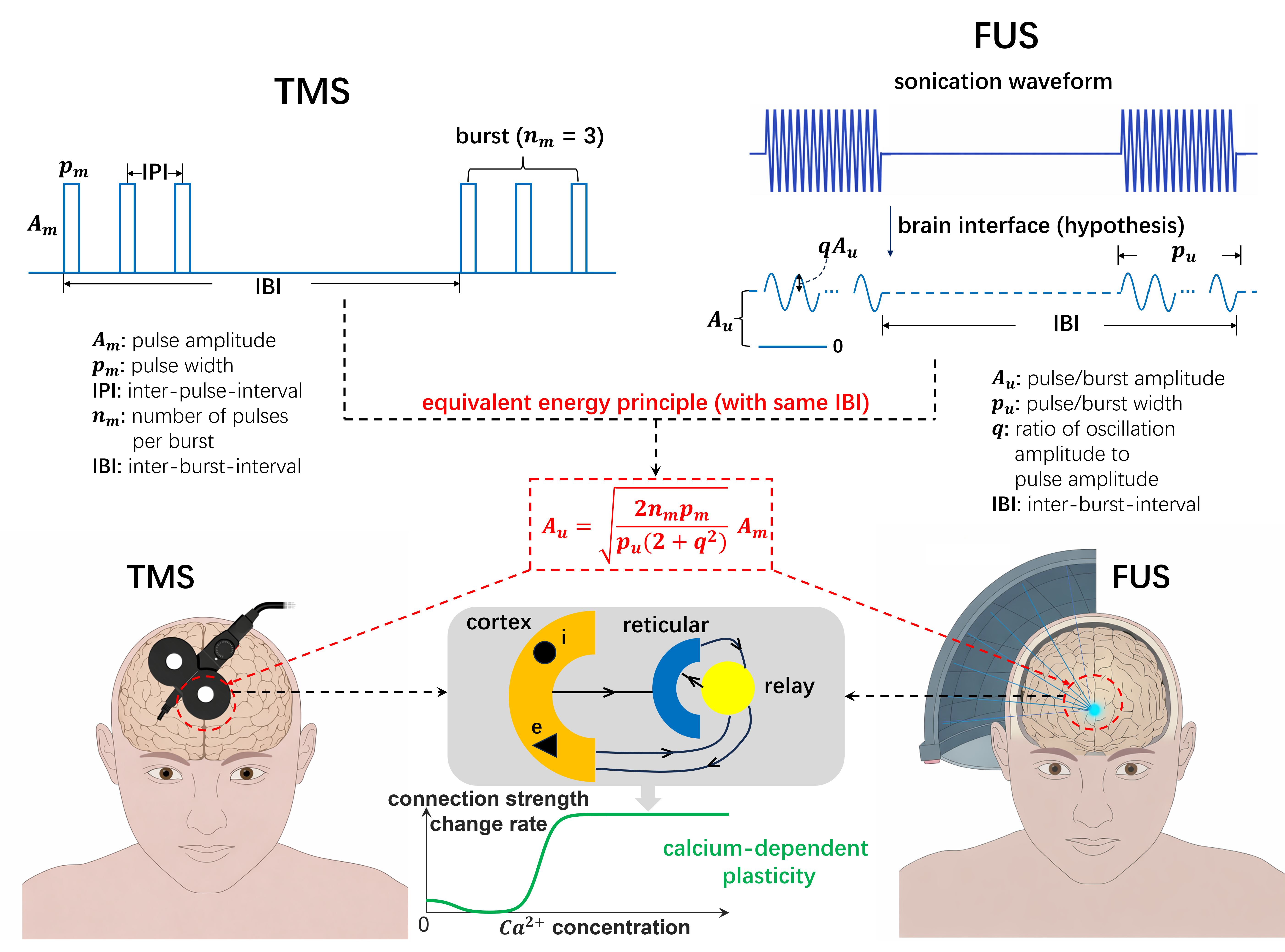
We extended the Fung-Robinson calcium-dependent plasticity model [1] by embedding it in a multi-population corticothalamic circuit generating alpha oscillations, implemented in NFTsim. For rTMS validation, a within-subject TMS-EEG experiment (N=21; 5 visits) tested 5 iTBS protocols varying inter-burst frequency and pulses-per-burst, measuring motor-evoked potentials (MEPs) and resting-state EEG alpha power. For rTFUS, a novel equivalent-energy principle (Fig.1) scaled continuous FUS burst amplitudes to deliver equivalent energy to corresponding rTMS waveforms, enabling direct model comparison across modalities. Predictions were compared against published rTFUS motor plasticity data across varying inter-burst frequencies and durations [4,5].

Results
Weaker iTBS protocols (3Hz/3PPB, 5Hz/2PPB) produced paradoxically stronger LTP-like MEP facilitation than standard iTBS (5Hz/3PPB), while the strongest protocol (7Hz/3PPB) robustly sign-flipped to LTD. MEP and resting-state alpha power showed a consistent inverse relationship across all protocols, supporting EEG as a plasticity biomarker outside the motor system. The corticothalamic model reproduced correct MEP directionality in 5/5 protocols and rank-ordering in 4/5, and captured alpha directionality in 4/5.Applying the equivalent-energy principle to rTFUS, the same model replicated published cTB-FUS plasticity results and accounted for the LTD/LTP sign-flip in cTB-TMS (40s vs. 80s) but not cTB-FUS, explained by waveform shape alone [4,5].

Discussion
These findings support a 'less is more' principle: gentler stimulation paradoxically yields stronger plasticity effects, with over-stimulation causing sign-reversal to LTD. The consistent MEP-alpha inverse relationship opens the possibility of using scalp EEG as a protocol-agnostic plasticity readout. The equivalent-energy principle provides a principled bridge between rTMS and rTFUS modelling, enabling the same calcium-dependent corticothalamic framework to account for both modalities without re-parameterization. Together these results establish a foundation for in silico exploration of the largely unmapped rTMS/rTFUS protocol space, with direct implications for optimizing clinical neuromodulation [2,3].

Figure 1. Equivalent energy principle for aligning rTMS and rTFUS plasticity models. rTMS delivers discrete pulse bursts; rTFUS delivers continuous bursts filtered by the skull interface to sub-1kHz sinusoids. The principle constrains parameters so both modalities deliver equal energy at the same carrier frequency, enabling unified mean-field modelling of calcium-dependent plasticity.


References
  1. Fung & Robinson (2014). Neural field theory of synaptic metaplasticity with applications to theta burst stimulation. J Theor Biol, 340, 164–176. https://doi.org/10.1016/j.jtbi.2013.09.021
  2. Kadak K, et al. (2026, submitted). Less is more: gentle protocols induce stronger facilitatory effects than standard iTBS through calcium-dependent metaplasticity.
  3. Tian Y, et al. (2026, submitted). Equivalent energy principle and calcium-dependent plasticity theory unify TMS and FUS effects.
  4. Zeng K, et al. (2024). Motor cortex plasticity by theta burst transcranial ultrasound. Ann Neurol, 91(2), 238–252. https://doi.org/10.1002/ana.26294
  5. Gamboa OL, et al. (2010). Reversal of theta burst after-effect with prolonged stimulation. Exp Brain Res, 204(2), 181–187. https://doi.org/10.1007/s00221-010-2293-4

Acknolwedgments
We acknowledge funding from the Krembil Foundation, Labbatt Foundation, UofT EMHSeed, and Fields Institute for Mathematical Sciences, that supported this work. 
Speakers
Sunday July 12, 2026 11:30am - 11:50am ADT
Ballroom B1

11:50am ADT

O3: Compartmentalized learning through coupled electrochemical adaptation in cortical pyramidal neurons
Sunday July 12, 2026 11:50am - 12:10pm ADT
Beatriz Barros1,2, Raquel Figueiredo1,2Renato Duarte*1, 2


1Center for Neuroscience and Cell Biology (CNC-UC), University of Coimbra, Portugal
2Centre for Innovative Biomedicine and Biotechnology (CiBB), University of Coimbra, Portugal
*Email: [email protected]

Introduction
A single cortical neuron simultaneously expresses Hebbian STDP proximally and cooperative plasticity distally, couples excitatory and inhibitory weight changes, and maintains homeostatic stability across timescales spanning seconds to days. Computational models treat these phenomena separately, yet the underlying molecular cascades are shared, shaped by local dendritic morphology and chemical composition. This convergence means that compartment-specific learning rules, local E/I balance, and multi-timescale homeostasis are not independent, but mechanistically coupled through intracellular dynamics. What emerges computationally from this coupling, and how it reshapes our understanding of single-neuron learning, remains an open question.

Methods
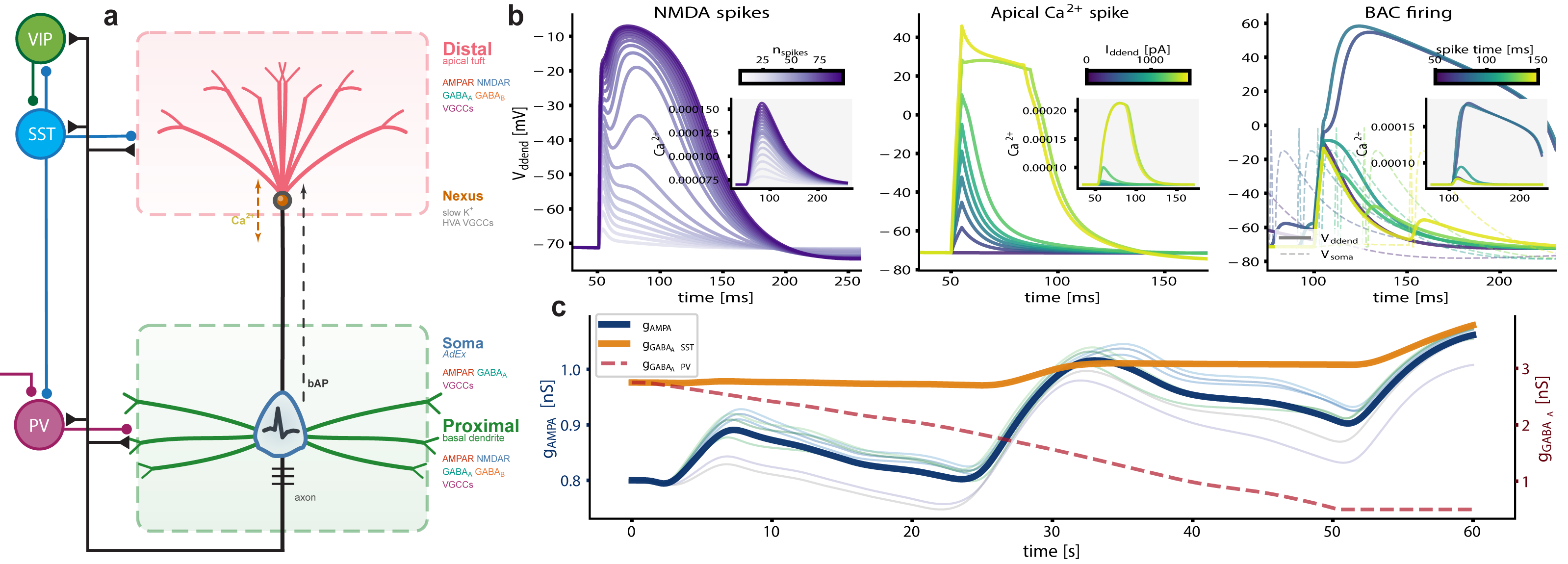
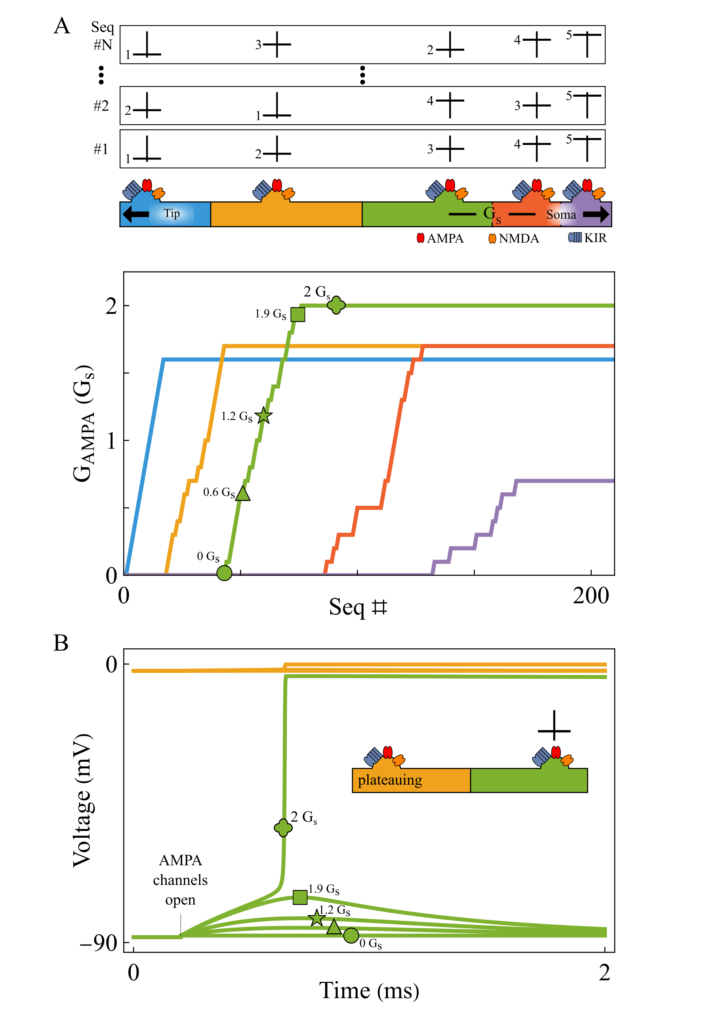
We build on a three-compartment neuron model [2], augmented with active, electrogenic dendritic processes (Fig. 1). Local calcium currents feed a slow dendritic integrator that drives calcium-dependent plasticity [3] at every synapse. Compartment-specific learning emerges naturally: bAP-dominated proximal calcium produces Hebbian STDP, while VGCC/NMDAR-dominated distal calcium yields cooperative plasticity. Inhibitory synapses read the same calcium with inverted thresholds, coupling E/I balance without explicit homeostatic targets. We extend this via stargazin phosphorylation [4], anchoring AMPAR trafficking, Kv7.2-mediated intrinsic excitability, and synaptic scaling in a three-tier cascade spanning seconds to days.

Results
A single plasticity rule, operating on compartment-resolved calcium trace, produces Hebbian STDP proximally (bAP-dominated) and cooperative, timing-independent plasticity distally (VGCC/NMDAR-dominated), matching recent in vivo observations [1]. Shared calcium maintains coupled E/I balance locally, without explicit homeostatic targets, and allows accurate stimulus representation from the response to localized perturbations. The stargazin cascade reproduces multiphasic homeostatic dynamics with intrinsic plasticity preceding synaptic scaling. We show that the apparent diversity of cortical plasticity rules is an emergent phenomenon, a consequence of intracellular dynamics and proceed to investigate its functional consequences.

Discussion
Compartment-specific signaling produces qualitatively different learning rules from the same mechanism, reframing credit assignment in cortical circuits [1] and emphasizing intracellular signaling as a primary locus of learning and memory. Stimulus associations, selectivity, and stability emerge from dendritic biophysics and are co-modulated with shared electrochemical substrates. Beyond the biophysical details, the framework we present here raises broader questions: can local, compartmentalized balance serve as a natural learning objective? And how does coupling different adaptation mechanisms shape information representation and memorization, with intracellular dynamics acting as a stack-like memory?

Figure 1. Augmented tripod neuron with compartment-specific electrogenic events and coupled E/I plasticity. (a) Circuit schematic with compartment-resolved receptors and interneuron targeting. (b) NMDA plateaus, apical Ca²⁺ spikes, and BAC firing with dendritic calcium transients (insets). (c) Shared calcium couples excitatory and inhibitory weight dynamics, actively maintaining E/I balance.References
[1] Wright, W. J., Hedrick, N. G., & Komiyama, T. (2025). Distinct synaptic plasticity rules operate across dendritic compartments in vivo during learning. Science, 388(6744), 322-328.
[2] Quaresima, A., Fitz, H., Duarte, R., van den Broek, D., Hagoort, P., & Petersson, K. M. (2023). The Tripod neuron: a minimal structural reduction of the dendritic tree. The Journal of Physiology, 601(15), 3265-3295.
[3] Graupner, M. & Brunel, N. (2012). Calcium-based plasticity model explains sensitivity of synaptic changes to spike pattern, rate, and dendritic location. PNAS, 109(10), 3991-3996.
[4] Rodrigues, M. V. et al. (2024). Type I TARPs regulate Kv7.2 potassium channels and susceptibility to seizures. bioRxiv, 2024.08.09.607194.

Acknowledgments
This work was supported by national funds through FCT—Foundation for Science and Technology, I.P., under the project HetSyn (2023.13758.PEX).

Speakers
avatar for Renato Duarte

Renato Duarte

Assistant Researcher, Center for Neuroscience and Cell Biology (CNC), University of Coimbra
Sunday July 12, 2026 11:50am - 12:10pm ADT
Ballroom B1

12:10pm ADT

O4: Rewarding Control: Feedback-Phase-Dependent 2Hz Medial Frontal Transcranial Alternating Current Stimulation Shifts the Expected Value of Control
Sunday July 12, 2026 12:10pm - 12:30pm ADT
Authors: Robert Louis Treuting1, Eric Rawls*1,2
Affiliations: 1Department of Psychology, University of North Carolina Wilmington, Wilmington, NC, USA, 2Department of Psychiatry and Behavioral Sciences, University of Minnesota Twin Cities, Minneapolis, MN, USA
*Email: [email protected]

Introduction
Medial frontal delta activity is a plausible control signal because the Reward Positivity (RewP) tracks outcome evaluation and prediction errors, and our prior simultaneous EEG-fMRI work localized key signed prediction-error contributions of the RewP to medial frontal cortex [1,2]. Here we tested whether 2 Hz transcranial alternating current stimulation (tACS) targeting this generator changes motivated cognitive control. We predicted that stimulation would alter latent decision parameters in an Expected Value of Control Stroop rather than merely speeding responses [2,3].

Methods
Seven healthy participants completed a reward×efficacy Stroop with congruent/incongruent targets across sham and active 2 Hz medial frontal tACS sessions. Accuracy was analyzed with binomial generalized linear mixed models and reaction time with Gamma models. We then fit a hierarchical Wiener diffusion model in brms to decompose behavior into drift rate (mu), boundary separation (bs), and nondecision time (ndt). In a second model restricted to active trials, sine and cosine terms indexed the phase of stimulation at the prior trial’s feedback.

Results
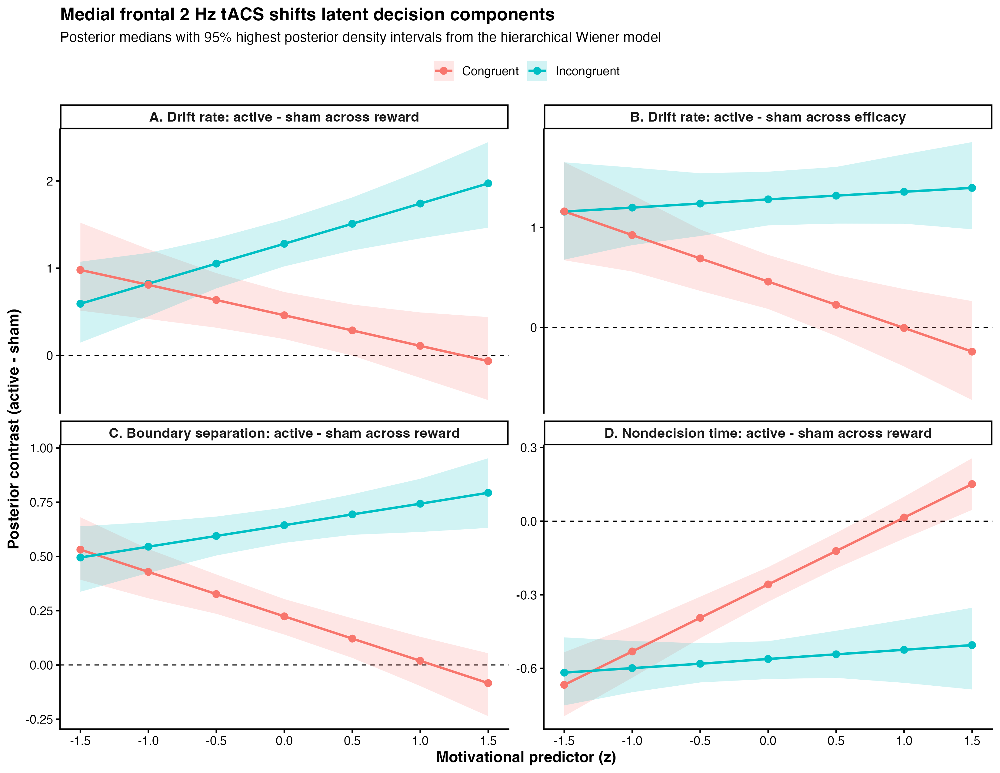
Active stimulation improved accuracy (χ²(1)=8.76, p=0.003) without a main reaction-time benefit, arguing against nonspecific speeding. In the diffusion model, stimulation shifted all three latent components: drift, boundary, and nondecision time. Posterior contrasts showed robust drift benefits throughout incongruent trials and selective benefits in congruent trials when reward or efficacy was low (Fig. 1). In active trials, prior-feedback phase predicted subsequent drift (feed_cos=0.39 [0.19, 0.59]; feed_sin=-0.26 [-0.48, -0.05]), consistent with causal modulation of a RewP-linked medial frontal mechanism for updating expected value of control from recent outcomes [1,2].

Discussion
These results suggest that medial frontal delta stimulation does not simply energize behavior. Instead, it changes how outcome information is converted into the next trial’s control state. Computationally, active 2 Hz tACS improved evidence accumulation while also reshaping caution and peripheral processing.

Figure 1. Active minus sham posterior contrasts from the hierarchical Wiener diffusion model. Top panels show drift-rate changes across reward and efficacy, separated by congruency. Bottom panels show boundary-separation and nondecision-time changes across reward. Points indicate posterior medians; ribbons show 95% highest posterior density intervals.

References
[1] Rawls, E., Demro, C., Teich, C. D., Zhang, J., Wang, A., Heilbronner, S. R., Mueller, B. A., Sponheim, S. R., & MacDonald, A. W., III. (2026). The search for RewP: Dissociating cortical generators of electrophysiological signed and unsigned prediction errors using simultaneous EEG-fMRI [Manuscript in preparation].
[2] Frömer, R., Lin, H., Dean Wolf, C. K., Inzlicht, M., & Shenhav, A. (2021). Expectations of reward and efficacy guide cognitive control allocation. Nature Communications, 12, 1030.
[3] Ratcliff, R., & McKoon, G. (2008). The diffusion decision model. Neural Computation, 20(4), 873–922.

Acknowledgments
We thank the Brain, Data, and Causality Lab and our University of Minnesota collaborators for the prior simultaneous EEG-fMRI work that motivated this study, and for foundational discussion of RewP source modeling and expected value of control.

Speakers
Sunday July 12, 2026 12:10pm - 12:30pm ADT
Ballroom B1

12:30pm ADT

Program Commitee Meeting
Sunday July 12, 2026 12:30pm - 2:00pm ADT

Sunday July 12, 2026 12:30pm - 2:00pm ADT
TBA

2:00pm ADT

FO2: Norepinephrine Restores Cortical Dynamics and Enables Machine Learning–Based Severity Mapping in a Multiscale Model of Parkinson’s Disease
Sunday July 12, 2026 2:00pm - 2:30pm ADT
Jeeyune Jung*1,3,   Adam Newton1,3,  Donald Doherty1,3,  Hong-Yuan Chu2,3,  Samuel Neymotin4 , William Lytton1,3
1. Department of Physiology and Pharmacology, SUNY Downstate Health Sciences University, Brooklyn, NY, USA 
2. Department of Pharmacology and Physiology, Georgetown University Medical Center, Washington, DC, USA 
3. Aligning Science Across Parkinson’s (ASAP) Collaborative Research Network, Chevy Chase, MD, USA
Center for Biomedical Imaging & Neuromodulation, Nathan Kline Institute, Orangeburg, NY, USA

*Email: [email protected]


Introduction
Parkinson’s disease (PD) involves not only basal ganglia dopamine loss but also cortical dysfunction, including excessive beta synchronization, abnormal beta–gamma coupling, altered bursting, and impaired corticospinal recruitment. Early locus coeruleus degeneration may reduce cortical norepinephrine (NE), disrupting cell-type-specific gain control in pyramidal tract (PT) and intratelencephalic (IT) neurons. We tested whether experimentally constrained NE modulation restores cortical excitability and network dynamics in an advanced MitoPark motor cortex model and whether NE-sensitive cortical biomarkers support severity mapping and prediction of dopamine-therapy response.


Methods
Whole-cell patch-clamp recordings from Layer 5 PT and IT neurons quantified NE (10 µM)-induced changes in firing–current relationships. Conductance-based single-cell models were fit to baseline and NE responses and embedded in a biophysically detailed laminar M1 network model in NEURON/NetPyNE. Parkinsonian simulations incorporated reduced PT5B intrinsic excitability and reduced thalamocortical drive, with disease-stage-dependent NE scaling from PK/PD modeling. From the resulting simulations, we extracted cortical biomarkers including PT5B firing, IT5B beta-synchronized bursting, IT/PT imbalance, beta power, beta-burst duration, beta–high gamma phase-amplitude coupling, and avalanche slope, which were then used for severity mapping. 

Results
NE exerted opposite intrinsic effects across Layer 5 pyramidal subtypes: PT firing increased, whereas IT repetitive firing decreased. In the Parkinsonian network, NE-dependent conductance changes partially rescued pathological dynamics: PT5B firing increased by ~60–70%, IT5B bursting declined, the IT/PT activity ratio shifted toward control-like values, and pathological beta–high gamma phase-amplitude coupling decreased by ~40%. Across the biomarker set, NE shifted cortical dynamics toward the control regime. In the machine-learning framework, greater deviation from control tracked greater disease severity and predicted progressively shorter and weaker levodopa benefit as NE support declined.
Discussion
These findings identify NE-sensitive intrinsic gain control as a mechanistic bridge between single-cell excitability and pathological cortical state in PD. Loss of noradrenergic modulation may directly contribute to corticospinal under-recruitment, hypersynchronous beta activity, and broader cortical biomarker abnormalities, whereas restoring NE-dependent PT/IT balance may complement dopamine-based therapy. This multiscale framework links cellular mechanisms, network dysfunction, severity mapping, and predicted treatment response, positioning noradrenergic modulation as a promising strategy for advanced PD.

References
1. Dura-Bernal, S., et al. (2023). Multiscale model of primary motor cortex circuits predicts in vivo cell-type-specific, behavioral state-dependent dynamics. Cell Reports.
2. Chu, H. Y., et al. (2024). Dysfunction of motor cortices in Parkinson’s disease. Cerebral Cortex.
3. Doherty, D. W., et al. (2025). Enhanced beta power emerges from simulated parkinsonian primary motor cortex. npj Parkinson’s Disease.

Acknowledgments
This work was supported by the Aligning Science Across Parkinson’s (ASAP) Collaborative Research Network. We thank colleagues for providing experimental data used to constrain the model. Computational resources were provided by SUNY Downstate Health Sciences University.


Sunday July 12, 2026 2:00pm - 2:30pm ADT
Ballroom B1

2:30pm ADT

O5: Impact of EAAT2 Dysfunction on AMPA/NMDA-Mediated Excitability in Neuronal Activity
Sunday July 12, 2026 2:30pm - 2:50pm ADT
Hannah van Susteren1,*, Guillaume Girier2,*, Michel J.A.M. van Putten3,4 , Jaroslav Hlinka2, Helmut Schmidt2, Hil G.E. Meijer1


1 Department of Applied Mathematics, University of Twente, Enschede, the Netherlands
2 Institute of Computer Science, Czech Academy of Sciences, Prague, Czech Republic
3 Department of Neurology and Clinical Neurophysiology, University of Twente, Enschede, the Netherlands
4 Medisch Spectrum Twente, Enschede, the Netherlands
* These authors contributed equally to this work.


Email: [email protected]


Introduction
Epilepsy is among the most prevalent neurological disorders. The astrocytic excitatory amino acid transporter (EAAT2) plays a key role in regulating excitability, by controlling extracellular glutamate levels and glutamate receptor activation [1,2]. Reduced EAAT2 expression has been reported in several epilepsy patients [1,3]. However, the impact of neuron-astrocyte interactions on hyperexcitability on single cell level is underexplored. We developed a biophysical model of a presynaptic neuron and astrocyte to explore the relation between astrocytic EAAT2-mediated glutamate clearance, presynaptic glutamate receptors and bursting activity.

Methods
We build on our previous work [4,5], where we consider a presynaptic neuron and an astrocyte in a finite extracellular space (ECS). This model describes sodium, potassium, chloride dynamics as well as calcium-dependent exocytosis and glutamate-glutamine (GG) recycling. For this study, we add a potassium bath with diffusion to the ECS to induce neuronal bursting (Fig. 1A). Additionally, we implement the presynaptic glutamate receptors AMPA and NMDA, which are important in regulating hyperexcitability. Lastly, we study the impact of the antiseizure drugs perampanel and memantine by simulating the effect of these AMPA and NMDA receptor antagonists.

Results
We induce neuronal bursting by increasing extracellular potassium in the bath. We first examine how AMPA and NMDA permeabilities affect burst frequency (Fig. 1C), where frequency refers to spike frequency during the last burst or during tonic firing, to fit the NMDA/AMPA ratio to experimental data [6]. Higher permeabilities increase neuronal firing and intracellular calcium, triggering a feedback loop that enhances neuronal glutamate release. Reducing EAAT permeability raises burst frequency and induces tonic firing (Fig. 1B). Finally, AMPA and NMDA antagonists, perampanel and memantine [7], reduce firing despite elevated extracellular glutamate, with perampanel showing a more significant reduction in firing frequency (Fig. 1D). 

Discussion
Our results show that reduced EAAT expression, as observed in several epilepsy patients, results in increased extracellular glutamate and overstimulation of excitatory glutamate receptors. Furthermore, we show that the AMPA and NMDA receptor permeabilities affect burst frequency. Receptor antagonists such as perampanel and memantine are able to reduce firing. In conclusion, our detailed neuron–astrocyte model provides insight into the effects of reduced EAAT expression and receptor antagonists on hyperexcitability.
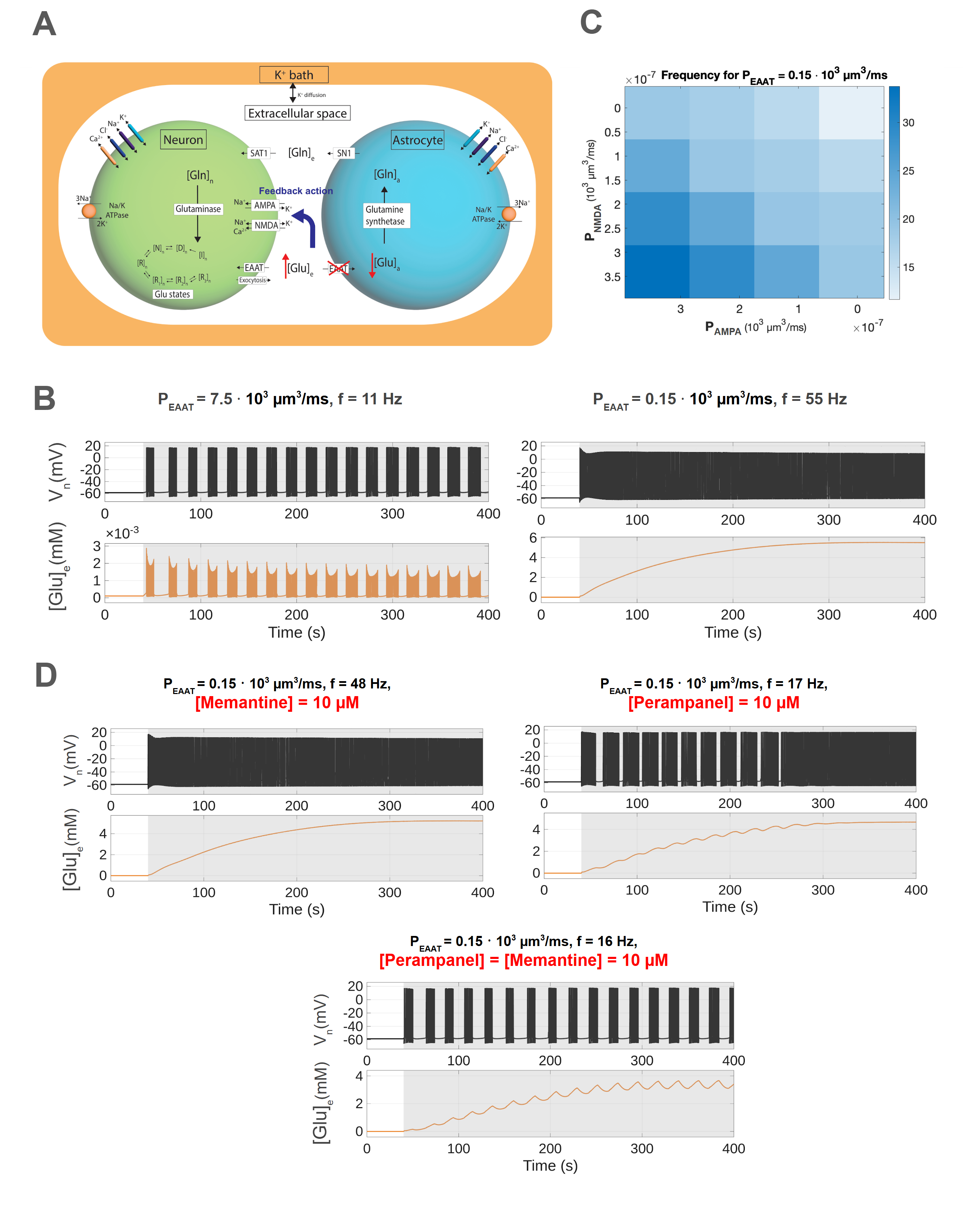
Figure 1. A: Three-compartment model illustrating the GG-cycle during EAAT2 knockout. B: The membrane potential, spike frequency f and ECS glutamate at different EAAT2 permeabilities. C: Spike frequency within bursts as a function of NMDA and AMPA receptor permeability.  D: Neuronal activity at fixed EAAT2 permeability (PEAAT=0.15 * 103  µm3/ms) under antagonist conditions.

References
[1] Green, J. L., dos Santos, W. F., & Fontana, A. C. K. (2021). Biochemical Pharmacology, 10.1016/j.bcp.2021.114786
[2] Scimemi, A., Tian, H., & Diamond, J. S. (2009). The Journal of Neuroscience, 10.1523/JNEUROSCI.4845-09.2009
[3] Barker-Haliski, M., & White, H. (2015). Cold Spring Harbor perspectives in medicine, 10.1101/cshperspect.a022863
[4] van Susteren, H., Rose, C. R., van Putten, M. J., & Meijer, H. G. (2025). bioRxiv, 10.1101/2025.11.10.687543
[5] Kalia, M., et al. (2021).  PLOS Computational Biology, 10.1371/journal.pcbi.1009019
[6] Watt, A. J., Sjöström, P. J., Häusser, M., Nelson, S. B., & Turrigiano, G. G. (2004).  Nature neuroscience, 10.1038/nn1220
[7] Chen, T.-S., Huang, T.-H., Lai, M.-C., & Huang, C.-W. (2023).  Biomedicines, 10.3390/biomedicines11030783

Acknowledgments
HVS, HGEM, MJAMVP funded from the DFG, FOR2795 ‘Synapses under stress’ to CRR (Prof. Dr. Christine R. Rose) (Ro2327/13-2 and 14-2).
GG, HS, and JH were supported by the ERDF-Project Brain dynamics, No. CZ.02.01.01/00/22\_008/0004643, a Lumina-Quaeruntur fellowship (LQ100302301), and the long-term strategic development financing of the Institute of Computer Science (RVO:67985807).

Speakers
HV

Hannah van Susteren

PhD student, University of Twente
Sunday July 12, 2026 2:30pm - 2:50pm ADT
Ballroom B1

2:50pm ADT

O6: Multiscale modeling of neural markers for adaptive deep brain stimulation in Parkinson’s Disease
Sunday July 12, 2026 2:50pm - 3:10pm ADT

Alberto Mazzoni1
,2,*, Federico Fattorini1,2, Nicolò Meneghetti1,2


1The Biorobotics Institute, Scuola Superiore Sant’Anna, Pisa, Italy
2Department of Excellence for Robotics and AI, Scuola Superiore Sant’Anna, Pisa, Italy


*Email: [email protected]


Introduction
Parkinson’s disease (PD) is a common neurodegenerative disorder causing severe impairments. Drug-resistant patients are treated with Deep Brain Stimulation (DBS) of the basal ganglia (BG), with current efforts focused on adaptive DBS. The most relevant biomarkers for adaptive DBS is the power in the beta ([12, 30] Hz) and gamma ([30, 100] Hz) range, yet the mechanisms underlying these rhythms remain unclear. Computational models can provide mechanistic insights into pathophysiology and test new stimulation treatments. Using spiking and morphological models, we show how beta and gamma resonances emerge from BG interactions, how DBS reshapes these dynamics, and how they are reflected in subthalamic nucleus local field potentials (LFPs).

Methods
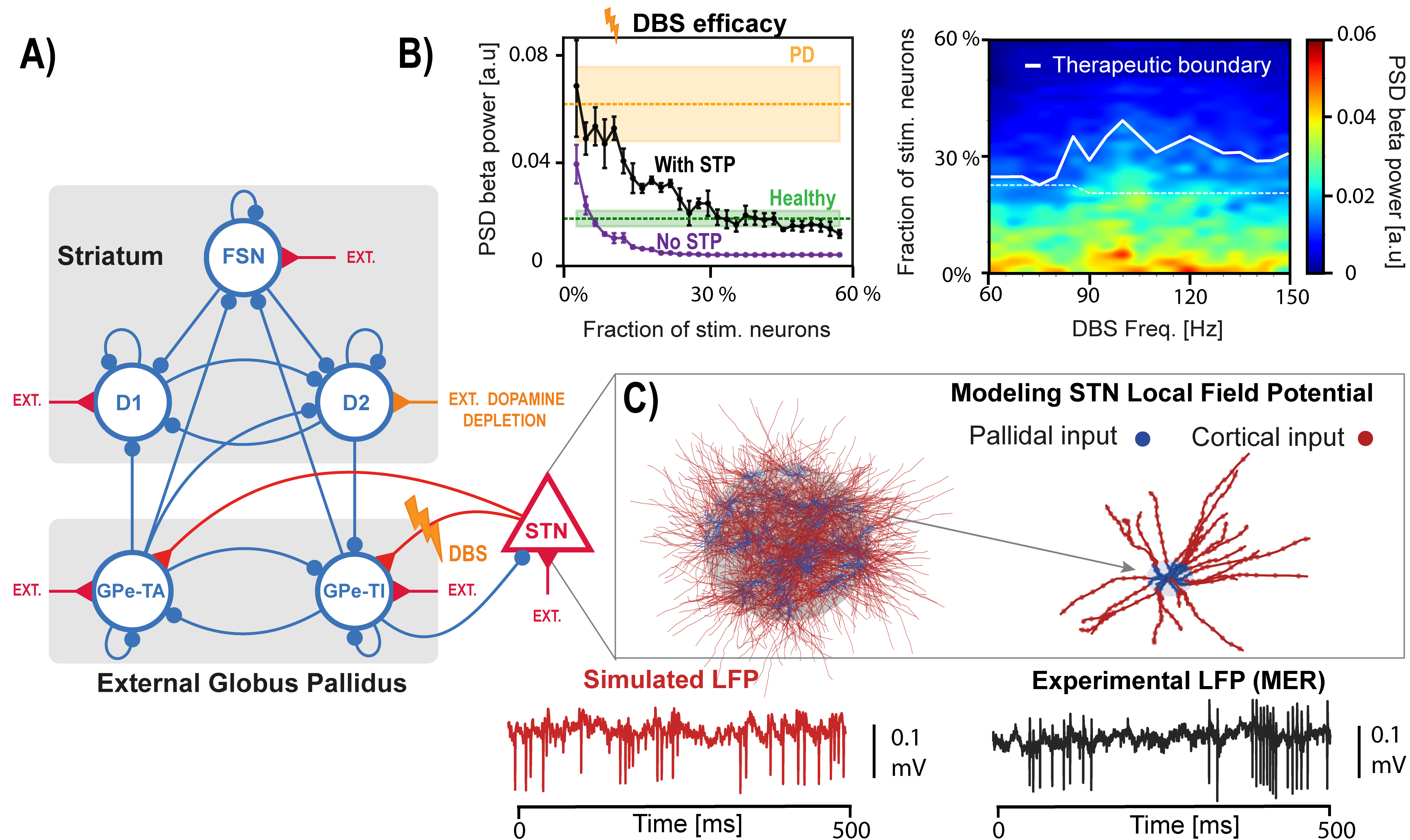
We implemented a spiking model of the basal ganglia with six neuronal populations: three within the striatum, two within the external globus pallidus, and the subthalamic nucleus (STN) (Fig. 1A) [1]. Dopamine depletion was simulated by modulating striatal inputs. We dissected the network mechanisms underlying beta and gamma resonances and we simulated STN DBS, considering short-term synaptic plasticity (Fig. 1B) [2]. To simulate the signals recorded by DBS electrodes we developed a population of morphological STN neurons model and computed LFPs associated with network activity through volume conduction theory (Fig. 1C).

Results
We show how beta oscillations arise from two independent loops in the BG model that strongly synchronize when dopamine is depleted [1]. STN DBS disrupts these oscillations, although without synaptic plasticity it requires unrealistically low stimulation levels (Fig 1B left). The model also supports the hypothesis that gamma-range stimulation can be as effective as the clinical standard of ~130 Hz used in PD (Fig. 1B right) [2]. Gamma oscillations emerge through recurrent inhibition in pallidal and striatal populations. Different from cortical ones, STN LFPs are largely noise-dominated due to weak correlations and symmetric neuronal morphology, becoming informative only when strong beta synchronization is present.

Discussion
We characterized in a spiking model of BG beta and gamma rhythmogenesis and their alteration due to PD-related dopamine depletion and DBS. Moreover, we investigated the origin of STN LFPs driving adaptive DBS, by integrating spiking and morphological modeling. Overall, we provide a multiscale framework for better understanding Parkinsonian dynamics and DBS mechanisms, showing how network modeling can clarify treatment mechanisms and guide improved stimulation strategies.

Figure 1. A) Spiking network model of the basal ganglia.  B) Left: efficacy of STN DBS as a function of the fraction of stimulated neurons, with and without short-term plasticity (STP). Right: effect of stimulation frequencies on beta spectral power.  C) Top: STN population and morphological neuron model receiving cortical and pallidal inputs. Bottom: simulated and recorded local field potentials (LFPs).
References

1. Ortone, A., Vergani, A. A., Ahmadipour, M., Mannella, R., & Mazzoni, A. (2023). Dopamine depletion leads to pathological synchronization of distinct basal ganglia loops in the beta band. PLOS Computational Biology, 19(4), 1–31. https://doi.org/10.1371/journal.pcbi.1010645
2. Ahmadipour, M., Fattorini, F., Meneghetti, N., & Mazzoni, A. (2026). In silico model of basal ganglia deep brain stimulation in Parkinson’s disease captures range of effective parameters for pathological beta power suppression. PLOS Computational Biology, 22(2), e1013280. https://doi.org/10.1371/journal.pcbi.1013280
Acknowledgments
This work was supported by the Italian Ministry of University and Research, under the complementary actions to the NRRP ‘Fit4MedRob - Fit for Medical Robotics’ Grant (# PNC0000007).


Speakers
Sunday July 12, 2026 2:50pm - 3:10pm ADT
Ballroom B1

3:10pm ADT

O7: Decreased alpha/theta temporal ExSEnt of left prefrontal cortex: a robust biomarker of dementia
Sunday July 12, 2026 3:10pm - 3:30pm ADT
Sara Kamali*1, Fabiano Baroni1, Pablo Varona1

1Department of Computer Engineering, Autonomous University of Madrid, Madrid, Spain

*Email: [email protected]

Introduction
Dementia involves progressive cognitive decline, with Alzheimer’s disease (AD) and frontotemporal dementia (FTD) as major subtypes. Electroencephalography (EEG) provides a noninvasive and accessible measure of brain dynamics and able to capture pathological slowing, often reflected by increased theta-to-alpha power ratio (TAR) [1]. Reduced signal complexity has also been reported in dementia. We tested whether Extrema-Segmented Entropy (ExSEnt), which separates temporal and amplitude irregularity, improves discrimination and interpretability beyond conventional electroencephalography biomarkers [2].

Methods
We analyzed resting-state EEG from 88 subjects, including 36 with AD, 23 with FTD, and 29 controls [3]. After preprocessing and independent component analysis, we classified brain sources into four clusters: right and left prefrontal cortices (R/LPFC) and right and left visual associations (R/LVA). Then, we computed a set of classical spectral and complexity features. We also measured the ExSEnt metrics, which quantifies the entropy of extrema-based segment durations, amplitudes, and their pair. To find the stable features for dementia detection, we performed stability selection with elastic-net logistic regression and nested leave-one-subject-out validation [4].

Results
ExSEnt improved discrimination mainly in the LPFC, where balanced accuracy increased from 71.5% to 82.6%. In that region, ExSEnt features showed high selection stability and replaced conventional measures as dominant predictors in 3 out of 4 brain areas. The most informative variables were temporal and joint temporal-amplitude entropy measures in alpha and theta bands, with negative coefficients indicating reduced irregularity and reduced dynamical variability in dementia. Other regions showed weaker or inconsistent gains, suggesting a localized effect rather than a global one. 

Discussion
ExSEnt provides a compact and interpretable measure for EEG-based dementia classification. The results suggest that dementia is associated not only with spectral slowing but also with reduced diversity of extrema timing and temporal-amplitude variability in LPFC dynamics. Because ExSEnt is explainable by design, when combined with stability selection and sparse linear classification, it yields robust biomarkers with direct physiological interpretation. These findings support ExSEnt as a promising candidate for explainable dementia screening and motivate future validation against cognitive severity measures.

References
[1] Partanen, J. V., Soininen, H., & Riekkinen, P. J. (1986). Does an ACTH derivative (Org 2766) prevent deterioration of EEG in Alzheimer's disease?. Electroencephalography and clinical neurophysiology, 63(6), 547-551.
[2] Kamali, S., Baroni, F., & Varona, P. (2025). ExSEnt: Extrema-Segmented Entropy Analysis of Time Series. arXiv preprint arXiv:2509.07751.
[3] Miltiadous, A., Tzimourta, K. D., Afrantou, T., Ioannidis, P., Grigoriadis, N., Tsalikakis, D. G., ... & Tzallas, A. T. (2023). A dataset of scalp EEG recordings of Alzheimer’s disease, frontotemporal dementia and healthy subjects from routine EEG. Data, 8(6), 95.
[4] Meinshausen, N., & Bühlmann, P. (2010). Stability selection. Journal of the Royal Statistical Society Series B: Statistical Methodology, 72(4), 417-473.

      Acknowledgments
      Funded by PID2024-155923NB-I00 and CPP2023-010818.
      Speakers
      Sunday July 12, 2026 3:10pm - 3:30pm ADT
      Ballroom B1

      3:30pm ADT

      O8: Frequency-dependent modulation of cortical traveling waves during general anesthesia
      Sunday July 12, 2026 3:30pm - 3:50pm ADT
      Duan Li*, Anthony G. Hudetz

      Center for Consciousness Science, Department of Anesthesiology, University of Michigan, Ann Arbor, MI

      *Email: [email protected]

      Introduction
      General anesthetics profoundly reshape neuronal activity and functional interactions, yet how they alter the spatiotemporal organization of the cortex is incompletely understood. Traveling waves provide a framework for examining coordinated neural activity across the cortex, but their modulation by anesthesia remains underexplored. Recently we showed that cortical dynamics under anesthesia exhibit spontaneous transitions among discrete states, including a paradoxical state characterized by low delta power and high complexity in deep anesthesia[1]. To investigate how traveling waves vary across cortical states, we recorded hemispheric ECoG at multiple anesthetic depths and analyzed wave dynamics as a function of cortical state and frequency.

      Methods
      ECoG was recorded from the right hemisphere with chronically implanted 32-site flexible polymer arrays (4×8 grid) at four desflurane concentrations (6-0%) for 1 h each. Cortical states were identified using PCA of power spectrograms followed by density-based clustering across concentrations. Within each state, traveling wave episodes were detected based on stabile spatial phase gradient patterns [2] between consecutive time points [3] in the delta, theta, and gamma bands. Wave occurrence and pattern richness were quantified, the latter defined as the entropy of SVD eigenvalues of the episode similarity matrix. Plane waves were identified and further classified as feedforward (posterior-to-anterior) or feedback (anterior-to-posterior).

      Results
      Seven states were identified. S1-S5 broadly tracked anesthetic depth but were not tied to a specific level. S6 corresponded to burst suppression, and S7 indicated a paradoxical state mostly during deep anesthesia. In S1, theta waves were mainly feedback-directed, whereas gamma were feedforward-directed. As anesthesia deepened, delta waves became more frequent but showed reduced pattern diversity. In contrast, the occurrence and diversity of theta and gamma patterns were largely preserved. Although gamma feedforward dominance persisted, theta feedback dominance was suppressed during deep anesthesia. In S7, reductions in delta complexity and theta organization were only partially reversed, despite reduced delta power and wake-like complexity.

      Discussion
      General anesthesia differentially modulates cortical traveling waves across frequency bands. Loss of theta feedback dominance paired with preserved gamma feedforward propagation suggests disrupted top-down integration despite maintained bottom-up information flow. Reduced diversity of delta waves during deep anesthesia suggests a shift toward more stereotyped, low-information dynamics. Importantly, traveling waves do not fully recover in the paradoxical state suggesting that spectrally activated deep anesthetic states lack coordinated spatiotemporal organization necessary for conscious processing. These findings suggest that traveling wave dynamics provide complementary insight into brain state dynamics beyond spectral power or complexity.

      References
      1. Li, D., & Hudetz, A. G. (2025). Dynamic electrocortical states and paradoxical complexity during desflurane anesthesia. bioRxiv. https://doi.org/10.1101/2025.10.13.682019
      2. Muller, L., Piantoni, G., Koller, D., Cash, S. S., Halgren, E., & Sejnowski, T. J. (2016). Rotating waves during human sleep spindles organize global patterns of activity that repeat precisely through the night. eLife, 5, e17267. https://doi.org/10.7554/eLife.17267
      3. Das, A., Zabeh, E., Ermentrout, B., & Jacobs, J. (2024). Planar, spiral, and concentric traveling waves distinguish cognitive states in human memory. bioRxiv. https://doi.org/10.1101/2024.01.26.577456

      Acknowledgments
      Research was supported by the National Institute of General Medical Sciences of the National Institutes of Health under award number R01-GM056398 and the Center for Consciousness Science, Department of Anesthesiology, University of Michigan Medical School, Ann Arbor, Michigan, USA. The authors express their gratitude to Dr. Shiyong Wang for his assistance in performing the experiments.

      Speakers
      DL

      Duan Li

      Associate Research Scientist, University of MIchigan
      Sunday July 12, 2026 3:30pm - 3:50pm ADT
      Ballroom B1

      3:50pm ADT

      Coffee break
      Sunday July 12, 2026 3:50pm - 4:20pm ADT

      Sunday July 12, 2026 3:50pm - 4:20pm ADT
      Ballroom Salon

      4:20pm ADT

      Poster Session 1
      Sunday July 12, 2026 4:20pm - 6:20pm ADT

      Sunday July 12, 2026 4:20pm - 6:20pm ADT
      Ballroom B2

      4:20pm ADT

      P001: Blind identification of sleep spindles through trispectral modulation analysis
      Sunday July 12, 2026 4:20pm - 6:20pm ADT
      Introduction
      Sleep spindles —oscillatory bursts of 12-16 Hz associated with non-REM (NREM) sleep— are a key marker of sleep-related neuroplasticity. The gold standard for sleep spindle detection is commonly taken to be visual inspection by a trained sleep specialist. No blind, purely data-driven, methods have yet been demonstrated that are capable of identifying spectral and temporal properties of spindles independently of human judgment. Because inter-rater agreement among experts is modest, the lack of an objective criterion for identifying spindles leaves many open questions about their true prevalence and nature. Here we describe strictly data-driven identification and detection of spindles using information in the trispectrum.

      Methods
      A subdomain of the trispectrum was used to identify the presence and characteristics of modulated carrier oscillations and their envelopes by way of an interpretable representation (modulogram) [Kovach et al., 2026]. A decomposition of the trispectrum (HOSD) [Kovach and Howard, 2019] allowed separation of modulated oscillations from each other and background noise, and their characterization by a recovered feature waveform, conveying average spectral and temporal characteristic of oscillatory bursts. These methods were applied towards identifying spindles in two open databases containing polysomnography samples and expert annotation of spindles: MASS [Lacourse et al., 2020] (N=100) and DREAMS [Devuyst et al., 2011] (N=8).

      Results


      1) Tricoherence between 11Hz and 16 Hz (FDR Q ≪ 0.05) provided a highly robust and specific correlate of sleep spindles in every sample.
      2) Oscillatory bursts identified with HOSD agreed well with expert spindle annotation (median AUC > 0.85), albeit at a lower amplitude threshold resulting in many more detections.
      3) A high proportion of these additional detections were validated as true positives by 4 blinded sleep specialists.
      4) N2 is distinguished by high-amplitude (> 12 dB) spindles while low amplitude oscillatory bursts in the spindle band are prevalent in all NREM stages.
      5) HOSD feature identification reveals descending frequency ( median −3.9 Hz/s, IQR 1.6) as a characteristic of spindle waveforms (signed rank test, P≪0.001).


      Discussion


      HOSD applied to the trispectral modulogram provides a reliable means to spindle identification and detection that is (1) independent of human judgment, (2) highly robust as gauged against available reference data sets (3) capable of revealing novel insights into properties of spindle waveforms and their association with sleep state.

      References


      Devuyst, S., et al. (2011). Automatic sleep spindles detection—overview and development of a standard proposal assessment method. In 2011 Annual international conference of the IEEE engineering in medicine and biology society, pp. 1713–1716. IEEE.
      Kovach, C. K., et al. Interpreting the trispectrum as the cross-spectrum of the wigner–ville distribution. IEEE Signal Processing Letters 33, 221–225.
      Kovach, C. K. and M. A. Howard (2019).  Decomposition of higher-order spectra for blind multiple-input deconvolution, pattern identification and separation. Signal Processing 165, 357 – 379.
      Lacourse, K., et al. (2020). Massive online data annotation, crowdsourcing to generate high quality sleep spindle annotations from eeg data. Scientific data 7 (1), 190.

      Acknowledgement


      \n\nNIH (Grant Number: 3UH3NS113769, R01NS117753 and R01DC004290)\n\nDOD (Grant Number: W81XWH-19-1-0637)
      Sunday July 12, 2026 4:20pm - 6:20pm ADT
      Ballroom B2

      4:20pm ADT

      P002: How Can Spiking Networks Remain Resilient Under Degeneration?
      Sunday July 12, 2026 4:20pm - 6:20pm ADT
      Introduction
      Progressive loss of synapses and neurons can reshape circuit activity before complete network failure. Yet it remains unclear why some spiking networks preserve weakly active, irregular dynamics under structural damage while others drift toward abnormal firing, variability or synchrony [1,2]. This problem is difficult because structural loss, baseline dynamics and excitatory-inhibitory organization interact. We address two questions: which factors support resilience during degeneration, and can activity changes be predicted from network structure?


      Methods
      We simulated an empirical layer-4 cortical microcircuit [3] and matched synthetic networks: Erdős-Rényi, small-world, scale-free, and two inhibition-promoting variants. All networks were calibrated to comparable baseline spiking activity. Degeneration was applied in two families, synaptic and neuronal, each with five pruning rules spanning random, peripheral, central and broadcaster-targeted damage. For each network, we related firing rate, variability and synchrony to global and subpopulation-resolved weighted structural descriptors.


      Results
      Inhibition-promoting architectures, including subpopulation-constrained and inhibitory-hub networks, resisted degeneration at moderate inhibitory strength, whereas generic synthetic networks drifted more strongly. Stronger inhibition could stabilize all network classes, showing that architecture changes the inhibitory gain required for resilience rather than defining an absolute resilient category. Effective synaptic weight organized within-class activity trends, while weight-aware E/I interaction features captured cross-architecture differences and predicted activity changes.


      Discussion
      These results suggest that circuits may be especially vulnerable when degeneration weakens inhibitory control or disrupts where inhibition is positioned. They also offer a possible interpretation of maladaptive sprouting: adding connections semi-randomly may restore connection number while blurring the fine inhibitory organization needed for stability. Conversely, therapies that boost inhibition or improve its recruitment could help stabilize activity under structural loss. 


      References
      [1] van Vreeswijk C, Sompolinsky H. Chaos in neuronal networks with balanced excitatory and inhibitory activity. Science. 1996.

      [2] Brunel N. Dynamics of sparsely connected networks of excitatory and inhibitory spiking neurons. Journal of Computational Neuroscience. 2000.
      [3] Landau ID, Egger R, Dercksen VJ, Oberlaender M, Sompolinsky H. The impact of structural heterogeneity on excitation-inhibition balance in cortical networks. Neuron. 2016.

      Acknowledgement
      This work was supported by the PEPR Sant´e Num´erique program (France 2030), project “Brain Health Trajectories (BHT)”, implemented by the Agence Nationale de la Recherche (ANR) under grant number ANR-22-PESN-0012-BHT. 

      Sunday July 12, 2026 4:20pm - 6:20pm ADT
      Ballroom B2

      4:20pm ADT

      P003: How long we live? Insights into neural ageing using fractional harmonic oscillator
      Sunday July 12, 2026 4:20pm - 6:20pm ADT
      Introduction

      Neural signals exhibit systematic changes across the lifespan. In particular, the 1/f slope of the neural power spectral density (PSD), a measure of power decay with frequency, shows age-dependent decline from infancy to old age. Another PSD feature, the peak power of gamma oscillations (20-66 Hz), elicited by visual stimulus, varies non-monotonically with age, while increasing from childhood to adolescence and decreasing later. While these neural features show promise as physiological markers of ageing, researchers could not capture their age-related variation together using a single model. Moreover, signal’s non-linearity, often measured by Higuchi fractal dimension (HFD), exhibits inconsistent changes with ageing.

      Methods

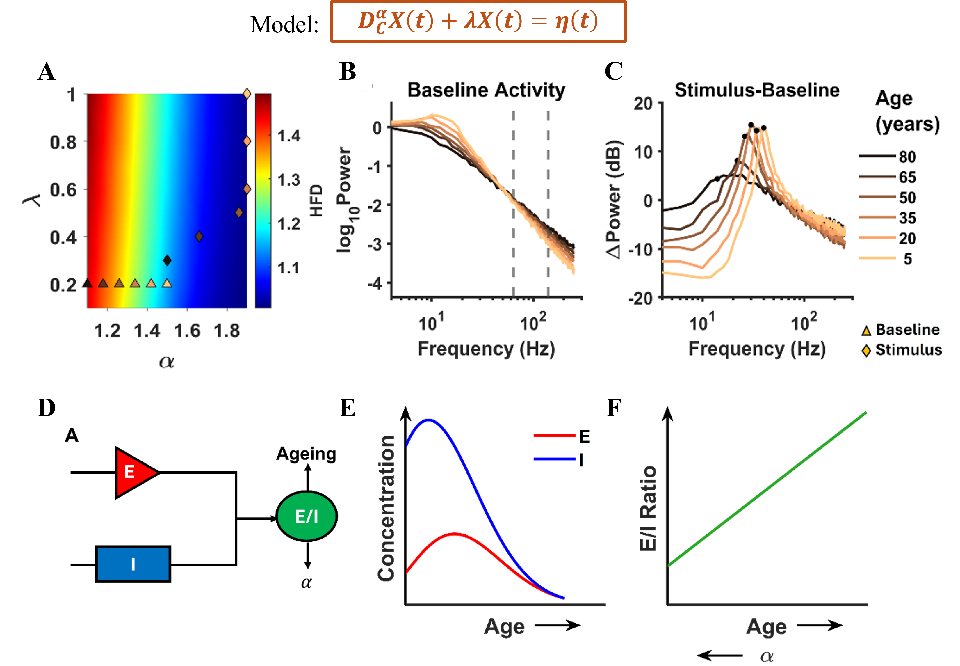
      Here, we model the neural signals via stochastic fractional harmonic oscillator (sFHO) (Fig.1 top), that captures the non-Markovian and fractal nature of the signals [1]. It intrinsically includes memory through its non-integer derivatives ‘alpha’. We use the observed monotonic decline of slope with age to get inferences about HFD from the model (Fig. 1A and B). The model explains age-related changes in power and centre frequency of stimulus-induced gamma oscillations (Fig. 1C) and resolves the conflicting findings of HFD. Moreover, using just mathematics, it predicts that in order to have a decline in EEG gamma power in old age, the gamma power should increase from childhood to adolescence.

      Results

      To understand the underlying neural mechanism, I hypothesize a relation of excitation-inhibition (E-I) ratio with the non-integer derivative and age (Fig. 1D). I show how the E-I ratio could be increasing with age monotonically (fig. 1F) despite a non-monotonic change in the individual concentration of excitation and inhibition neurotransmitters (Fig. 1E). Taking a bold step forward, I use this framework to estimate human life expectancy in existing electroencephalogram (EEG) and electrocorticogram (ECoG) datasets of healthy adults and epileptic patients as 76.9 and 69.7 years respectively that was consistent with population statistics.

      Discussion

      The present model captures the changes in PSD features from infancy to old age, in place of focusing only either on childhood-related growth or degeneration in late stages of life, thereby, providing a unified framework. It could help in constraining neural mechanisms governing ageing and has huge potential for future individual lifespan estimation and disease-risk assessment.


      Figure 1. Age-related inferences: Top: Model equation. (A) HFD variation with α and λ. (B), (C) The PSDs and ΔPower (in dB) corresponding to triangles and diamonds respectively. (D) Illustration of E/I dependence on ageing and α. (E) The concentration of excitatory and inhibitory neurotransmitters varying non-monotonically with age. (F) Corresponding monotonic E/I ratio with age and α.
      References
      1. Aggarwal, S. (2025). Decoding human lifespan from neural noise and explaining age-related changes in fractal dimension and gamma oscillations using fractional harmonic oscillator (p. 2025.09.13.675905). bioRxiv. https://doi.org/10.1101/2025.09.13.675905

      Acknowledgement

      The author expresses gratitude towards Prof. Banibrata Mukhopadhyay, Department of Physics, IISc, Prof. Supratim Ray, Centre for Neuroscience, IISc and Dr. Surya Prakash for scientific discussions and guidance that enriches the quality of this work.
       
      Speakers
      Sunday July 12, 2026 4:20pm - 6:20pm ADT
      Ballroom B2

      4:20pm ADT

      P004: Beyond Optimality: Neural Mechanisms of Heuristic Decision-Making
      Sunday July 12, 2026 4:20pm - 6:20pm ADT
      Introduction
      Humans and animals deploy diverse strategies within their ecological environments. Much of neuroscience has focused on normative frameworks in which agents optimize decisions — Bayesian inference, speed–accuracy tradeoffs, gradient-based learning [1,2]. Yet real agents frequently rely on heuristics specifically adapted to their ecology: cognitive shortcuts that save time and resources, and can outperform optimal strategies in complex environments [3]. Despite their influence in psychology and behavioral economics as fast, automatic "System 1" processes, heuristics remain underrepresented in neuroscience. How neural systems implement and switch between heuristics and deliberative strategies remains a central challenge.

      Methods
      We study heuristics in perceptual decision-making using a discrimination task where one “over-represented” stimulus evokes stronger population activity. Monkeys were shown to solve this task by relying on a simple heuristic: summing activity to detect the over-represented stimulus, instead of optimally integrating activity weighted by task relevance [3]. We implement a model neural network trained with either gradient descent or Oja learning rule under over-represented and equally-represented conditions. We then assess, as in [3], whether optimal readout or heuristic strategies were implemented using: (1) accuracy imbalance between stimuli under inactivation; (2) choice probability correlating single-neuron activity with stimulus choice.

      Results
      While networks trained with gradient descent learn optimal strategies, networks trained with the Oja rule reproduce empirical signatures of heuristics when the activity is uncentered. Using Oja, networks learn to extract the first principal component of population activity as readout weights [4]. When activity is uncentered, the mean dominates, driving Oja toward a constant readout weight vector "summation" solution. When stimuli become equally-represented, this solution fails and the network centers activity through slow integration of mean activity, yielding a non-heuristic solution consistent with theoretical and experimental predictions. Our framework provides a mechanistic model of switching between heuristic and optimal strategies.

      Discussion
      Understanding how the brain switches between fast heuristics (System 1) and deliberate cognition (System 2) has broad implications for psychology, neuroscience, and economics [5]. Our framework suggests heuristics are not mere shortcuts but ecologically rational strategies — tuned to environmental statistics and implemented through simple neural computations. This reframing has consequences for how we study decision-making across species. Beyond basic science, these insights can inform neuroAI systems that integrate rapid heuristic strategies with precise reasoning, offering a path toward more generalizable, energy-efficient models.

      References

      1. Körding, K. P., & Wolpert, D. M. (2004). Bayesian integration in sensorimotor learning. Nature, 427(6971), 244–247. https://doi.org/10.1038/nature02169
      2. Richards, B. A., & Kording, K. P. (2023). The study of plasticity has always been about gradients. The Journal of Physiology, 601(15), 3141–3149. https://doi.org/10.1113/JP282747
      3. Laamerad, P., Krause, M. R., Guitton, D., & Pack, C. C. (2025). Inactivation of primate cortex reveals inductive biases in visual learning. Current Biology, 35(19), 4699–4713.e6. https://doi.org/10.1016/j.cub.2025.08.027
      4. Oja, E. (1982). A simplified neuron model as a principal component analyzer. Journal of Mathematical Biology, 15(3), 267–273. https://doi.org/10.1007/BF00275687
      5. Kahneman, D. (2011). Thinking, fast and slow. Farrar, Straus and Giroux.


      Acknowledgement
      This work was supported by IVADO Projet Exploratoire (Explo24CO-3750823649).
      Sunday July 12, 2026 4:20pm - 6:20pm ADT
      Ballroom B2

      4:20pm ADT

      P005: A Brain-Wide Atlas of Intrinsic Neural Timescales in Mice
      Sunday July 12, 2026 4:20pm - 6:20pm ADT
      Introduction

      Intrinsic neural timescales quantify how long neurons integrate information, a fundamental metric of brain organization [1]. Yet accurate brain-wide mapping has been limited by two methodological challenges: binned autocorrelation methods underestimate timescales in low-firing neurons, and single-exponential models obscure multi-component temporal structure. We addressed both by combining iSTTC (intrinsic Spike Time Tiling Coefficient) [2] with multi-exponential modeling [3]. Applied to 89,047 neurons across 266 mouse brain regions (IBL 2025 dataset), this framework enables timescale estimation in low-firing neurons and captures multi-component temporal structure, providing broad coverage across 86% of mouse brain regions.

      Methods

      We analyzed spontaneous spiking activity from 580,598 units across 427 sessions and 131 mice in the IBL 2025 Brainwide Map Release. Units meeting quality criteria (≥100 spikes, declining autocorrelation, R^2 ≥ 0.5) were retained (89,047 units). Timescales were estimated using iSTTC, and each autocorrelation fitted with 1-4 exponential components, with optimal complexity selected by BIC. The amplitude-weighted effective timescale \u200bτ_eff captures the overall integration window of a neuron, weighted by the contribution of each exponential component. To test the anatomical gradient, we fitted a Bayesian hierarchical model with random intercepts for region, mouse, session, and probe.

      Results

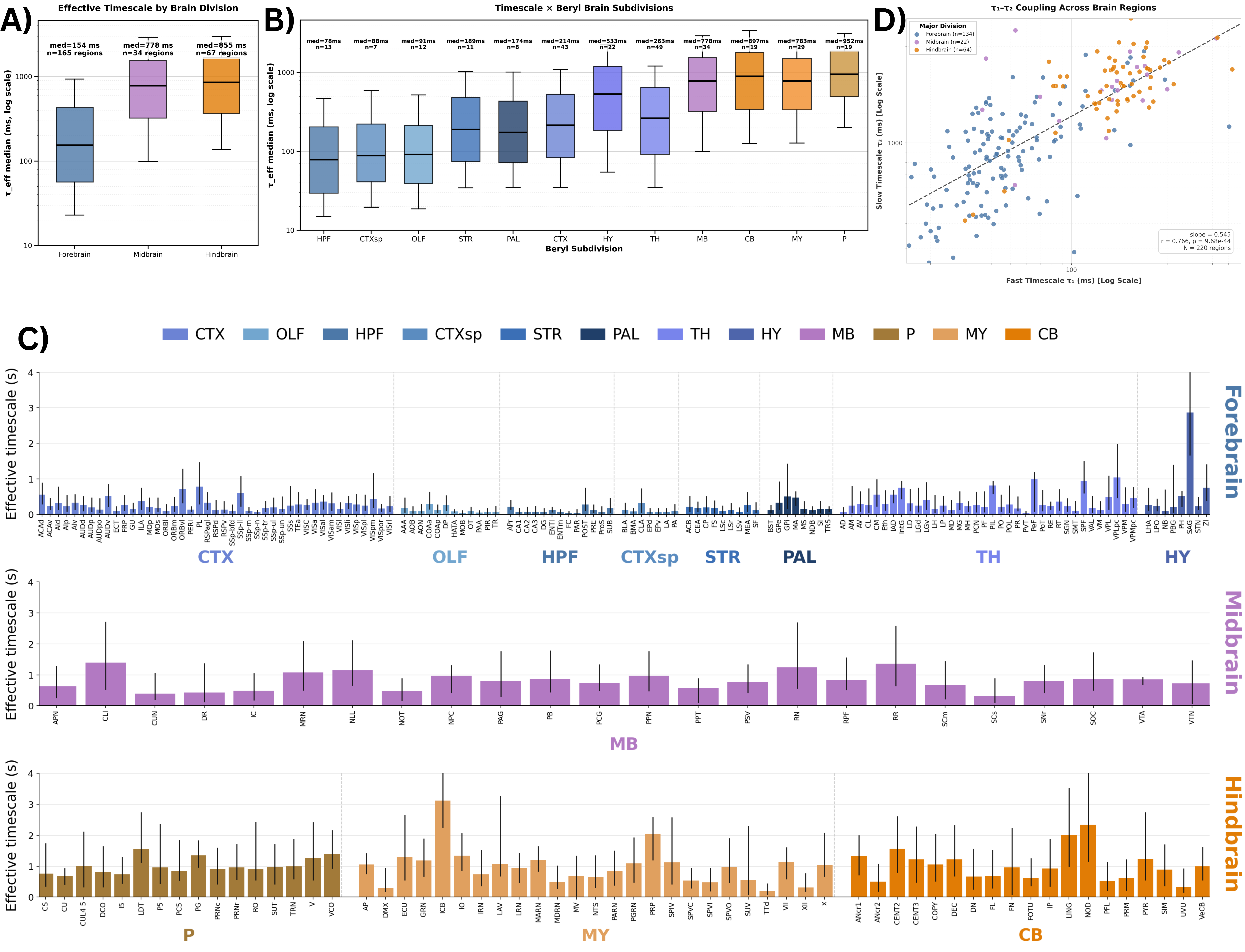
      Median τ_eff spanned nearly two orders of magnitude across regions (37.9-3,115 ms), following a rostro-caudal gradient: forebrain 213 ms (IQR = 120-304 ms), midbrain 765 ms (IQR = 482-968 ms), hindbrain 956 ms (IQR = 723-1183 ms). This gradient was observed in 100% of individual mice (median Spearman ρ = 0.76, p < 10⁻³⁰). A Bayesian hierarchical model controlling for mouse, session, and probe confirmed brain region as the dominant variance source (24.1%), with substantial within-region heterogeneity remaining (median IQR = 588 ms). 73.9% of neurons required multi-component models: τ₂ co-varied sublinearly with τ₁ across regions (r = 0.766, p = 9.68 × 10⁻⁴⁴, slope = 0.545).

      Discussion

      Anatomical position along the rostro-caudal axis is a strong organisational principle of intrinsic neural timescales, holding robustly across 220 regions. The longer timescales of midbrain and hindbrain may reflect their roles in integrating homeostatic, motor, and state-related signals over extended periods. The majority of neurons (73.9%) are better described by multiple timescale components; τ₁ may reflect intrinsic neuronal properties, while τ₂, extending to several seconds, points to recurrent network interactions or neuromodulation. Within-region variance (median IQR=588 ms) likely reflects cell type, laminar position, and local connectivity, motivating future integration with anatomical cell-type data.

      Brain-wide map of intrinsic neural timescales. (A) τ_eff by major brain division. (B) τ_eff across 12 brain subdivisions. (C) Median τ_eff per region; error bars show 10th–90th percentiles. Regions grouped by division, ordered alphabetically; colors denote subdivision. (D) Fast (τ₁) vs slow (τ₂) timescales across regions.
      References

      1. Murray, J. D., et al. (2014). A hierarchy of intrinsic timescales across the primate cortex. Nat Neurosci, 17(12), 1661-1663. https://doi.org/10.1038/nn.3862

      2. Pochinok, I., Hanganu-Opatz, I. L., & Chini, M. (2026). iSTTC: A robust method for accurate estimation of intrinsic neural timescales from single-unit recordings. PLOS Comput Biol, 22, e1013385. https://doi.org/10.1371/journal.pcbi.1013385

      3. Shi, Y. L., et al. (2025). Brain-wide organization of intrinsic timescales at single-neuron resolution. bioRxiv. https://doi.org/10.1101/2025.08.30.673281

      4. Beiran, M., & Ostojic, S. (2019). Contrasting the effects of adaptation and synaptic filtering on the timescales of dynamics in recurrent networks. PLOS Comput Biol, 15(11), e1007462. https://doi.org/10.1371/journal.pcbi.1007462


      Acknowledgement
      Supported by Neuromatch Impact Scholars Program. Data from International Brain Laboratory 2025 Brainwide Map Release. We thank the IBL consortium for open data access and Jason Manley for supervision. Analysis builds on iSTTC methodology (Pochinok et al. 2026) and multi-exponential fitting framework (Shi et al. 2025).

      Sunday July 12, 2026 4:20pm - 6:20pm ADT
      Ballroom B2

      4:20pm ADT

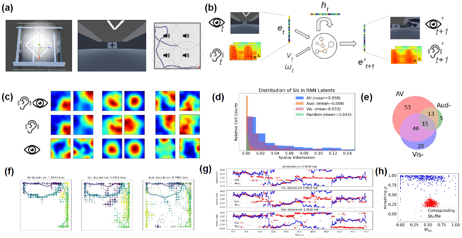
      P006: Disentangling Sensory Drivers of Spatial Codes with Recurrent Audiovisual Models in VR
      Sunday July 12, 2026 4:20pm - 6:20pm ADT
      Introduction
      Spatial navigation relies on integrating multimodal cues[1], yet both in vivo and in silico hippocampal research overwhelmingly focuses on vision [2,3,6]. While recent work showed mice entorhinal cortex contain both unimodal and multimodal cells[3], how different modalities are weighted and integrated remains poorly understood. We develop a modelling pipeline with an agent traversing a multimodal VR environment. A recurrent neural network was trained to perform a next-state prediction task[2]. We hypothesised that the network would develop place cell-like units that utilise both modalities, and that integrating modalities would result in more robust spatial representations, with each sense contributing additively to the cognitive map.

      Methods
      An agent traversed a 2D arena with audiovisual cues in Unity3D[4]. Binaural audio broadcasted with head-related transfer functions[5] and visual frames were encoded via autoencoders into low-dimensional embeddings. We trained an RNN[2] to perform next-state prediction using its current sensory states and motion, under three conditions: audiovisual, visually-lesioned and auditorily-lesioned. Latent units were classified into place units using empirical metrics such as spatial information scores. These tunings were used to perform maximum-likelihood decoding of the agent’s position and orientation. The relative contributions of sensory inputs were effectively decomposed using a linearly weighted combination of their unimodal responses.

      Results
      The audiovisual model produced 127 spatially tuned place cells, significantly more than visually-driven (33 cells) and auditorily-driven (81 cells) ones. Furthermore, the audiovisual model yielded the lowest trajectory decoding error (0.151 m) compared to visual-only (0.879 m) and auditory-only (0.293 m) ones, with the highest spatial information content. Unimodal units that respond to a single modality were identified, as well as multimodal units that remap when both are present. Finally, by approximating multimodal ratemaps as linear combinations of unimodal maps, we found that most place cells integrate modalities additively, exhibiting intermediate visual weightings (μ=0.405) and relying more on auditory cues.

      Discussion
      While derived in silico, these results offer a framework for biological navigation. The model suggests the hippocampus may additively processes multisensory streams to reduce uncertainty rather than switching between senses. Notably, auditory cues proved dominant in our VR setup, likely because visual landmarks lose salience at a distance or vanish when facing walls. Consequently, multimodal units anchor to the most reliable available cues—in this case, sound. We further hypothesise these units will dynamically remap or reweigh sensory reliance if a primary modality degrades. Ultimately, this model provides a normative theory for multisensory integration, generating precise, testable predictions for planned in vivo ferret recordings.

      (a) Virtual environment with visual cues and sound sources; (b) Model architecture and pipeline; (c) Spatial ratemap examples in audiovisual, and lesioned (-) conditions; (d) Distribution of spatial information contents; (e) Number of place units identified; (f) ML decoding of position; (g) ML decoding of head direction; (h) Distribution of visual weights (x) and resulting correlations (y); compar
      References

      Jeffery, K. J. (2007). Integration of the sensory inputs to place cells: what, where, why, and how?. Hippocampus
      [1] Levenstein, D., ..., & Richards, B. (2024). Sequential predictive learning is a unifying theory for hippocampal representation and replay. bioRxiv
      [2] Nguyen, D., ... , & Gu, Y. (2024). The medial entorhinal cortex encodes multisensory spatial information. Cell reports
      [3] George, T. M., ..., & Barry, C. (2024). RatInABox, a toolkit for modelling locomotion and neuronal activity in continuous environments. Elife
      [4] Cuevas-Rodríguez, ... & Reyes-Lecuona, A. (2019). 3D Tune-In Toolkit: An open-source library for real-time binaural spatialisation. PloS one
      [5] Banino, A., ... & Kumaran, D. (2018). Vector-based navigation using grid-like representations in artificial agents. Nature


      Acknowledgement
      We thank Barry Lab, Bizley La, the Department of UCL Cell and Developmental Biology, the Ear Institute for this work.
      This work was supported by the UKRI Biotechnology and Biological Sciences Research Council [grant
      number BB/T008709/1].
      Sunday July 12, 2026 4:20pm - 6:20pm ADT
      Ballroom B2

      4:20pm ADT

      P007: Spatial attention shapes the pupillary light response
      Sunday July 12, 2026 4:20pm - 6:20pm ADT
      Introduction
      Traditionally, the pupillary light response is viewed as a global reflex stabilizing retinal illumination by integrating luminance across the visual field [1]. However, recent work suggests pupil responses are also modulated by spatial mechanisms linked to attention and eye movement planning. When global luminance is held constant, directing attention to brighter regions produces stronger constriction, indicating location-specific luminance weighting [2]. Moreover, the pupil can begin adjusting to upcoming saccade target luminance before gaze shifts, suggesting anticipatory modulation linked to presaccadic attention [3]. Together, these findings suggest pupil dynamics reflect both global and gaze-dependent local luminance signals.


      Methods
      51 participants (ages 20-25) were recorded with an EyeLink-1000 eye-tracker while freely viewing 10 naturalistic movies. First, luminance was extracted at the pixel level frame-by-frame using a photometric calibration. Analysis 1: gaze-contingent retinal luminance was mapped onto a 1°×1° spatial grid and regularized regression was used to estimate spatial luminance sensitivity of the pupil across the visual field. Analysis 2: intersaccadic intervals (ISI) between saccades were identified and linear mixed-effects models tested whether late-ISI pupil diameter predicted upcoming saccade goal luminance, controlling for current fixation, opposite-direction (control location), and global luminance.


      Results

      Discussion
      These findings advance understanding of how pupil dynamics encode spatial and temporal visual information during natural viewing. Central weighting (Analysis 1) likely reflects attentional allocation. The anticipatory effect at upcoming saccade targets (Analysis 2) suggests presaccadic attention modulates pupillary responses before gaze arrival, consistent with oculomotor structures such as the superior colliculus and frontal eye fields influencing pupil control [4]. Limitations include use of instantaneous pupil measurements without accounting for pupillomotor delay (~200-300ms) in analysis 2. Ongoing work aims to incorporate temporal lags in analysis 2 and test generalizability across diverse tasks.


      References
      1. Watson, A. B., & Yellott, J. I. (2012). A unified formula for light-adapted pupil size. Journal of vision, 12(10), 12. https://doi.org/10.1167/12.10.12
      2. Binda, P., & Murray, S. O. (2015). Spatial attention increases the pupillary response to light changes. Journal of vision, 15(2), 1. https://doi.org/10.1167/15.2.1
      3. Mathôt, S., van der Linden, L., Grainger, J., & Vitu, F. (2015). The pupillary light response reflects eye-movement preparation. Journal of experimental psychology. Human perception and performance, 41(1), 28–35. https://doi.org/10.1037/a0038653
      4. C. Wang, & D.P. Munoz, Neural basis of location-specific pupil luminance modulation, Proc. Natl. Acad. Sci. 115 (41), 10446-10451, https://doi.org/10.1073/pnas.1809668115


      Acknowledgement
      The research was undertaken thanks in part to funding from the Connected Minds Program, supported by Canada First Research Excellence Fund, Grant #CFREF-2022-00010, and the Natural Sciences and Engineering Research Council of Canada (NSERC).

      Sunday July 12, 2026 4:20pm - 6:20pm ADT
      Ballroom B2

      4:20pm ADT

      P008: Movement representations for classification and perception: posture dominates over dynamics
      Sunday July 12, 2026 4:20pm - 6:20pm ADT
      Introduction
      Motion perception is the remarkable ability of the visual system to recognize complex human movements effortlessly. Computational movement analysis seeks representations that mirror this efficiency while remaining interpretable. The motor modularity hypothesis proposes that movements are composed of weighted primitives [1], yet whether theory-driven decompositions outperform alternative approaches remains untested. We compared Temporal Movement Primitives (TMPs), Legendre polynomial coefficients, and autoencoder embeddings to ask which movement features enable motion classification and assess if the chosen movement features align with how human observers discriminate actions, paralleling perceptual research on form versus motion cues [2].


      Methods
      We analyzed videos of 16 daily activities from the MoVi dataset [3] and extracted joint-angle trajectories using MMPose, with segmentation via visual inspection. We represented these trajectories in three ways: 1- TMP weights from Bayesian decomposition with Gaussian process priors for varying numbers of primitives (1- 20); 2- Legendre polynomial coefficients for varying maximum degrees (0–10); 3- latent vectors from an encoder-decoder network. To determine which features drive discrimination, we assessed cross-validated classification accuracy, optimal primitive count and polynomial degree, reconstruction quality, and interpretability. To isolate dynamics from posture, we repeated analyses after subtracting mean joint positions per trial.


      Results
      Legendre coefficients achieved 96% cross-validated classification accuracy across 16 classes, outperforming TMP weights (91%) and autoencoder features (85%). Optimal TMP count was 5 primitives, and the optimal Legendre degree was 0, revealing postural configuration, not temporal dynamics, is the primary discriminative feature. After posture removal, degree-2 polynomials captured remaining discriminative dynamics. Classification and movement generation dissociated: when generating movements from averaged category weights, TMPs preserved dynamics, producing natural motion, whereas Legendre-generated movements retained original posture but with unclear motion. L1 regularization identified 10 joints carrying the most discriminative information.


      Discussion
      Posture dominance for activity classification aligns with biological motion perception, showing form cues often suffice for action-type recognition. The dissociation between classification and generation shows discriminative and generative adequacy are distinct properties: Legendre coefficients excel at categorization, TMPs preserve temporal structure for synthesis, and autoencoders achieve optimal dimensionality reduction from 240 (5 primitives × 48 coordinates) to 32. Beyond classification, these results reveal that movement is organized into separable postural and dynamic components, opening avenues to explore minimum temporal duration for motion perception, whether partial cycles suffice, and how accuracy scales with available dynamics.


      References
      1. Knopp, B., Velychko, D., Dreibrodt, J., & Endres, D. (2019b). Predicting perceived naturalness of human animations based on generative movement primitive models. ACM Transactions on Applied Perception, 16(3), 1–18. https://doi.org/10.1145/3355401
      2. Lange, J., & Lappe, M. (2006). A Model of Biological Motion Perception from Configural Form Cues. Journal of Neuroscience, 26(11), 2894–2906. https://doi.org/10.1523/jneurosci.4915-05.2006
      3. Ghorbani, S., Mahdaviani, K., Thaler, A., Kording, K., Cook, D. J., Blohm, G., & Troje, N. F. (2021). MoVi: A large multi-purpose human motion and video dataset. PLoS ONE, 16(6), e0253157. https://doi.org/10.1371/journal.pone.0253157 


      Acknowledgement
      This work was supported by the Natural Sciences and Engineering Research Council (NSERC) of Canada. The research was undertaken thanks in part to funding from the Connected Minds Program, supported by Canada First Research Excellence Fund, Grant #CFREF-2022-00010. Also, we thank the creators of the MoVi dataset for making their data publicly available.

      Sunday July 12, 2026 4:20pm - 6:20pm ADT
      Ballroom B2

      4:20pm ADT

      P009: Optimal Self-Organization in Mean-Field Multi-Agent Neuronal Networks
      Sunday July 12, 2026 4:20pm - 6:20pm ADT
      Introduction
      Neuronal networks (NNs) are predominantly modeled as dynamical systems, requiring ad hoc analyses to explore the interplay between population codes (PCs) and computation [3,4]. By viewing PCs as distributions over the NN state space [2] we develop a framework for modeling emergent computation through the lens of multi-agent (MA) optimal control (OC). In it, neurons regulate their local parameters to collectively control the PC and (in turn) optimize a cost function. A mean-field (MF) limit is derived for a large class of NNs, whence we prove theorems establishing global optima as laws of self-organization. Such models are built on a rich mathematical literature, with great potential for further theoretical results and learning algorithms.

      Methods
      Mean-field game/control theory is an analytical tool for characterizing/learning optimal decision-making in complex strategic systems. MF limits are useful because they ‘average out’ microscale fluctutations to distill macroscale behavior, dramatically simplifying controls while providing powerful approximations for large, finite populations. However, traditional MF theories fail for general networks. We resolve this limitation and maximize the class of compatible models/cost functions while preserving a detailed theoretical characterization. The setup in [1] closely resembles what we propose, but lacks control. Moreover, the MF is taken to model a PC by characterizing the NN up to each neuron’s identity.

      Results
      For a large class of computational tasks where neurons contribute to the PC anonymously, we characterize OCs without any a priori restriction on their structure or the information available to each neuron. Specifically, under an OC: (i) neurons are decentralized (acting independently given their local state and each subpopulation’s MF) such that the emergent parameter regime is realized through a process of self-organization, and (ii) neurons of the same species deploy an identical strategy, reinforcing its biological plausibility; neurons of the same type will behave identically under fixed conditions. As a concrete example, we construct a multi-population Hodgkin Huxley network designed to express a binary decision via its MF PC.

      Discussion
      Reformulating NNs as MF MA systems unlocks a wealth of analytical tools, learning algorithms and theoretical guarantees ripe for neuroscience applications. This work is a crucial first step in bridging the gap. Our technical results are complemented by the conjecture that PCs are expressed through the MF, which in turn emerges from optimal laws of self-organization at the microscale. Unlike many cost function-based NNs, these are global optima, affording greater normative potential. In exchange, models require target computations to be expressed as explicit cost functions assessing the PC directly, which has received little attention to-date. Future work will focus on developing this aspect further, along with simulation/learning studies.

      References
      1. Baladron, J., Fasoli, D., Faugeras, O., & Touboul, J. (2012). Mean-field description and propagation of chaos in networks of hodgkin-huxley and fitzhugh-nagumo neurons. Journal of Mathematical Neuroscience, 2, 10. doi: 10.1186/2190-8567-2-10
      2. Beck, J. M., Latham, P. E., & Pouget, A. (2011). Marginalization in neural circuits with divisive normalization. The Journal of Neuroscience, 31(43), 15310–15319. doi: 10.1523/JNEUROSCI.1706-11.2011
      3. Denève, S., & Machens, C. K. (2016). Efficient codes and balanced networks. Nature Neuroscience, 19(3), 375–382. doi: 10.1038/nn.4243
      4. Wong, K.-F., & Wang, X.-J. (2006). A recurrent network mechanism of time integration in perceptual decisions. The Journal of Neuroscience, 26(4), 1314–1328. doi: 10.1523/JNEUROSCI.3733-05.2006

      Acknowledgement
      We acknowledge the support of the Natural Sciences and Engineering Research Council of Canada (NSERC).
      \nNous remercions le Conseil de recherches en sciences naturelles et en génie du Canada (CRSNG) de son soutien.
      Sunday July 12, 2026 4:20pm - 6:20pm ADT
      Ballroom B2

      4:20pm ADT

      P010: Computing Motor Error with Inhibition: A Minimal Excitatory-Inhibitory Circuit Motif
      Sunday July 12, 2026 4:20pm - 6:20pm ADT
      Introduction
      Motor control requires continuous comparison between desired and actual states, yet how error signals are computed at the cellular level is not well understood. Optimal Feedback Control theory presents what computations the brain might perform during movement but not how neurons implement them [1,2]. Inhibition in sensorimotor cortex is typically framed as maintaining excitatory-inhibitory balance, shaping activity patterns, or providing gain control [3], not as computing error signals. We propose a minimal excitatory-inhibitory (E-I) circuit motif in which inhibition implements subtraction, providing a mechanistic account of error computation.


      Methods

      Results
      Individual E-I pairs produce rectified subtraction and track sinusoidal inputs up to 5 Hz with a gain above 0.5 before attenuating at higher frequencies, comfortably exceeding the approximately 2.4 Hz bandwidth imposed by muscle-tendon dynamics [4]. At the population level, linear decoders achieve R-squared greater than 0.85 for position error and velocity during centre-out reaching tasks.


      Discussion

      References
      [1] Scott, S. H. (2004). Optimal feedback control and the neural basis of volitional motor control. Nat Rev Neurosci, 5(7), 532-546.
      [2] Todorov, E. & Jordan, M. I. (2002). Optimal feedback control as a theory of motor coordination. Nat Neurosci, 5(11), 1226-1235.
      [3] Isaacson, J. S. & Scanziani, M. (2011). How inhibition shapes cortical activity. Neuron, 72(2), 231-243.
      [4] Crevecoeur, F. & Scott, S. H. (2014). Beyond Muscles Stiffness: Importance of State-Estimation to Account for Very Fast Motor Corrections. PLoS Comput Biol, 10(10), e1003869.


      Acknowledgement
      This work was supported by a Natural Sciences and Engineering Research Council of Canada (NSERC) Discovery Grant to G. Blohm.

      Sunday July 12, 2026 4:20pm - 6:20pm ADT
      Ballroom B2

      4:20pm ADT

      P011: Dendrites Learn to Detect Input Sequences Through Local Plasticity
      Sunday July 12, 2026 4:20pm - 6:20pm ADT
      Introduction

      Neurons can encode information not only by which cells fire, but also by the order in which they fire: n active neurons can represent n! sequences. Hippocampal place cells fire in such sequences during navigation and replay these sequences during rest. During replay, synapses can be activated sequentially from tip to soma along dendrites [1]. A dendrite can selectively advance depolarization in response to tip-to-soma inputs, making it sequence selective [2]. But this selectivity operates within a limited range of AMPA conductance that changes with synaptic spacing, so we ask whether local plasticity can tune these conductances. Our plasticity rule may offer a mechanism for the emergence of plateaus like those observed in BTSP [3].


      Methods
      Voltage-dependent NMDA and KIR channels make each dendritic segment bistable: stable at rest and at plateau [4]. A synaptic input briefly opens AMPA channels, depolarizing the segment and potentially transitioning it from rest to plateau. For plateau advancement to be sequence selective, AMPA conductance must be just strong enough that an input transitions a segment from rest to plateau only with the support of depolarizing current from its plateauing tip-side neighbor. We therefore strengthen AMPA conductance when a segment fails to plateau despite its tip-side neighbor plateauing, and weaken AMPA conductance when it plateaus despite its tip-side neighbor resting. We tested whether this rule operates across different synaptic spacings.

      Results
      Repeated tip-to-soma and shuffled input presentations robustly tune each segment’s AMPA conductance into its sequence-selective range. Starting from weak AMPA conductances, analogous to AMPA-silent synapses, inputs initially fail to advance the plateau. Under our plasticity rule, the AMPA conductance of the segment adjacent to the established plateau increases until the segment begins plateauing, after which tuning progresses to the next segment (Fig. 1A). Thus, segments are tuned sequentially from tip to soma, and the number of presentations increases linearly. Because each synapse independently converges on the conductance range it requires, the rule remains effective across heterogeneous synaptic spacings.


      Discussion
      Because plateau initiation occurs at a bifurcation, gradual AMPA tuning produces an abrupt transition: a segment initially fails to plateau, then suddenly starts plateauing once AMPA conductance enters the sequence-selective range (Fig. 1B). This sudden plateauing may offer a mechanistic interpretation of BTSP, in which plateaus emerge abruptly after repeated trials: during the initial trials, dendritic plateaus may remain local, but after tuning is complete they may propagate to the soma. Glutamate uncaging experiments could test whether repeated tip-to-soma stimulation selectively strengthens AMPA conductances from tip to soma. Future work should identify a local signal reporting whether the tip-side neighbor plateaued.

      Local AMPA plasticity tunes dendritic sequence selectivity. Repeated tip-to-soma and shuffled inputs progressively strengthen AMPA conductance from tip to soma (A). When a segment (green) receives input while its tip-side neighbor is plateauing (yellow), weak AMPA initially fails to trigger a plateau. As AMPA increases, the segment abruptly transitions to plateau at a bifurcation (B).References
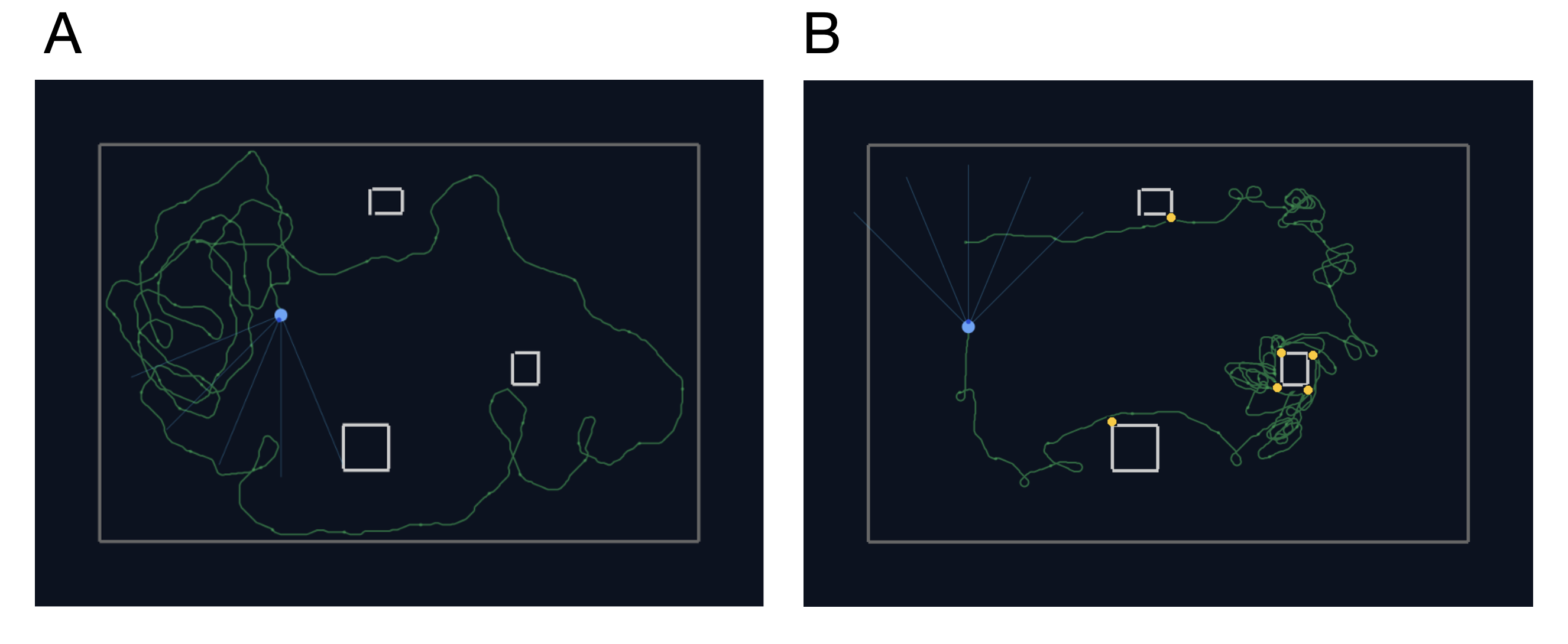
      1. Ishikawa, T., & Ikegaya, Y. (2020). Locally sequential synaptic reactivation during hippocampal ripples. Science Advances, 6(7), Article eaay1492. https://doi.org/10.1126/sciadv.aay1492
      2. Boahen, K. (2022). Dendrocentric learning for synthetic intelligence. Nature, 612(7938), 43–50. https://doi.org/10.1038/s41586-022-05340-6
      3. Bittner, K. C., Milstein, A. D., Grienberger, C., Romani, S., & Magee, J. C. (2017). Behavioral time scale synaptic plasticity underlies CA1 place fields. Science, 357(6355), 1033–1036. https://doi.org/10.1126/science.aan3846
      4. Sanders, H., Berends, M., Major, G., Goldman, M. S., & Lisman, J. E. (2013). NMDA and GABAB (KIR) conductances: The “perfect couple” for bistability. The Journal of Neuroscience, 33(2), 424–429. https://doi.org/10.1523/JNEUROSCI.1854-12.2013


      Acknowledgement

      This study was supported by the Stanford Bioengineering Department and Masason Foundation. 


      Sunday July 12, 2026 4:20pm - 6:20pm ADT
      Ballroom B2

      4:20pm ADT

      P012: Integrating Neuroimaging and Omics to Characterize Parkinson’s Disease Progression
      Sunday July 12, 2026 4:20pm - 6:20pm ADT
      Introduction
      Parkinson's disease (PD) is a progressive neurodegenerative disorder characterized by dopaminergic neuronal loss and heterogeneous clinical trajectories with motor and non-motor symptoms [1]. Predicting disease progression requires biomarkers capturing both brain alterations and underlying molecular mechanisms. Neuroimaging reveals structural brain changes [2], while transcriptomic and proteomic analyses characterize molecular processes involved in disease pathology [3]. However, these modalities are often studied separately. Here we integrated neuroimaging and omics data to investigate how molecular signatures relate to brain alterations during PD progression.


      Methods
      We analyzed data from the Parkinson’s Progression Markers Initiative (PPMI) [4], a longitudinal study including de novo PD patients, prodromal individuals, and healthy controls. Subjects were selected if both brain imaging and molecular measurements were available, including cerebrospinal fluid proteomics or whole-blood RNA-seq data collected at different disease stages. Differential gene and protein expression analyses were performed, followed by functional enrichment analysis. In parallel, quantitative features such as contrast ratios and volumes of neuromelanin-rich areas were extracted from 2D gradient echo (GRE) brain images with magnetization transfer (MT). Associations between omics and imaging-derived features were then evaluated.

      Results
      A subset of genes and proteins showed significant associations with imaging features reflecting brain alterations typically observed in Parkinson’s disease. These findings suggest that disease progression is reflected by both molecular and imaging signatures. Further analysis revealed that progression does not follow a simple linear trajectory but instead involves distinct stages. Moreover, patterns of molecular and structural changes differed between sexes, highlighting heterogeneity in disease progression and suggesting potential sex-specific mechanisms.

      Discussion
      These findings highlight the value of integrating imaging and omics data to better characterize PD progression. Linking molecular signatures with structural brain alterations may improve our understanding of disease mechanisms and support the development of multimodal biomarkers for monitoring disease evolution.


      References
      [1]: Kalia, L. V., & Lang, A. E. (2015). Parkinson’s disease. The Lancet, 386(9996), 896–912.
      [2]: He, H., et al. (2020). Progressive brain changes in Parkinson’s disease: A meta-analysis of structural magnetic resonance imaging studies. Brain Research Bulletin, 164, 272–279.
      [3]: Sharma, S., & Dhamija, R. K. (2025). The quest for Parkinson’s disease biomarkers: Traditional and emerging multi-omics approaches. Molecular Biology Reports, 52, Article 831.
      [4]: Parkinson’s Progression Markers Initiative (PPMI). https://www.ppmi-info.org

      Acknowledgement
      Data were obtained from the Parkinson’s Progression Markers Initiative (PPMI) database (www.ppmi-info.org). PPMI – a public-private partnership – is funded by the Michael J. Fox Foundation for Parkinson’s Research and funding partners.

      Sunday July 12, 2026 4:20pm - 6:20pm ADT
      Ballroom B2

      4:20pm ADT

      P013: Relationships Between Connectivity and Longitudinal Memory Decline Using Network Analysis and Partial Least Squares
      Sunday July 12, 2026 4:20pm - 6:20pm ADT
      Introduction

      There has been recent evidence to suggest that autistic adults have higher risk of neurodegenerative disease and dementia compared to non-autistic adults [1]. Previous studies have found some age-related brain differences between autistic and non-autistic adults, but how these differences may contribute to increased dementia risk remain unclear. We used a combination of network analysis and partial least squares to identify functional and structural connectivity patterns that correlate with long-term memory decline in both autistic (n=40) and non-autistic (n=33) adults using data from a longitudinal study.


      Methods

      We obtained T1, diffusion, and functional MRI data. Brain networks with 96 regions of interest were constructed using the CONN [2] and TVB-UKBB [3] pipelines for functional and structural connectivity from participants’ first scan. Long-term memory change was measured using the slope of a mixed effects model for the delayed recall (A7) score of the Auditory Verbal Learning Test evaluated 2-5 times across 2-9 years of follow-up. Networks were thresholded and quantified with the Brain Connectivity Toolbox in MATLAB [4]. Network measures were residualized using age and sex as covariates. Behavioral partial least squares was used to identify multivariate correlations between network measures and long-term memory outcomes [5].


      Results

      For the non-autistic adults, there was a significant latent variable relationship between structural connectivity and long-term memory change; weaker structural connectivity in classic memory regions (e.g. hippocampus) correlated with greater memory decline. The autistic adults showed significant latent variable relationships between functional connectivity and long-term memory change; weaker, less interconnected, and less organized functional connectivity across the whole brain correlated with greater memory decline. For both cases, there was a significant difference between groups for the latent variable relationship, demonstrating differing relationships between connectivity and memory.


      Discussion
      Both the autistic and non-autistic adults showed significant relationships between connectivity and memory decline. We found that, for the non-autistic adults, memory decline was related to structural connectivity patterns that involved classic memory regions (e.g. hippocampus). For the autistic adults, memory decline was related to functional connectivity patterns across the whole brain. These findings suggest the potential for unique MRI based biomarkers to identify increased risk of accelerated memory decline in autistic adults.


      References

      1. Starkstein, S., Gellar, S., Parlier, M., Payne, L., & Piven, J. (2015). High rates of parkinsonism in adults with autism. J. Neurodev. Disord.
      2. Whitfield-Gabrieli, S., & Nieto-Castanon, A. (2012). Conn: a functional connectivity toolbox for correlated and anticorrelated brain networks. Brain connectivity.
      3. Frazier-Logue, N., Wang, J., Wang, Z., Sodums, D., Khosla, A., Samson, A. D., ... & Shen, K. (2022). A robust modular automated neuroimaging pipeline for model inputs to TheVirtualBrain. Front. Neuroinform.
      4. Rubinov, M., & Sporns, O. (2010). Complex network measures of brain connectivity: uses and interpretations. Neuroimage.
      5. Krishnan, A., Williams, L. J., McIntosh, A. R., & Abdi, H. (2011). Partial Least Squares (PLS) methods for neuroimaging: a tutorial and review. Neuroimage.


      Acknowledgement
      We would like to acknowledge funding sources for our project, the National Institute on Aging [P30 AG072980], the National Institute of Mental Health [R01MH132746; K01MH116098], the Department of Defense [AR140105], and the Arizona Biomedical Research Commission [ADHS16-162413].

      Sunday July 12, 2026 4:20pm - 6:20pm ADT
      Ballroom B2

      4:20pm ADT

      P014: Modeling box jellyfish obstacle avoidance behavior with evolutionary optimization and small feedforward neural networks
      Sunday July 12, 2026 4:20pm - 6:20pm ADT
      Introduction
      Box jellyfish (Tripedalia cystophora) are animals without a centralized brain [1]. Despite their small decentralized nervous system, they can perform visually guided obstacle avoidance behavior (OAB) [2], which is crucial for survival in their natural habitat. Recent work has shown that box jellyfish are even capable of associative learning and identified the learning center to be the rhopalial nervous system (RNS) [2]. These abilities raise the question which level of neural complexity is required to perform such actions. Here we investigate the innate OAB with a minimal sensorimotor architecture including a multilayer perceptron (MLP) optimized with a biologically plausible learning algorithm not including gradient descent.


      Methods
      We developed a rectangular two-dimensional simulation platform containing walls and obstacles with varying luminance values. Agents, each steered by an MLP, receive these values by a vector depending on the directional visual sensors (Fig.1) along with a physical sensor indicating a previous collision. Inputs are fed into the MLP to make the movement decision, resulting in a movement trajectory. Agents are rewarded when randomly placed food items are retrieved and penalized for collisions, resulting in a fitness value. Weights of the MLP are optimized by evolutionary search using fitness, following a neuroevolutionary paradigm used for autonomous navigation and neural control systems [3,4]. Agents are then tested in different environments. 


      Results
      In different runs with various environmental and fitness conditions, agents consistently developed OAB strategies by trying to minimize collisions while continuing to forage (Fig. 1). We study the quality of OAB when training parameters, including training time and training arenas, are varied and find that agents show best behavior for an intermediate amount of training time and in arenas where strong contrasts between wall elements where present. Overall, trajectories showed qualitative and quantitative similarities to the innate behavior of true box jellyfish. In particular, learning to avoid high-contrast objects does not lead to avoidance of objects with uniform luminosity irrespective of their distance [2].


      Discussion
      Our results show that evolutionary training enables small MLPs to successfully control OAB in agents mimicking box jellyfish. Whereas MLPs are feedforward neural networks, the biological RNS is a recurrent neural network [1], and therefore, future work will integrate more biologically plausible neural network architectures. Furthermore, in a next step, we will examine how the successfully learned innate OAB leads to associative learning by using our highly customizable setup in circular arenas with differing wall contrasts, similar to the experiments performed in [2]. Ultimately, our results will enable us to derive minimal requirements for neural architectures underlying associative learning, allowing for comparisons across organisms [5].

      Example trajectories (green) of an agent (light blue circle with blue rays indicating visual sensors) in an arena with three high-contrast obstacles. A: The agent forages where no obstacles are present. It also frequents the part of the arena with obstacles, but never collides with them. B: Trajectory for an agent with less training times, leading to frequent collisions (yellow dots).References
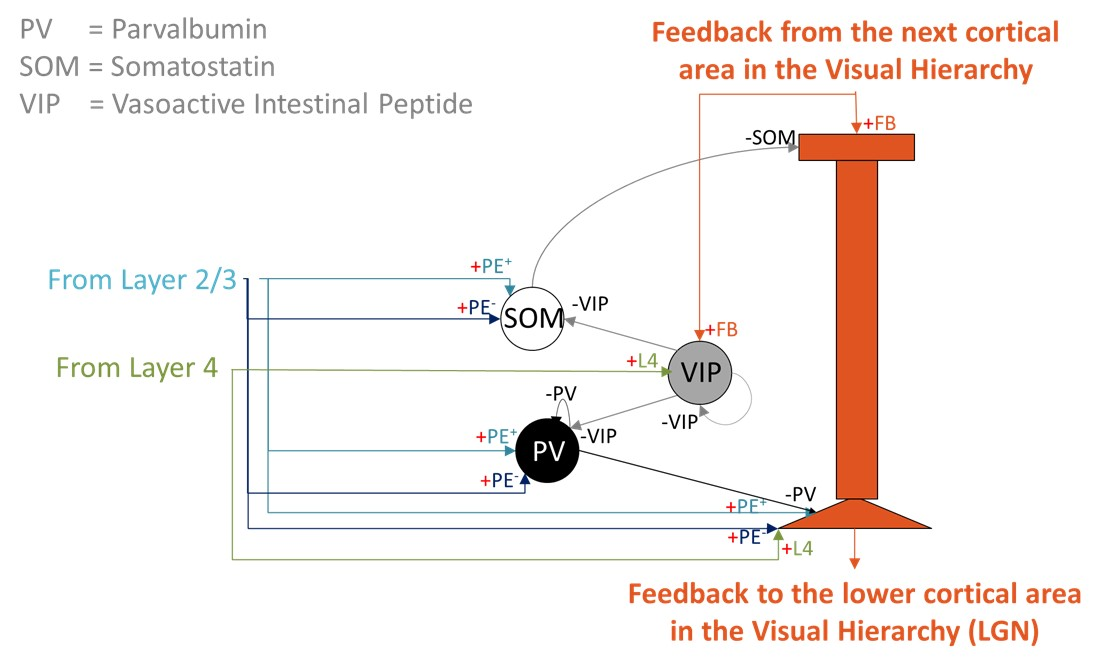
      1. Nielsen, Sofie K.D. et al. (2021). Journal of Comparative Neurology. https://doi.org/10.1002/cne.25148
      2. Bielecki, J. et al. (2023). Curr. Biol. https://doi.org/10.1016/j.cub.2023.08.056
      3. Floreano, D., & Mondada, F. (1998). Neural Networks. https://doi.org/10.1016/S0893-6080(98)00082-3 

      4. Whitley, D. et al. (1993). Machine Learning. https://doi.org/10.1023/A:1022674030396
      5. Zhou, Baohua et al. (2022). elife.  https://doi.org/10.7554/eLife.72067


      Acknowledgement
      We would like to thank Christoph Speckgens and Hermann Kohlstedt for helpful discussions. Funded by the Deutsche Forschungsgemeinschaft (DFG, German Research Foundation) – Project-ID 434434223 – SFB 1461. 
      Speakers
      avatar for Wilhelm Braun

      Wilhelm Braun

      Junior Research Group Leader, Kiel University (CAU Kiel), Faculty of Engineering, Department of Electrical and Information Engineering
      Early nervous systems, functional neuronal networks, stochastic neural dynamics, animal behavior, reinforcement learning, network reconstruction
      Sunday July 12, 2026 4:20pm - 6:20pm ADT
      Ballroom B2

      4:20pm ADT

      P015: A biologically grounded spiking model of feedback-gated prediction broadcasting in cortical Layer 5
      Sunday July 12, 2026 4:20pm - 6:20pm ADT
      Introduction

      Predictive coding proposes that cortical circuits continually compare incoming sensory signals with top-down expectations and propagate mismatches across a hierarchy to refine perception and behaviour [1,2]. Although influential, many existing models remain abstract and do not explain how deep cortical layers transform superficial prediction errors into broadcast predictions. They often omit spiking dynamics, laminar specialization, interneuron diversity, and compartmental dendritic processing [3,4]. Here we present a biologically grounded model of cortical Layer 5 in primary visual cortex that links signed prediction errors in Layer 2/3 to feedback-gated prediction broadcasting through dendritic coincidence and inhibitory control.

      Methods

      We extended our hierarchical spiking predictive-coding framework by building on our previous Layer 4 sensory encoding model and Layer 2/3 prediction-error circuit [5,6]. Layer 5 pyramidal neurons were modelled as two-compartment spiking units with somatic input from PE+ neurons, encoding features present in feedforward input but absent from feedback, and PE- neurons, encoding features predicted by feedback but absent from feedforward input. Apical dendrites received top-down feedback. A local VIP-PV-SOM microcircuit gated dendritic Ca2+ spikes and burst output (Fig. 1) [3,4,7]. Connectivity followed Gabor-based feature tuning, and simulations tested aligned strong, aligned weak, and mismatched feedback conditions.

      Results

      The model reproduced distinct Layer 5 output regimes across conditions. When feedback was aligned with sensory evidence, VIP-mediated disinhibition enabled apical Ca2+ spikes, loosened dendritic excitation-inhibition balance, and drove strong burst firing in pyramidal neurons. With aligned but weaker feedforward input, bursting persisted at lower rates. In contrast, mismatched feedback-maintained SOM/PV inhibition, suppressed dendritic amplification, and produced predominantly tonic firing. Decoding Layer 5 population activity reconstructed a refined sensory prediction by integrating complementary signals from both PE+ and PE- populations, linking local error signals to updated top-down output.

      Discussion

      These findings identify Layer 5 as a conditional broadcast stage in hierarchical predictive coding, where dendritic coincidence detection determines whether local evidence is sufficient to support a top-down prediction. By combining laminar circuit organization, interneuron diversity, feature-selective connectivity, and spiking dynamics, the model links cortical physiology with predictive computation in a mechanistic way. It also generates experimentally testable predictions about feedback-gated bursting, dendritic coincidence, compartment-specific inhibition, and precision-weighted prediction in cortical circuits, while providing a compact framework for biologically inspired and neuromorphic inference systems.

      Layer 5 predictive-coding microcircuit. A two-compartment Layer 5 pyramidal neuron integrates somatic input from Layer 2/3 prediction-error populations (PE+ and PE-) and Layer 4 feature neurons with apical feedback from higher cortical areas. A local VIP-PV-SOM motif regulates apical Ca2+ spikes and burst output.
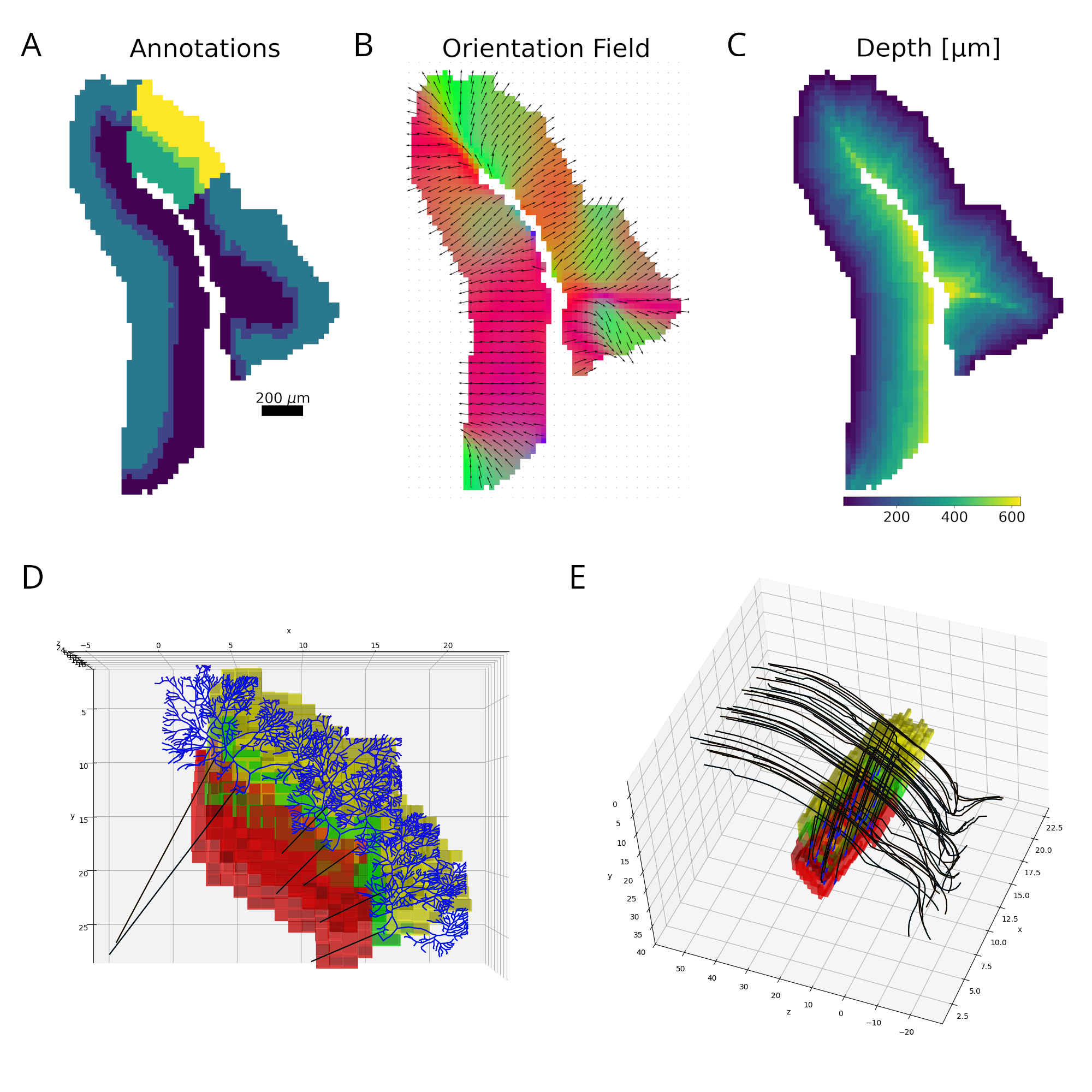
      References


      1. Rao, R. P. N., & Ballard, D. H. (1999). Nature Neuroscience, 2, 79-87.
      2. Keller, G. B., & Mrsic-Flogel, T. D. (2018). Neuron, 100, 424-435.
      3. Hertäg, L., & Clopath, C. (2022). PNAS, 119, e2115699119.
      4. Mikulasch, F. A., et al. (2023). Trends in Neurosciences, 46, 45-59.
      5. Nemati, E., Davey, C. E., Meffin, H., & Burkitt, A. N. (2025). bioRxiv. 10.1101/2025.10.20.683584.
      6. Nemati, E., Davey, C. E., Meffin, H., & Burkitt, A. N. (2025). bioRxiv. 10.1101/2025.11.01.686040.
      7. Larkum, M. (2013). Trends in Neurosciences, 36, 141-151.


      Acknowledgement


      ANB and HM acknowledge support by the Australian Government through the Australian Research
      Council’s Discovery Projects funding scheme [DP220101166].
      EN acknowledges support from a Melbourne Research Scholarship, and the Diane Lemaire and Dee
      & John Collier Travel Scholarships at the University of Melbourne.
      Sunday July 12, 2026 4:20pm - 6:20pm ADT
      Ballroom B2

      4:20pm ADT

      P016: Vestibular predictions during maternal gait help shape development of neural of timekeeping
      Sunday July 12, 2026 4:20pm - 6:20pm ADT
      Introduction
      Humans develop beat perception and rhythm synchronization remarkably early, suggesting that prenatal experience may play a formative role. The neural basis behind this remains poorly understood. We propose that maternal gait during pregnancy helps shape the development of neural timekeeping by pairing rhythmic auditory events with correlated smooth vestibular input that the fetus learns to anticipate.


      Methods
      We developed a biologically grounded recurrent neural network with parallel auditory and vestibular pathways. One version of the network contained generic excitatory and inhibitory units; another incorporated a diversity of units modeled from cortical neurons. The models were trained via single-step backpropagation with auditory pulses paired with sinusoidal vestibular waveforms mimicking maternal locomotion. The model was trained to predict the input five timesteps in advance — representing vestibular predictions during maternal gait — across a range of tempos. Vestibular input was gradually removed as training performance improved, encouraging the network to rely on internally generated predictions given only auditory pulses.

      Results
      We explored the effects of networks incorporating multiple biologically realistic cell types, which outperformed single-type networks on synchronization tasks. The dual auditory-vestibular architecture further improved both synchronization and continuation performance compared to either network on its own. Weighted tempo sampling, based on training loss, reduced drift toward preferred tempos during continuation and could represent musical training during life.


      Discussion
      These results demonstrate that a biologically inspired predictive network can be trained through a plausible developmental curriculum to internalize and maintain rhythmic structure across different tempos. This model offers a platform for investigating the neural basis of timekeeping and how early sensory experience — beginning in utero and refined by musical training — may scaffold the rhythm synchronization abilities universal to humans. 


      References
      Yousefabadi, M., & Cannon, J. (2025). Maternal Gait Contributes To Development Of Beat Perception And Urge To Move To Music In A Predictive Processing Network Model. Zenodo. https://doi.org/10.5281/zenodo.17247501

      Acknowledgement
      This research was funded in part by the Natural Sciences and Engineering Research Council of Canada

      Sunday July 12, 2026 4:20pm - 6:20pm ADT
      Ballroom B2

      4:20pm ADT

      P017: Adapting the reconstruction of the cerebellar cortex to the shape of the brain
      Sunday July 12, 2026 4:20pm - 6:20pm ADT
      Introduction
      Current knowledge on the cellular composition and local connectivity of the cerebellar cortex has enabled the reconstruction of detailed microcircuit models [1, 2]. However, up to now, none of these models take into account the real convoluted shape of the cerebellar cortex. We aim at reconstructing and simulating atlas-mapped mouse cerebellar regions, capturing the relationship between structure, dynamics, and function. 
      We have developed a pipeline to reconstruct the mouse cerebellar cortex embedded into the Allen Mouse Brain Atlas (AMBA) [3]. Using the Brain Scaffold Builder (BSB) framework [1], we placed, oriented, bent and connected the neurons. The generated circuit can be simulated and validated against experimental findings.

      Methods
      We extracted a column of the mouse declive (vermal part of the Lobule VI) from the AMBA (Fig. 1A). We placed cells based on literature densities [1], including the unipolar brush cells [4], and proposed a new strategy to place Purkinje cells based on linear density [5] (Fig. 1D). To connect the cells, we computed the orientation and depth [6] of each voxel (Fig. 1BC). These fields were used to bend the cells’ neurites following the local curvature (Fig. 1E). We applied voxel intersection on these bended cells [1]. We assigned point-neuron electrical parameters for each cell type and synaptic parameters for each connection type [7]. We compared this model to our previous nonspecific and regular-paralleliped circuit (canonical circuit) [1].

      Results
      Our pipeline employed constraints for each neuron type, and the produced circuit indeed preserved the morphological properties of the canonical circuit, such as maintaining fibers parallel for granule cells (Fig. 1E). More importantly, the pipeline guaranteed a coherent connectome, which matched the synaptic convergences/divergences of the canonical circuit. We proved that, without proper bending and scaling, the number of synapses would be underestimated, especially for longer intersomatic distances.
      Finally, we simulated that circuit using the BSB interfacing with the NEST simulator [8] in resting state and under stimulus. The signal propagation and population-specific firing properties were well reproduced, as in the canonical circuit.

      Discussion
      The developed pipeline is able to leverage atlas data to estimate the heterogeneous spatial properties of the cerebellum, embedding them into circuit reconstructions. The atlas registration will also facilitate the integration of our model into larger brain circuits [9]. The morphology bending algorithm will be soon enhanced in order to adapt the spatial distribution of neurites to match the expected densities of fibers in the considered regions. 
      We plan to leverage the Blue Brain Cell Atlas pipeline [6] to reconstruct the whole declive as well as different regions of the cerebellar cortex, to study how the heterogeneity of their local properties gives rise to differences in their structure and function at the macroscale level.

      Figure 1. Reconstruction pipeline. A. Declive layers shown in colors with the selected column highlighted. B. Orientation field showing the local axons’ main axis. Colors represent the vectors’ norm. C. Distance to the outside border, following the orientation field. D. E. Purkinje and granule cells´ morphology scaled and bent according to the declive shape.

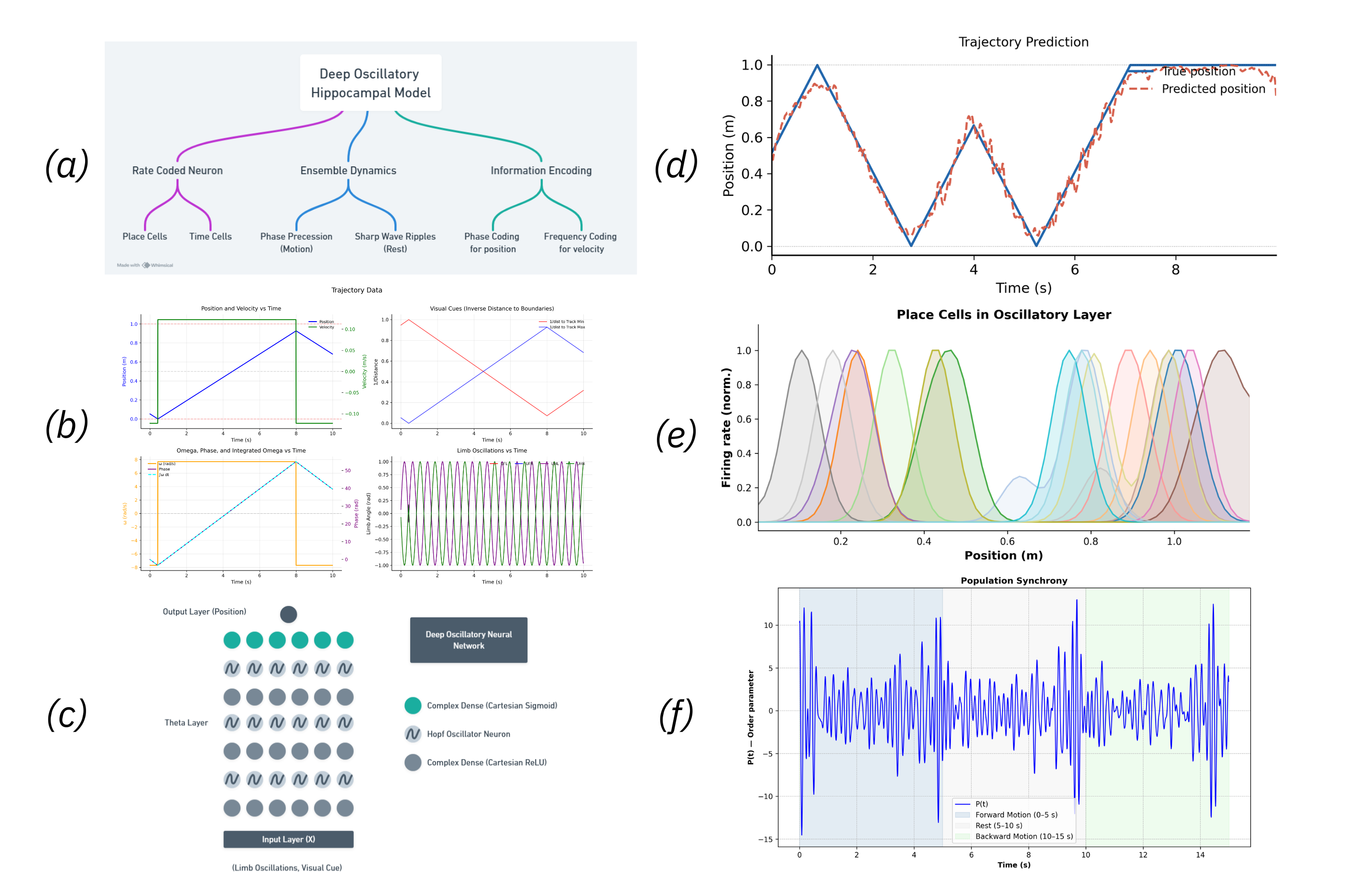
      References
      1. https://doi.org/10.1038/s42003-022-04213-y
      2. https://doi.org/10.1038/s41598-025-25727-5
      3. https://doi.org/10.1111/j.1601-183X.2009.00552.x
      4. https://doi.org/10.1007/s00429-013-0531-9
      5. https://doi.org/10.1002/jnr.24206
      6. https://doi.org/10.1371/journal.pcbi.1010739
      7. https://doi.org/10.3389/fncom.2019.00068
      8. https://doi.org/10.4249/scholarpedia.1430
      9. https://doi.org/10.1523/ENEURO.0111-17.2017
      Sunday July 12, 2026 4:20pm - 6:20pm ADT
      Ballroom B2

      4:20pm ADT

      P018: Digital Twins in stroke and spreading depression
      Sunday July 12, 2026 4:20pm - 6:20pm ADT
      Introduction
      Multiscale modeling (MSM) permits us to better understand how ischemia and spreading depolarization (SD) together damage neurons. However, it is a long step from there to producing clinical tools to counter or prevent stroke. Such tools must include clinical data from neurology and cardiology, endocrinology and other clinical specialties, as well as from other biomedical sciences, and must engage with body sensors (data integrators -DIs -for sensor information consolidation) and with the patient him or herself. We are developing digital twins (DTs) to incorporate these elements to extend personalized care. DTs will incorporate MSMs and DIs with large language models (LLMs) to communicate with the patient and with clinicians.


      Methods
      We have developed LLM to interact with patients and now combine them with our MSMs that include neural and vascular elements. MSM simulates & constrains detailed reaction--diffusion, electrophysiolo- gy, circuit models. LLM correlates literature and simulation details to identify simulation boundaries.


      Results

      Discussion
      DT medical personalization can help distinguish multiscale parameters, enabling patient-specific predictions and suggest therapy testing. Pairing of MSM detailed models with LLMs allows ingesting large electronic medical record (EMR) and archival research text to structured knowledge, further augmented with DI access to personal (digital watch and monitors) and clinical tools. Brain ischemia is a bridge disease since mutli-organ (cardiac, brain, vessel, lung) ; detailed clinical correlates and preventive strategies. microscale; multi-physics;  multi-specialty: neurology, vascular, cardiac, endocrine.


      References
      none

      Acknowledgement
      Supported by NIH R01MH086638
      Speakers
      avatar for Adam Newton

      Adam Newton

      Research Scientist, SUNY Downstate Health Sciences University

      avatar for Robert McDougal

      Robert McDougal

      Associate Professor, Yale University
      Looking for a postgrad or postdoc position implementing simulation methods? I'm hiring.I'm an Associate Professor in the Health Informatics division of Biostatistics, and a developer for NEURON and ModelDB. Computationally and mathematically, I'm interested in dynamical systems modeling... Read More →
      Sunday July 12, 2026 4:20pm - 6:20pm ADT
      Ballroom B2

      4:20pm ADT

      P019: A Unified Deep Oscillatory Network Model of the Hippocampal Sharp Wave Ripples
      Sunday July 12, 2026 4:20pm - 6:20pm ADT
      Introduction
      The well-known link between neural dynamics of spatial navigation and hippocampus is reflected in characteristic phenomena like neurons encoding spatial and temporal variables, and oscillatory dynamics such as phase precession in locomotion and sharp-wave ripples at rest. Existing computational models like oscillatory interference models, continuous attractor network and deep learning models either account for oscillatory behaviors or spatial coding within a rate-coded framework, capturing only a subset of features not addressing rest or temporal dynamics [1,2,3]. We propose an oscillatory hippocampus model that comprehensively captures these constructs, providing a unified framework to study translation of neural activity into navigation.


      Methods
      A complex valued deep oscillatory neural network is trained to estimate position coordinates of a 1D trajectory from limb oscillations and environmental visual cues of a quadruped (animal) that alternates between motion and rest [4]. The oscillatory layers in the network include a central layer with an intrinsic theta band (4-8 Hz) enabling study of hippocampal spatial navigation. The network’s complex hidden layer activations are analyzed to study the encoding of spatiotemporal information. Statistic measures are applied to the mean firing rates across spatial and temporal bins to identify place and time cells. Oscillatory behaviors are shown using Hebbian learning and regression analyses on the complex oscillatory layer activations.


      Results
      Place cells identified from the complex activations were found to tile the traversed trajectory. Time cells were observed to encode elapsed time during the task, independent of state of motion or rest. Position and velocity were encoded through oscillator population dynamics - position reflected in the mean phase and velocity in the mean frequency of the oscillator population. Sharp-wave ripple–like events generated via Hebbian learning exhibited higher amplitudes at periods of rest, indicating increased synchrony among oscillators. These findings are consistent with existing experimental observations, offering new insights into how spatiotemporal information can be represented through the joint encoding of frequency, phase, and amplitude.


      Discussion
      Spatial and temporal representations emerged naturally as the model learned to map sensorimotor inputs to position. Rate-coded properties were evident at the level of individual neurons, and oscillatory phenomena at the level of neuronal populations. The internal oscillatory dynamics are interpretable through the parameters of amplitude, phase and frequency. These results suggest that the proposed model offers a unified framework that can capture spatiotemporal representations during motion and rest. Its ability to encode information in interpretable oscillatory variables enables investigation of broader hippocampal functions - navigation, associative memory, and working memory, across diverse task structures and environmental conditions.

      Figure 1. (a) Model Flowchart, (b) Input Data, (c) Oscillatory Neural Network Diagram, (d) Trajectory Prediction, (e) Place Cells - different colors correspond to different neurons, (f) Sharp Wave Ripples
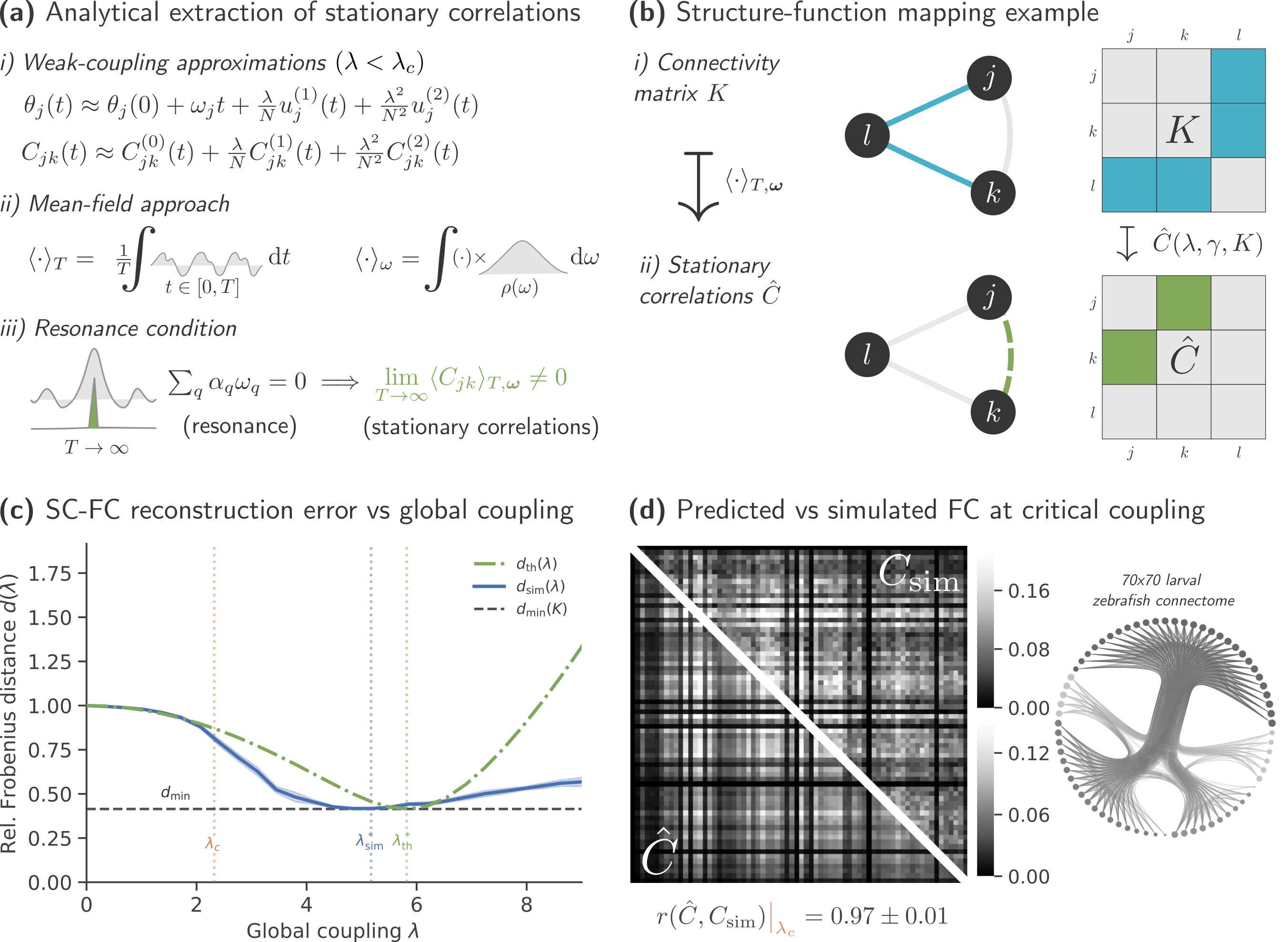
      References
      1. O’Keefe, J., & Recce, M. L. (1993). Phase relationship between hippocampal place units and the EEG theta rhythm. Hippocampus, 3(3), 317–330.https://doi.org/10.1002/hipo.450030307
      2. Burgess, N., Barry, C., & O’Keefe, J. (2007). An oscillatory interference model of grid cell firing. Hippocampus, 17(9), 801–812.https://doi.org/10.1002/hipo.20327
      3. Buzsáki, G. (2015). Hippocampal sharp wave–ripple: A cognitive biomarker for episodic memory and planning. Hippocampus, 25(10), 1073–1188.https://doi.org/10.1002/hipo.22488
      4. Rohan, N. R., Vigneswaran, C., Ghosh, S., Rajendran, K., Gaurav, A., & Chakravarthy, V. S. (2025). Deep oscillatory neural network. Scientific Reports, 15(1), 40968.

      Acknowledgement
      My supervisor Prof. V. Srinivasa Chakravarthy, 
      Mentors from Computational Neuroscience Lab and the Dept. of Medical Sciences and Technology
      More importantly, my parents.
      Sunday July 12, 2026 4:20pm - 6:20pm ADT
      Ballroom B2

      4:20pm ADT

      P020: Prefrontal and Parietal Local Field Potentials Employ Different Visuospatial Codes for Reach: A Complex-Valued Network Classification Approach
      Sunday July 12, 2026 4:20pm - 6:20pm ADT
      Introduction
      Understanding how cortical oscillations coordinate spatial memory and motor planning is a central challenge in systems neuroscience. We tested whether phase–amplitude dynamics in cortical local field potentials (LFPs) encode distributed versus region-specific signals for spatial memory and planning under varying visuospatial conditions.


      Methods
      We developed a Complex-Valued Neural Network (CVNN) model [1, 2] to decode landmark-dependent spatial states from LFPs recorded in the posterior ventrolateral prefrontal cortex (pVLPFC, 128 channels) and intraparietal sulcus (IPS, 32 channels) of a female rhesus monkey performing memory-guided reaching tasks in which visual landmarks were stable, shifted 8° in one of eight directions, or absent [3, 4]. Preprocessed LFPs were transformed into complex-valued time series using the Hilbert transform to preserve phase and amplitude information [5].


      Results
      We trained separate CVNN models on IPS or pVLPFC signals which classified the three landmark conditions with >90% training accuracy and more than 51% overall validation accuracy, significantly above chance (33%). However, validation performance revealed inter-regional specialization: the IPS model performed best for no-landmark trials (88.35% ± 6.99), whereas the pVLPFC model showed superior performance for shifted-landmark trials (71.73% ± 8.59). We then trained dual-stream models combining pVLPFC and IPS recordings. The single-region results were confirmed via region occlusion analysis after training: removing pVLPFC improved no-landmark classification, while removing IPS improved shifted-landmark classification.


      Discussion
      These findings suggest that IPS specializes in maintaining spatial representations for reach plans in egocentric coordinates, whereas pVLPFC shows enhanced encoding in the presence of visual landmarks, especially in the dynamic landmark-shift conditions, indicating complementary computational roles in maintaining and updating spatial representations for reach.


      References
      1. N. Benvenuto and F. Piazza, "On the complex backpropagation algorithm," in IEEE Transactions on Signal Processing, vol. 40, no. 4, pp. 967-969, April 1992, doi: 10.1109/78.127967.
      2. G. M. Georgiou and C. Koutsougeras, "Complex domain backpropagation," in IEEE Transactions on Circuits and Systems II: Analog and Digital Signal Processing, vol. 39, no. 5, pp. 330-334, May 1992, doi: 10.1109/82.142037.
      3. Lin, J., Wang, H., Sun, S., Yan, X., & Crawford, J. D. (2023). Influence of a visual landmark shift on memory-guided reaching in monkeys. Journal of Vision, 23(9), 4828–482 https://doi.org/10.1167/jov.23.9.4828.
      4. Lin, J. Y. X. (2024). Influence of a visual landmark shift on memory-guided reaching in the monkey.
      5. Freeman, W. J. (2007). Hilbert transform for brain waves. Scholarpedia, 2(1), 1338.

      Acknowledgement
      This research was funded by the Connected Minds Program, supported by the Canada First Research Excellence Fund.

      Sunday July 12, 2026 4:20pm - 6:20pm ADT
      Ballroom B2

      4:20pm ADT

      P021: Local dendritic voltage provides a reliable read-out of global synaptic activity
      Sunday July 12, 2026 4:20pm - 6:20pm ADT
      Introduction
      Neurons receive synaptic inputs across a spatially extended dendritic tree [1]. Recent work has shown that neuronal excitability is independent of the size of the dendritic tree when distributed dendritic, instead of somatic, inputs are considered [2]. Such dendritic normalisation has also been shown to improve the speed and robustness of learning [3]. The question remains, however, whether the same principle applies to local dendritic voltages across the entire neuron, and whether this might be computationally useful.


      Methods
      We derive analytical results using the cable equation [4] in passive dendritic structures, and validate our results using simulations of passive and active cells, including detailed and biophysically validated multicompartmental models, in the Matlab Trees Toolbox package [5], T2N [6], and the NEURON environment [7].

      Results
      We first show analytically that the steady state voltage response of a dendritic cable receiving distributed inputs is completely independent of dendrite size and measurement location; a dendrite acts like a ‘bucket’ filling with synaptic ‘water’. We investigate how far perturbations due to stochastic inputs impact the ‘bucketness’ of a cell, and find that the local dendritic voltage at every location in the dendrite typically reflects the strength of global inputs. We confirm that calcium concentrations are much longer-lived and more local than voltages. We finally show that the interaction between calcium and voltage could provide a substrate for robust learning by reinterpreting long-term plasticity rules [8,9].

      Discussion
      Dendritic voltages are surprisingly global and quickly equalise deviations in synaptic inputs. In contrast, calcium transients can provide a long-lived record of local afferents. The interplay between these two indicators provides a continuous, biophysically grounded, learning signal at every point in a dendritic tree. Our results provide a foundation for further studies into the many ways dendrites provide a space for complex computations at the single neuron level.

      References
      1.\tChklovskii D. Neuron. 2004;43(5):609–17. 10.1016/j.neuron.2004.08.012
      2.\tCuntz H, Bird A, et al. Neuron. 2021;109(22):3647-3662.e7. 10.1016/j.neuron.2021.08.028 PMID: 34555313.
      3.\tBird AD, Jedlicka P, Cuntz H. PLOS Comp Bio. 2021;17(8):e1009202. 10.1371/journal.pcbi.1009202
      4.\tRall W. Ann NY Acad Sci. 1962;96(4):1071–92. 10.1111/j.1749-6632.1962.tb54120.x
      5.\tCuntz H, Forstner F, et al. PLOS Comp Bio. 2010;6(8):e1000877. 10.1371/journal.pcbi.1000877
      6.\tBeining M, Mongiat L, et al. eLife. 2017;6:e26517. 10.7554/eLife.26517
      7.\tHines M, Carnevale N. Neural Comput. 1997;9(6):1179–209. PMID: 9248061.
      8.\tBienenstock EL, Cooper LN, Munro PW. J Neurosci. 1982;2(1):32–48. 10.1523/JNEUROSCI.02-01-00032.1982 PMID: 7054394.
      9.\tOja E. J Math Biology. 1982;15(3):267–73. 10.1007/BF00275687

      Acknowledgement
      None.
      Sunday July 12, 2026 4:20pm - 6:20pm ADT
      Ballroom B2

      4:20pm ADT

      P022: Electrophysiological and computational analysis of burst generation in Drosophila class III cold nociceptors
      Sunday July 12, 2026 4:20pm - 6:20pm ADT
      Introduction
      In Drosophila larvae, Class III (CIII) primary sensory neurons detect nociceptive cold temperatures, with about half responding to rapid cooling with transient bursting (1,2). Cold responses have been linked to activation of thermosensitive TRP channels, including TRPM, PKD2, and NOMPC (1,3). We previously showed that lowering extracellular Cl⁻ enhances spiking and promotes bursting in CIII neurons, consistent with a depolarizing shift of the Cl⁻ reversal potential. Here, we test whether pharmacological or ionic perturbations that produce appropriate membrane depolarization are sufficient to create bursting mechanisms in CIII neurons at room temperature, revealing a mechanism that does not rely on TRP channel activation.

      Methods
      Intracellular recordings were obtained from CIII neurons in Drosophila larvae under pharmacological and ionic manipulations. Experimental conditions included reduced extracellular Cl⁻ (6 mM; 134 mM control), elevated extracellular K⁺ (15 mM; 3 mM control), Ca²⁺ removal, and tetrodotoxin (TTX, 20 nM) to block voltage-gated Na⁺ channels. Direct current injection was used to characterize transitions between silence, spiking, and bursting across conditions. In parallel, a biophysical computational model of the CIII neuron was developed and constrained by experimental measurements. Model parameters were tuned to reproduce passive electrical properties and validated by comparison with experimentally observed activity patterns.

      Results
      In control, current injection (5–20 pA) produced tonic spiking. In low-Cl⁻ saline, the same stimulation  induced bursting in 90% of neurons (18/20). Elevated extracellular K⁺ promoted bursting in all neurons examined (6/6), indicating that global depolarization facilitates burst generation. Removal of extracellular Ca²⁺ did not eliminate bursting, suggesting that Ca²⁺ influx is not strictly required for burst generation under these conditions. In contrast, tetrodotoxin (20 nM) abolished both spikes and the underlying depolarizing potentials. Biophysical modeling reproduced these transitions and suggested that the voltage-gated Na⁺ current plays a prominent role in sustaining the depolarizing envelope supporting burst generation.

      Discussion
      These results demonstrate that CIII neurons can generate the full spectrum of activity patterns—silence, tonic spiking, and bursting—without activation of thermosensitive TRP channels. Depolarizing manipulations such as reduced extracellular Cl⁻, elevated K⁺, or current injection reliably promoted bursting. These findings suggest that practically all CIII neurons are intrinsically burst-capable when operating within an appropriate depolarized regime. Biophysical modeling reproduced the observed transitions and dissected the contributions of ionic gradients and membrane conductances, providing a mechanistic framework in which Na⁺ channel dynamics contribute prominently to the generation of bursting activity.

      References
      1. Turner, H. N., et al. (2016). The TRP Channels Pkd2, NompC, and Trpm Act in Cold-Sensing Neurons to Mediate Unique Aversive Behaviors to Noxious Cold in Drosophila. Current Biology,  26(23): 3116-3128. https://doi.org/10.1016/j.cub.2016.09.038
      2. Maksymchuk, N., et al. (2022). Transient and Steady-State Properties of Drosophila Sensory Neurons Coding Noxious Cold Temperature. Frontiers in Cellular Neuroscience,16, 831803. https://doi.org/10.3389/fncel.2022.831803
      3. Himmel, N. J., et al., (2023). Chloride-dependent mechanisms of multimodal sensory discrimination and nociceptive sensitization in Drosophila. eLife, 12, e76863. https://doi.org/10.7554/eLife.76863

      Acknowledgement
      NIH grant R01NS115209 to DNC and GSC.

      Sunday July 12, 2026 4:20pm - 6:20pm ADT
      Ballroom B2

      4:20pm ADT

      P023: Low-Dimensional Projections of Neural Population Activity in M1, PMd, and PMv Hand Representations Demonstrate Differences in Effector Dependence
      Sunday July 12, 2026 4:20pm - 6:20pm ADT
      Introduction
      The activity of the primary motor cortex (M1) across stages of a motor task evolves through attractors that capture the dominant activity when preparing and executing a task [1, 2]. This suggests that dimensionality reduction (DR) plays a key role in how M1 controls movements. In primates, in addition to M1, motor commands are generated by a network of frontal areas, the premotor cortex. Neurons in the premotor ventral (PMv) and dorsal (PMd) cortices discharge in relation to various parameters of movements and send projections to M1 [3]. We extend DR techniques to PMv, PMd, and M1 to characterize variation in neural population activity in context of reaching and grasping movements and identify the most explanatory neurons in these cortices.


      Methods
      Our data was collected from four rhesus macaque monkeys implanted with microelectrode arrays in the distal (hand) representation of M1, PMv, and PMd. We recorded isolated neurons spiking activity while monkeys performed a custom-made reach-to-grasp task. Following instruction cues, they reached with their left or right arm to grab a pellet or press on a plate using precision grasps in a vertical or horizontal orientation. For each neuron, we computed spike density estimates (SDE) by splicing peri-event windows and normalizing across all trials (like in [4]) for each hand-orientation combination. The condition-wise SDEs of all neurons were concatenated along the time dimension to perform principal component analysis (PCA) for each cortex.


      Results
      PCA of the neural population activity in each cortex demonstrates differences across conditions. For each cortex, the first 3 principal components capture over 90% of the variance of the neural population dynamics. Low-dimensional trajectories of neural population activity in M1 shows greater divergence in neural activity when varying the hand used than varying target orientation. However, these low-dimensional trajectories across conditions are more similar for the premotor areas, with PMv having the most similarity. Moreover, the principal angles between the subspaces of the principal components for the hand used show that the neuron weights are more consistent for PMv, demonstrating less effector dependence in PMv than in PMd or M1.


      Discussion
      The principal components (PCs) in each cortex indicate the weight assigned to each neuron which yields a sorting based on the explainability of the population dynamics. Combining this sorting with independent classification techniques of individual neurons allows for selection and classification of the most important neuron types in a population. Meanwhile, the differences in effector dependence and principal angles between M1, PMd, and PMv suggest a hierarchical structure of signals. Effector-independent PMv activity may structure the common movement parameters before PMd facilitates the more effector-dependent preparation. Low-dimensional representations such as PCA could explain this structure through the coupling of PCs across cortices.


      References
      [1] Churchland, M. M., et al. Stimulus onset quenches neural variability: a widespread cortical phenomenon. Nature neuroscience, 13(3), 369–378. https://doi.org/10.1038/nn.2501
      [2] Davare, M., et al. Dissociating the role of ventral and dorsal premotor cortex in precision grasping. The Journal of neuroscience, 26(8), 2260–2268. https://doi.org/10.1523/JNEUROSCI.3386-05.2006
      [3] Shenoy, K. V., Sahani, M. et Churchland, M. M. (2013). Cortical Control of Arm Movements: A Dynamical Systems Perspective. Annual Review of Neuroscience, 36, 337–359. https://doi.org/10.1146/annurev-neuro-062111-150509
      [4] Zimnik, A. A.-O., et al. Identifying Interpretable Latent Factors with Sparse Component Analysis. bioRxiv: the preprint server for biology, https://doi.org/10.1101/2024.02.05.578988

      Acknowledgement
      This research was supported by CIHR Grant No. 175069 and the FRQNT Strategic Clusters Program (Centre UNIQUE - Centre de recherche Neuro-IA du Québec).
      Sunday July 12, 2026 4:20pm - 6:20pm ADT
      Ballroom B2

      4:20pm ADT

      P024: Shared-input structure determines functional connectivity in neural oscillator networks
      Sunday July 12, 2026 4:20pm - 6:20pm ADT
      Introduction
      Functional connectivity (FC) describes statistical dependencies between the activity of neurons or groups of neurons [1]. Comparing FC with anatomical connectivity (SC) has emerged as a promising avenue to study how brain structure supports function [1,2]. Studies have reported a wide range of SC–FC correspondence values [1,2], highlighting the need for theoretical insights into these relationships [3]. We derive here a closed-form analytical mapping from SC to FC for any oscillator network, showing that shared presynaptic inputs govern coactivity and identifying an optimal regime of maximal SC–FC alignment, with a lower bound on SC reconstruction from FC. An empirical whole-brain larval zebrafish connectome is used for validation [4,5].


      Methods
      We develop an analytical framework linking SC to FC in coupled neural systems. We study coupled neural oscillators on a heterogeneous, weighted and directed network using the Kuramoto model [3]. A second-order perturbative expansion is obtained in the reduced coupling λ/N around the uncoupled regime, valid for arbitrary network size and topology. Time-averaged correlations are expanded and a stationary filter identifies finite contributions as T→∞ (Fig. 1a). Averaging over intrinsic frequencies drawn from a Cauchy–Lorentz distribution of width γ yields a closed-form prediction of FC. The coupling strength λ* maximising SC–FC alignment is obtained analytically by minimising a normalised Frobenius distance between predicted and simulated FC.


      Results
      Functional connectivity is determined by shared presynaptic inputs and not by direct synaptic connections. The stationary expansion retains only a second-order structure proportional to KKᵀ (Fig. 1b), yielding Ĉ = I + (5λ²)/(4γ²N²) · (KKᵀ - diag(KKᵀ)). First-order terms cancel, so direct connections do not contribute to FC, and the first anatomical fingerprint appears through shared-input structure. The optimal coupling λ*, derived solely from K, defines a theoretical lower bound on SC–FC reconstruction error (Fig. 1c). Simulations on an empirical whole-brain larval zebrafish connectome [4] show an excellent agreement between predicted and simulated FC (cosine similarity ≥ 0.97) across the valid coupling regime (Fig. 1d).


      Discussion
      Our closed-form expression reveals three results not accessible from simulation alone. First, stationary coactivity is determined by shared presynaptic inputs rather than by direct synaptic connections. Second, direct connections do not contribute to stationary coactivity in the canonical Kuramoto model, cautioning against using raw FC as a direct estimator of SC. Third, the explicit prefactor 5/(4γ²) obtained through a non-trivial analytical derivation, reveals that broader intrinsic-frequency dispersion weakens the structural imprint on FC, making reconstruction of SC from FC harder in heterogeneous neural populations.

      Figure 1. Predicting coactivity from anatomy in neural oscillators. (a) Derivation of predicted functional connectivity: phase trajectories are expanded, correlations averaged, and stationary terms selected. (b) Example for N=2 oscillators. (c) SC–FC reconstruction error follows theory up to synchronization (λc = 2.32). (d) Predicted and simulated FC remain highly similar (cosine similarity ≥ 0.97).
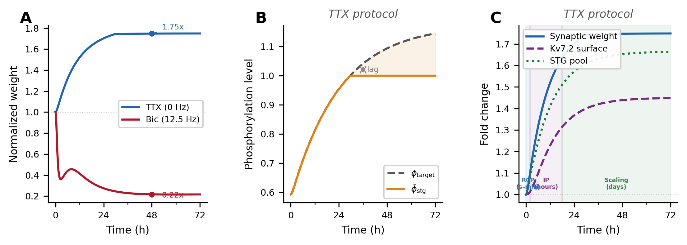
      References
      [1] Fotiadis, P., et al. (2024). Structure–function coupling in macroscale human brain networksNature Reviews Neuroscience, 25(10), 688–704.
      [2] Zamani Esfahlani, et al. (2022). Local structure-function relationships in human brain networks across the lifespanNature Communications, 13(1), 2053.
      [3] Pope, M., et al. (2021). Modular origins of high-amplitude cofluctuations in fine-scale functional connectivity dynamicsProceedings of the National Academy of Sciences, 118(46), e2109380118.
      [4] Kunst, M., et al. (2019). A Cellular-Resolution Atlas of the Larval Zebrafish BrainNeuron, 103(1), 21-38.e5.
      [5] Légaré, A., et al. (2025). Structural and genetic determinants of zebrafish functional brain networksScience Advances, 11(28), eadv7576.


      Acknowledgement
      We thank Benjamin Claveau, Antoine Légaré and Vincent Thibeault for helpful discussions, and Paul De Koninck’s lab for generating the data that initiated this project.

      Sunday July 12, 2026 4:20pm - 6:20pm ADT
      Ballroom B2

      4:20pm ADT

      P025: A Conductance-Based Whole-Brain Modeling Framework for Isolating Pharmacological Effects on Excitation-Inhibition Dynamics
      Sunday July 12, 2026 4:20pm - 6:20pm ADT
      Introduction
      The excitation-inhibition (E:I) ratio is a key biomarker in psychiatric conditions, and can be modulated by pharmacological interventions. Ketamine, an NMDA receptor antagonist, blocks NMDA receptors on inhibitory neurons, driving cortical disinhibition. Electrophysiologically, ketamine reduces the mismatch negativity (MMN) signal, which is a measure of sensory surprise within the brain's predictive coding framework [1]. Connectome-based neural-mass models excel at linking macroscopic electrophysiology to microcircuit mechanisms. Here, we extend a conductance-based neural mass model into a whole-brain framework to validate its capacity to capture ketamine's specific effects on NMDA receptor dynamics.


      Methods
      We modeled a previously published EEG dataset from 19 subjects recorded during a roving auditory MMN task under placebo and ketamine conditions [2] using a whole-brain modeling framework. We parcellated the brain into 200 distinct regions using Schaefer atlas. We developed an extension of the conductance-based neural-mass model introduced in [3] to simulate voltage (v) and gating (g) for AMPA, GABA, and NMDA receptors across pyramidal, excitatory, and inhibitory populations in the parcellated regions. We computed normalized gating by voltage interactions and applied principal component analysis (PCA) across different conditions to isolate and compare dominant temporal trajectories between pharmacological interventions.


      Results
      Analysis of the primary temporal trajectories (PC1) revealed distinct activation profiles across AMPA, GABA, and NMDA receptors following stimulus onset. Under placebo conditions, the network exhibited a robust MMN response. This was particularly evident in the normalized integrated synaptic activity (NMDA+AMPA+GABA), which produced a prominent deflection in both excitatory and pyramidal populations. Administration of ketamine markedly attenuated this effect across these key populations. Decomposing these network-level changes by receptor type demonstrated that ketamine primarily blunted the differential activation within GABA and NMDA signaling pathways.


      Discussion

      References
      1. Garrido, M. I., Kilner, J. M., Stephan, K. E., & Friston, K. J. (2009). The mismatch negativity: A review of underlying mechanisms. Clinical Neurophysiology, 120(3), 453–463. https://doi.org/10.1016/j.clinph.2008.11.029 
      2. Schmidt A, Bachmann R, Kometer M, Csomor PA, Stephan KE, Seifritz E, & Vollenweider FX. (2012). Mismatch negativity encoding of prediction errors predicts S-ketamine-induced cognitive impairments. Neuropsychopharmacology, 37(4), 865–875. https://doi.org/10.1038/npp.2011.261 
      3. Marreiros, A. C., Kiebel, S. J., Daunizeau, J., Harrison, L. M., & Friston, K. J. (2009). Population dynamics under the Laplace assumption. NeuroImage, 44(3), 701–714. https://doi.org/10.1016/j.neuroimage.2008.10.008 

      Acknowledgement
      We acknowledge the support of Canadian Institute of Health Research (CIHR-Project Grant) and Swiss Neuromatrix Foundation.
      Sunday July 12, 2026 4:20pm - 6:20pm ADT
      Ballroom B2

      4:20pm ADT

      P026: Compartment-specific calcium dynamics drive local, co-dependent excitatory and inhibitory plasticity in cortical networks
      Sunday July 12, 2026 4:20pm - 6:20pm ADT
      Introduction
      Learning requires neural circuits to remain adaptable while preserving learned representations—a fundamental trade-off known as the plasticity-stability dilemma. Dendritic arbors equipped with compartment-specific inhibition support local gating of excitatory plasticity, allowing multiple input streams to be integrated independently within a single neuron, without disrupting existing knowledge [1]. Co-dependent excitatory and inhibitory plasticity has been shown to account for quick, stable, and long-lasting memory storage in biological networks [2]. However, this co-dependence has been formalized through phenomenological spike-timing rules, leaving the underlying biophysical mechanisms unspecified.


      Methods
      Motivated by its central role in dendritic integration and long-term plasticity, we hypothesized that intracellular calcium orchestrates the local induction of excitatory and inhibitory plasticity. We extended a three-compartment cortical pyramidal cell model to include compartment-specific calcium dynamics from distinct sources (back-propagating action potentials, voltage-gated calcium channels, and NMDA receptors) and implemented co-dependent excitatory and inhibitory learning rules based on the calcium control hypothesis [3], driven by a shared local calcium signal. We embedded our augmented neuron model into a canonical cortical microcircuit model with cell type-specific connectivity and compartment-specific, differential inhibition.


      Results
      Our calcium-based learning rules yielded balanced networks with enhanced memory capacity and robustness to noise and continual learning. We identified compartment-specific fixed points for excitation-inhibition balance. Targeted perturbation of compartment-specific calcium dynamics resulted in selective memory retrieval with transient disruption of the local excitation-inhibition balance.


      Discussion
      Our findings support a biophysically plausible role for calcium compartmentalization in coordinating excitatory and inhibitory plasticity through local heterosynaptic interactions. The compartment-specific excitation-inhibition fixed points likely arise from the locality of calcium signals and their distinct sources, providing mechanistic insight into how cortical networks achieve compartment-specific control of learning-induced plasticity. Altogether, these results bridge synaptic biophysics and network-level computation while generating generalizable principles to inform the development of more efficient, biologically grounded adaptive systems.


      References
      1. Yang, G. R., Murray, J. D., & Wang, X.-J. (2016). A dendritic disinhibitory circuit mechanism for pathway-specific gating. Nature Communications, 7(1), 12815. https://doi.org/10.1038/ncomms12815
      2. Agnes, E. J., & Vogels, T. P. (2024). Co-dependent excitatory and inhibitory plasticity accounts for quick, stable and long-lasting memories in biological networks. Nature Neuroscience, 27(5), 964–974. https://doi.org/10.1038/s41593-024-01597-4
      3. Graupner, M., & Brunel, N. (2012). Calcium-based plasticity model explains sensitivity of synaptic changes to spike pattern, rate, and dendritic location. Proceedings of the National Academy of Sciences, 109(10), 3991–3996. https://doi.org/10.1073/pnas.1109359109

      Acknowledgement
      This work was supported by national funds through FCT—Foundation for Science and Technology, I.P., under the project HetSyn (2023.13758.PEX).
      Speakers
      avatar for Renato Duarte

      Renato Duarte

      Assistant Researcher, Center for Neuroscience and Cell Biology (CNC), University of Coimbra
      Sunday July 12, 2026 4:20pm - 6:20pm ADT
      Ballroom B2

      4:20pm ADT

      P027: Adaptive Homeostasis: Coupling Synaptic Plasticity, Homeostatic Scaling, and Intrinsic Plasticity
      Sunday July 12, 2026 4:20pm - 6:20pm ADT
      Introduction

      Synaptic plasticity underlies learning and memory. To prevent instability from unconstrained Hebbian modifications, neurons engage homeostatic processes to globally adjust synaptic weights and membrane excitability [1]. Despite co-occurring, Hebbian and homeostatic plasticity are traditionally modeled independently, leaving their molecular crosstalk unresolved [2]. Recent evidence identifies stargazin, a TARP, as a critical link: it undergoes phosphorylation during long-term potentiation (LTP) and dephosphorylation during homeostatic downscaling [3], and interacts with Kv7.2 subunits to modulate intrinsic excitability [4]. No existing model unifies this coupling across scales; we present a multi-resolution framework that bridges this gap.

      Methods
      We developed a multi-scale model extending resource competition principles [5] from single synapses to the whole neuron. At the biophysical level, calcium-dependent competition between kinases and phosphatases governs stargazin phosphorylation, which regulates AMPAR trafficking across synapses and Kv7.2 surface expression. A reduced formulation eliminates fast variables (calcium quasi-steady-state, AMPAR equilibrium) to yield a tractable per-synapse/per-branch description. A conceptual model further condenses dynamics into three coupled variables (weights, resources, excitability), enabling investigation of network-level consequences.


      Results

      Simulations reproduced the timescale separation between fast calcium transients, rapid LTP-driven AMPAR insertion, and gradual resource-constrained downscaling (Fig. 1). The biophysical model produces three compensatory tiers: AMPAR redistribution via pool competition (seconds–minutes), M-current adjustment via Kv7.2 trafficking (hours), and synaptic scaling via stargazin pool dynamics (days). Under 48-hour TTX and bicuculline protocols, the model reproduces bidirectional scaling consistent with experimental data [1]. The reduced and conceptual models preserve quantitative accuracy with fewer variables, enabling network-level investigation.


      Discussion

      Our findings provide a biophysical account of how neurons maintain stability while preserving the capacity for input-specific memory allocation. The three-tier model hierarchy (from molecular cascades to analytically tractable abstraction) enables both detailed validation against experimental data and the investigation of how homeostasis interacts with ongoing learning dynamics in network settings. The model highlights the necessity of multi-scale molecular crosstalk, positioning stargazin as a core integrator of synaptic plasticity and multi-scale homeostasis..  Embedding the conceptual model in recurrent circuits allows us to investigate how this multi-scale, compartmentalized integration constrains learning and computation. 

      Figure 1. Simulated homeostatic response to inactivity (TTX). (A) Multiplicative synaptic scaling: upscaling (1.75×) and downscaling (0.22×) at 48 h. (B) Stargazin phosphorylation (φ̄_stg) lags the homeostatic target (φ_target) due to enzymatic inertia. (C) Three compensatory tiers emerge sequentially: AMPAR redistribution (seconds–min), Kv7.2 adjustment (hours), and synaptic scaling (days).
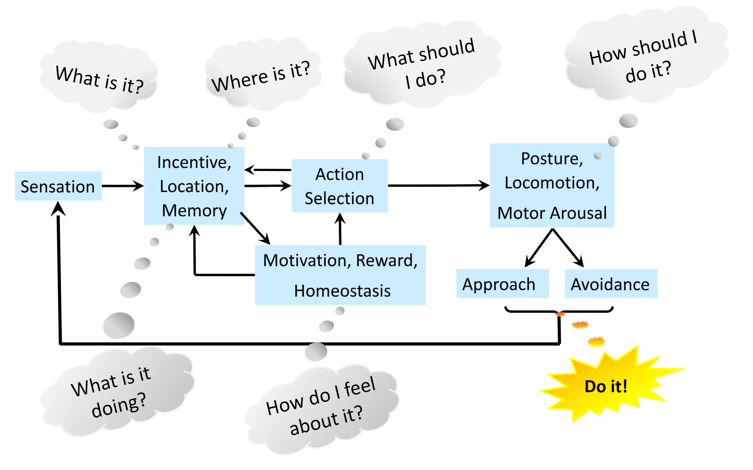
      References

      [1] Turrigiano, G. G., Leslie, K. R., Desai, N. S., Rutherford, L. C., & Nelson, S. B. (1998). Activity-dependent scaling of quantal amplitude in neocortical neurons. Nature, 391(6670), 892-896.
      [2] Turrigiano, G. G. (2017). The dialectic of Hebb and homeostasis. Philosophical Transactions of the Royal Society B, 372(1715), 20160258.
       [3] Louros, S. R., Caldeira, G. L., & Carvalho, A. L. (2018). Stargazin Dephosphorylation Mediates Homeostatic Synaptic Downscaling of Excitatory Synapses. Frontiers in Molecular Neuroscience, 11, 328. 
      [4] Rodrigues, M. V., et al. (2024). Type I TARPs regulate Kv7.2 potassium channels and susceptibility to seizures. bioRxiv.
      [5] Triesch, J., Vo, A. D., & Hafner, A. S. (2018). Competition for synaptic building blocks shapes synaptic plasticity. eLife, 7, e37836.


      Acknowledgement
      This work was supported by national funds through FCT—Foundation for Science and Technology, I.P., under the project HetSyn (2023.13758.PEX).

      Speakers
      avatar for Renato Duarte

      Renato Duarte

      Assistant Researcher, Center for Neuroscience and Cell Biology (CNC), University of Coimbra
      Sunday July 12, 2026 4:20pm - 6:20pm ADT
      Ballroom B2

      4:20pm ADT

      P028: Reliability of Eigenspectra Decay and Variance Scaling in 3T and 7T fMRI
      Sunday July 12, 2026 4:20pm - 6:20pm ADT
      Introduction
      A system organized to criticality is able to switch phases from small inputs[1]. Prior research indicates that neural systems are organized to near-criticality across multiple scales[2]. One modality for which criticality analysis is novel is fMRI. fMRI allows for the analysis of resting state brain networks (RSNs), groups of units that coactivate with one another and reflect specific cognitive processes[3,4]. 
      As a step towards characterizing the critical dynamics of brain networks, it is necessary to first assess the reliability with which critical metrics can be estimated. To identify network-specific factors from individual-specific ones, we compared metrics obtained for each RSN across scanners and across participants. 

      Methods
      The metrics of criticality examined are derived using a new method, phenomenological renormalization group (pRG) [5]. In this approach, units are paired based on correlation. These pairs are summed together and normalized. A correlation matrix is calculated between the new clusters. This process is repeated such that, at each iteration n of coarse-graining, the number of units, k represented by each cluster is 2n.
      Following coarse-graining, several metrics are calculated: μ, the power law exponent describing the covariance eigenspectra decay for a cluster of size K (λ ~ (r/K)) and α, the scaling exponent for cluster variance (σ2(K) ~ Kα). These analyses are performed for each network within each participant at each scan strength (3T, 7T). 

      Results
      pRG analysis in fMRI data yielded power-law relationships showing scaling of variance with cluster size and eigenvalues with rank. Within individuals, we compared exponents obtained from 7T and 3T scans for each network. While values of exponents varied across networks, we found a high degree of correlation between exponents in 3T and 7T data: 0.791 (μ) and 0.523 (α) for participant 1. For participant 2, correlation coefficients were 0.818 (μ) and 0.934 (α). 
      We examined whether exponents were similar between participants. When comparing exponents of distinct networks between participants 1 and 2, we found correlation coefficients of 0.368 (μ) and 0.5493 (α) for 3T and coefficients of 0.747 (μ) and 0.795 (α) for 7T.

      Discussion
      Our results indicate that RSNs possess critical dynamics that correlate with themselves across scanner strengths and individuals, indicating that RSNs have intrinsic dynamics likely reflecting different cognitive processes. 
      Interestingly, there is a large difference in α correlation coefficients (3T vs 7T) between participants 1 and 2. This may indicate that the stability of critical dynamics between scanner strengths varies across individuals. Also, the stronger correlations between participants at 7T compared with 3T are likely because of the stronger signal-to-noise ratio at 7T.
      Future directions are to assess reliability of criticality metrics in future participants and to characterize specific metrics of criticality within RSNs.

      References
      1. Fontenele, A. J., et al. (2019). Criticality between cortical states. Physical Review Letters, 122(20), 208101.
      2. Hengen, K. B., & Shew, W. L. (2024). Is criticality a unified set-point of brain function? (p. 2024.09.02.610815). bioRxiv. https://doi.org/10.1101/2024.09.02.610815
      3. Meshulam, L., et al. (2019). Coarse Graining, Fixed Points, and Scaling in a Large Population of Neurons. Physical Review Letters, 123(17), 178103. https://doi.org/10.1103/PhysRevLett.123.178103
      4. O’Byrne, J., & Jerbi, K. (2022). How critical is brain criticality? Trends in Neurosciences, 45(11), 820–837. https://doi.org/10.1016/j.tins.2022.08.007
      5. Rosazza, C., & Minati, L. (2011). Resting-state brain networks: Literature review and clinical applications. Neurological Sciences, 32(5), 773–785.

      Acknowledgement
      VM was supported through a PhD candidate research assistantship at the University of Minnesota. fMRI data was able to collected through use of Center for Magnetic Resonance Research resources at the University of Minnesota. Analysis was conducted using Minnesota Supercomputing Institute resources at the University of Minnesota. 
      Sunday July 12, 2026 4:20pm - 6:20pm ADT
      Ballroom B2

      4:20pm ADT

      P029: Modeling Emotional Contagion in Rats Using Dynamical State-Space Models
      Sunday July 12, 2026 4:20pm - 6:20pm ADT
      Introduction
      Emotional contagion, the sharing of another individual’s emotional state, is a key component of social behavior and empathy. Neural population dynamics underlying internal affective states are increasingly studied using latent dynamical models that reveal structured activity patterns associated with behavioral states (Nair et al., 2022). In rodents, socially relevant experiences can also shape memory and internal state representations (Veyrac et al., 2015). Here, we investigate neural and behavioral dynamics in observer rats witnessing conspecifics receiving footshocks.


      Methods
      Adult observer rats were implanted with Neuropixels probes to record large-scale neuronal population activity while simultaneously measuring locomotor speed and pupil diameter. Animals observed a demonstrator receiving footshocks in an adjacent compartment. Spike trains were converted to firing rates and analyzed using a recurrent Switching Linear Dynamical System (rSLDS), a state-space model that captures both discrete neural states and continuous latent dynamics underlying population activity.


      Results
      Shock observation produced significant increases in pupil dilation and reductions in locomotor speed, with MANOVA revealing significant inter-rat and inter-shock variability. The rSLDS identified discrete neural states that shifted around shock events and captured coordinated dynamics across neural and behavioral signals. Importantly, a distinct neural latent state emerged following shock observation and persisted longer than the behavioral responses, indicating sustained internal processing beyond immediate physiological changes.


      Discussion
      Our findings demonstrate that latent dynamical modeling reveals structured neural state transitions associated with socially transmitted distress. While neural and behavioral responses showed synchronized shifts around shock events, neural population activity displayed a prolonged state not fully explained by pupil dilation or immobility. These results support the presence of emotional contagion, suggesting that observer animals maintain a sustained neural representation of another individual’s distress.


      References
      Nair, A. et al. (2022).An approximate line attractor in the hypothalamus encodes an aggressive state. Cell, 185(25), 4841–4859.https://doi.org/10.1016/j.cell.2022.11.027
      Veyrac, A., Allerborn, M., Gros, A., Michon, F., Raguet, L., Kenney, J., Godinot, F., Thevenet, M., García, S., Messaoudi, B., Laroche, S., & Ravel, N. (2015). Memory of occasional events in rats: Individual episodic memory profiles, flexibility, and neural substrate. Journal of Neuroscience, 35(20), 7575–7586. https://doi.org/10.1523/JNEUROSCI.3941-14.2015


      Acknowledgement
      This work is supported by Dutch Brain Interface Initiative (DBI2), project number 024.005.022 of the research programmed Gravitation, which is financed by the Dutch Ministry of Education, Culture, and Science (OCW) via the Dutch Research Council (NWO)

      Sunday July 12, 2026 4:20pm - 6:20pm ADT
      Ballroom B2

      4:20pm ADT

      P030: Scalable Modular Architectures for Naturalistic Behavior and Cognitive Mapping in Biological and Artificial Intelligence
      Sunday July 12, 2026 4:20pm - 6:20pm ADT
      Introduction
      Biological intelligence is far more adaptive, autonomous and efficient than current artificial intelligence, despite recent advances. Neuroethological comparisons and computational simulations show that nervous systems across phyla share a conserved modular organization for information flow from sensation to behavior [1](Fig. 1). Differences between mammals and primitive soft-bodied invertebrates are largely in kind and amount of detail handled by different modules. 4 computations (What is it? Where is it? How do I feel about it? What should I do?) compose waking consciousness, while cognitive mapping enables intricate subjective experience, adding the question What is it doing? We explore these computations in agent-based simulations.

      Methods
      The agent-based simulation Cyberslug [2] was modeled on the core decision-making circuitry of a relatively simple animal, the predatory sea slug Pleurobranchaea californica, which retains character of the last common ancestor of bilaterally symmetric animals. Cyberslug is essentially an easily-scalable hybrid dynamical system which reproduces Pleurobranchaea’s decision-making in foraging. With small, biologically-plausible additions to Cyberslug, we developed the ASIMOV model with reward‑dependent plasticity for sensory valuation [3], as well as a Feature Association Matrix (FAM), a memory module inspired by hippocampal architecture [4]. Agents were tested in various simulated spatial environments with olfactory cues, coded in NetLogo.

      Results
      The modular neural organization of Pleurobranchaea was shown to be markedly similar to that of vertebrates and other invertebrates (e.g., insects, cephalopods) providing a conserved core circuitry for computational modeling and expansion [1,2]. Cyberslug reproduced adaptive cost‑benefit decision‑making in foraging [2]. ASIMOV extensions captured realistic sensory valuation, including addiction‑like dynamics [3]. Addition of the FAM showed how episodic memory and spatial cognitive mapping emerge from simple associative learning rules. This enabled the latest ASIMOV-FAM agent for efficient spatial navigation, one‑shot learning, and improved performance in sparse‑reward environments compared to standard reinforcement learning [4].

      Discussion
      Our results show that conserved modular architectures can organize the flow of information in animals and support naturalistic behavior, episodic memory, and cognitive mapping in artificial agents. Simple associative mechanisms analogous to hippocampal function were sufficient for sequence learning, spatial cognitive mapping, and recall, highlighting a plausible computational basis for subjective‑like experience and flexible intelligence. Our incremental elaboration of biologically grounded circuits produce increasingly complex cognition and dynamic behavior, providing a scalable computational framework for neuroethological studies, as well as further development of flexible, autonomous artificial intelligence (AI).

      Figure 1. Both simple and complex animals handle flow of information from sensation to behavior with a common modular nervous system organization. Stimuli characteristics, incentives and locations are integrated with memory, motivation and affect for decisive action selection, with 5 critical computations from “What is it?” to “How should I do it?”, and cognitive mapping adding “What is it doing?”.

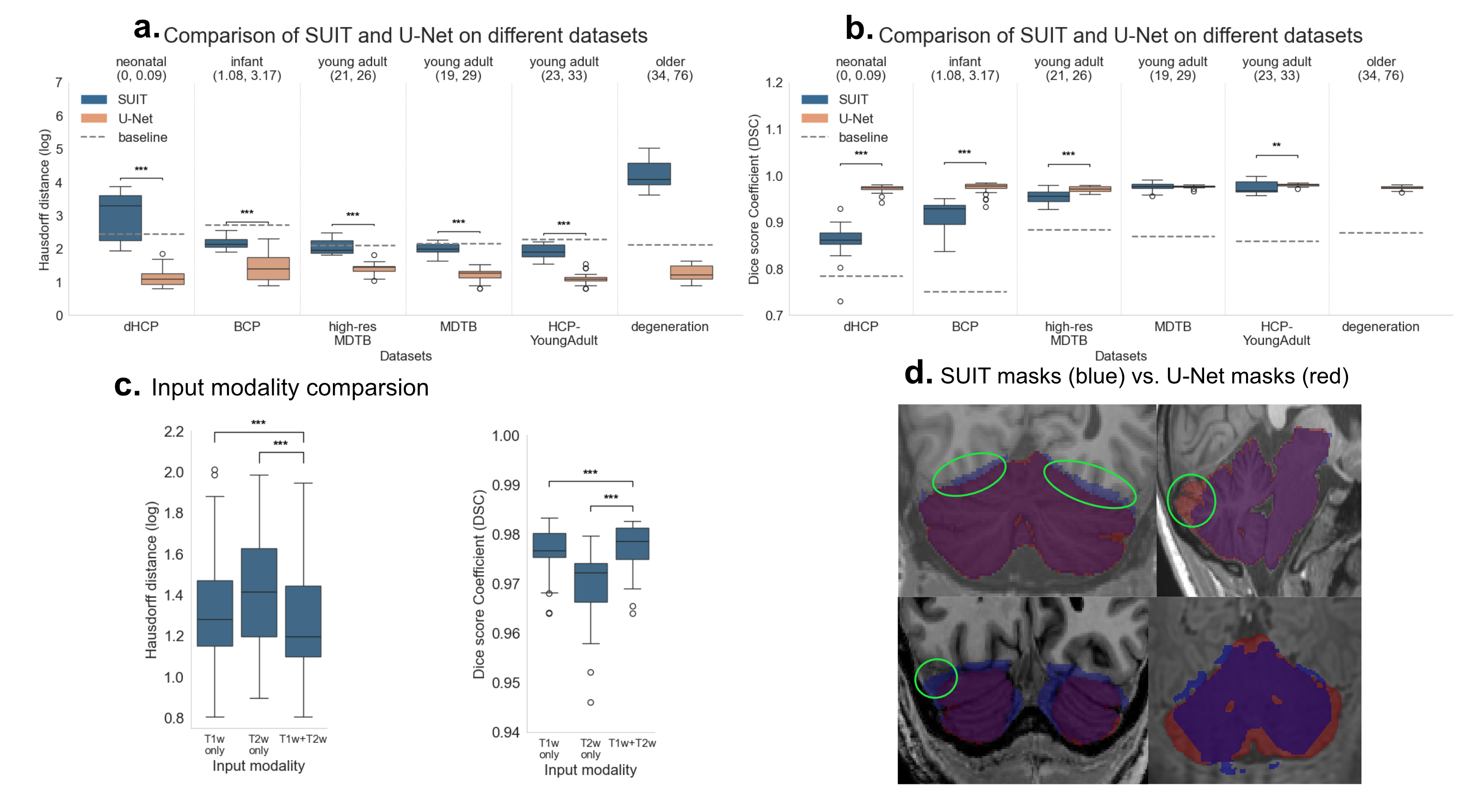
      References
      1. Gribkova, E. D., Lee, C. A., Brown, J. W., Cui, J., Liu, Y., Norekian, T., & Gillette, R. (2023). A common modular design of nervous systems originating in soft-bodied invertebrates. Frontiers in physiology, 14, 1263453.
      2. Brown, J. W., Caetano-Anollés, D., Catanho, M., Gribkova, E., Ryckman, N., ... & Gillette, R. (2018). Implementing goal-directed foraging decisions of a simpler nervous system in simulation. Eneuro, 5(1).
      3. Gribkova, E. D., Catanho, M., & Gillette, R. (2020). Simple aesthetic sense and addiction emerge in neural relations of cost-benefit decision in foraging. Scientific reports, 10(1), 9627.
      4. Gribkova, E. D., Chowdhary, G., & Gillette, R. (2024). Cognitive mapping and episodic memory emerge from simple associative learning rules. Neurocomputing, 595, 127812.


      Acknowledgement
      This work was supported by the Office of Naval Research (MURI grant N00014-19-1-2373).
      Sunday July 12, 2026 4:20pm - 6:20pm ADT
      Ballroom B2

      4:20pm ADT

      P031: Pathway-Specific Temporal Structure of Behavior-Predictive Modeling from Dopaminoceptive Striatal Activity
      Sunday July 12, 2026 4:20pm - 6:20pm ADT
      Introduction
      Forecasting behavioral actions from neuronal circuit activity requires selecting an appropriate prediction horizon and history window for the model. This choice depends on the signal timescale, circuitry, target behaviors, and task structure. In dopaminoceptive striatal circuits, D1R signals may carry both fast action-related and slower reward-expectation components [1]. Modeling based on inhibitory D2R signals is equally important but more challenging, because suppression predictors relate to events less directly, interact with state-dependent effects, and may therefore yield weaker or less interpretable forecasts. In this study, we present an extension of a previously reported D1R forecasting hyperparameter search to D2R signal analysis.


      Methods
      Data came from 9 D1-Cre and 11 A2A-Cre mice with fiber-photometry recordings of GCaMP6 signal from the ventrolateral striatum during head-fixed anticipatory licking protocol [2]. Ca2+, isosbestic channels and licking were sampled at 20 Hz. Signals were detrended for photobleaching, low-pass filtered, artifact-corrected by robust regression of the control channel, and normalized [3]. Licks and lick bursts were extracted as prediction targets. Predictors used fixed-basis representations of signal history [4], with grid search over preset history windows (L) and forecast horizons (H). With binomial logistic GLM, leave-one-mouse-out testing compared dopamine-channel and isosbestic-only models using Precision@1%.


      Results
      Across leave-one-mouse-out testing, D1R signaling improved high-confidence forecasting beyond the isosbestic control in a timescale-dependent manner: for single licks, the strongest gain occurred at short windows (best at L = 0.5 s, H = 0.25 s; ΔP@1% = 0.148), whereas burst-start prediction peaked at an intermediate forecast horizon (H = 1 s), with history peaks at L = 1 s (ΔP@1% = 0.091) and L = 8 s (ΔP@1% = 0.098), consistent with both rapid event-related and slower schedule-aligned dynamics. D2R signaling showed its strongest advantage for burst events at longer history and wider forecast windows (L = 3 s, H = 3 s; ΔP@1% = 0.204) and for single licks at the shortest windows (L = 0.25 s, H = 0.25 s; ΔP@1% = 0.111).

      Discussion
      We extended a temporal hyperparameter search developed for D1R fiber-photometry signals to D2R signals to compare the optimal forecast horizon and history length for excitatory and inhibitory predictors. For single-lick events, both pathways showed similar dynamics: behavior was best predicted over short horizons from recent signal history. For state-dependent events such as licking bursts, the models diverged. D1R signals peaked at a 1 s horizon with 1 s of history, consistent with consummatory or anticipatory state changes. D2R signals were most informative at a 3 s horizon with a 3 s history window, reflecting distinct underlying mechanisms. These findings further indicate the importance of temporal parameter selection for analysis.


      References
      1. Kim, H. R., et al. (2020). A unified framework for dopamine signals across timescales. Cell, 183(6), 1600–1616. https://doi.org/10.1016/j.cell.2020.11.013
      2. Toda, K., et al. (2017). Nigrotectal stimulation stops interval timing in mice. Current Biology, 27(24), 3763–3770. https://doi.org/10.1016/j.cub.2017.11.003
      3. Keevers, L. J., & Jean-Richard-dit-Bressel, P. (2025). Obtaining artifact-corrected signals in fiber photometry via isosbestic signals, robust regression, and dF/F calculations. Neurophotonics, 12(2), 025003–025003. https://doi.org/10.1117/1.NPh.12.2.025003
      4. Pillow, J. W., et al. (2005). Prediction and decoding of retinal ganglion cell responses with a probabilistic spiking model. Journal of Neuroscience, 25(47), 11003–11013. https://doi.org/10.1523/JNEUROSCI.3305-05.2005

      Acknowledgement
      The authors have no additional acknowledgments to report

      Sunday July 12, 2026 4:20pm - 6:20pm ADT
      Ballroom B2

      4:20pm ADT

      P032: AnalySim data and project sharing site: admin panel, notebook and CSV browser expansion
      Sunday July 12, 2026 4:20pm - 6:20pm ADT
      Introduction
      In this poster, we present the updates in the development of the AnalySim science gateway for data sharing and analysis of computational neuroscience projects. AnalySim is an open source project that runs as a web service that allows creating scientific projects and sharing them. An early testing version of the gateway is currently hosted at https://analysim.tech, supported by the NSF-funded ACCESS advanced computing and data resource. The gateway can be installed and run within a lab or larger organization. AnalySim facilitates data sharing, data hosting for publications, interactive visualizations, collaborative research, and crowdsourced analysis. It differs from Github by offering a notebook-oriented interface for a research audience.

      Methods
      AnalySim is built with a .Net backend API service with C# and an Angular frontend using Typescript. The data is saved in a relational database, PostGreSQL, including binary blob storage for files. The interface has a file browser, including a CSV viewing tool, and notebook editing and display capabilities. Hosted projects can have multiple notebook files, where one can be designated as the main project description. Projects can be shared with the public or kept private and shared only with collaborators. Notebooks can be in Python or Javascript backend; ObservableHQ online notebooks are also supported. The source code can be found at https://github.com/soft-eng-practicum/AnalySim along with instructions on how to install and run it.

      Results


      AnalySim has been a participant of the International Neuroinformatics Coordinating Facility (INCF) Google Summer of Code (GSoC) program since 2021. Participation in GSoC 2025 added major features. The user interface was improved to have a more consistent style, and new pages were added to support new functionality, together with less visible improvements in the backend. The changes were: (1) addition of an admin panel that allows browsing and deleting users, projects, datasets, and notebooks; (2) improving the CSV data browser; (3) improvements in the notebook list and using the default notebook as the project description.

      Discussion

      References
      N/A

      Acknowledgement
      We thank INCF and GSoC for supporting AnalySim. This work used Jetstream2 at Indiana University through allocation BIO220033 from the Advanced Cyberinfrastructure Coordination Ecosystem: Services & Support (ACCESS) program, which is supported by National Science Foundation grants #2138259, #2138286, #2138307, #2137603, and #2138296.
      Sunday July 12, 2026 4:20pm - 6:20pm ADT
      Ballroom B2

      4:20pm ADT

      P033: Greater segregation, not integration, accompanies increasing working memory load
      Sunday July 12, 2026 4:20pm - 6:20pm ADT
      Introduction


      Introduction
      The brain reorganizes its network architecture to meet cognitive demands [1], and the balance between segregated and integrated systems is thought to shift with cognitive load [2]. How system segregation changes as load increases, and whether those changes support performance, remains unresolved. This is the case particularly for working memory, where prior findings conflict on whether segregation or integration underpins successful function, and have mainly focused on differences between rest and task rather than incremental increases in task load [3].


      Methods


      Methods
      Using functional MRI in healthy adults, we measured system segregation across five canonical resting-state networks from rest (N=69) to an N-back working memory task (N=27). We also measured system segregation across four levels of task load (0- to 3-back). We correlated system segregation against working memory accuracy and used repeated measures analyses of variance (RM-ANOVA) to compare segregation across task loads. Reproducibility testing included measuring system segregation across structural and functional (Yeo, [4]) parcellations,  sparsity thresholds, and approximating segregation with modularity, a parcellation-independent measure. For a granular understanding, segregation was measured on a subnetwork level as well.


      Results


      Results
      At rest, greater system segregation predicted higher working memory accuracy after controlling for age, sex, and motion. Within the task, segregation increased with load, and both 3-back segregation and its change across load predicted 3-back accuracy. These load effects reproduced when using the parcellation-independent modularity measure, but not under the functional parcellations. This may be related to the divided representation of executive-cognitive regions in these functional networks, as subnetwork analyses revealed that load effects in our primary parcellation was  driven by increased segregation of the unified attention/executive network.


      Discussion


      Discussion
      These findings indicate that the relationship between segregation and cognition is state- and scale-dependent. Load-dependent strengthening of cognitive-network connectivity, rather than global integration, appears to support working memory performance, refining the common assumption that higher cognitive demand requires greater network integration.


      References


      [1] Park, H. J., & Friston, K. (2013). Structural and functional brain networks: from connections to cognition. Science, 342(6158), 1238411.
      [2] Cohen, J. R., & D\'Esposito, M. (2016). The segregation and integration of distinct brain networks and their relationship to cognition. Journal of Neuroscience, 36(48), 12083-12094.
      [3] Finc, K., Bonna, K., He, X., et al, & Bassett, D. S. (2020). Dynamic reconfiguration of functional brain networks during working memory training. Nature communications, 11(1), 2435.
      [4] Yeo, BT Thomas, Fenna M. Krienen, .. Roffman et al. "The organization of the human cerebral cortex estimated by intrinsic functional connectivity." Journal of neurophysiology (2011).

      Acknowledgement


      \nCanadian Institute of Health Research (CIHR) Project Grant [PJT-168878]* \nNSHA Fibromyalgia Research Fibromyalgia Grant* 
      Sunday July 12, 2026 4:20pm - 6:20pm ADT
      Ballroom B2

      4:20pm ADT

      P034: Rising threat, merging networks: dynamic heat pain lowers segregation and turns S2, a core pain region, into an integrative hub
      Sunday July 12, 2026 4:20pm - 6:20pm ADT
      Introduction


      How stronger threat reshapes large-scale brain network organization remains untested. Heightened arousal via sympathetic activation has been proposed to increase functional integration, but no study has directly tested this (1-3). We compared two equivalent heat pain stimuli, a static ramp-and-hold and a dynamically escalating profile, the latter more strongly engaging pain, threat, and reward-punishment circuitry (Sunavsky, in review).

      Methods


      In 30 participants, we extracted epochs spanning 39 TRs from different stimulus conditions and computed region-to-region functional connectivity across a 131-node parcellation. Matrices were proportionally thresholded (0.05 to 0.5) and characterized using graph metrics: system segregation, Louvain modularity, nodal degree, and participation coefficient (4). Conditions were compared with paired t-tests at each threshold, FDR-corrected across all nodes and thresholds.

      Results


      Dynamically escalating threat reduced system segregation relative to the static condition, consistent with greater between-network integration under heightened arousal. Whole-brain FDR-corrected analysis showed increased participation coefficient in bilateral secondary somatosensory cortex (S2, parietal operculum), indicating stronger cross-network connector behavior. Among regions more strongly activated by the dynamic stimulus, only S2 and nucleus accumbens also showed increased hubness (binarized degree). Modularity did not differ between conditions.

      Discussion


      Escalating threat shifts network organization toward integration rather than reconfiguring module structure, since segregation fell while modularity was preserved. The convergence of higher participation and degree at S2 identifies it as a key integrative hub recruited by dynamic threat, with the accumbens implicating reward-punishment circuitry. These findings provide direct evidence that arousal-linked threat promotes functional integration in the human brain.

      References


      1. Shine, J.M., Bissett, P.G., Bell, P.T., Koyejo, O., Balsters, J.H., Gorgolewski, K.J., Moodie, C.A., & Poldrack, R.A. (2016). The dynamics of functional brain networks: integrated network states during cognitive task performance. Neuron, 92(2), 544-554.
      2. Shine, J.M. (2019). Neuromodulatory influences on integration and segregation in the brain. Trends in Cognitive Sciences, 23(7), 572-583.
      3. Chan, M.Y., Park, D.C., Savalia, N.K., Petersen, S.E., & Wig, G.S. (2014). Decreased segregation of brain systems across the healthy adult lifespan. Proceedings of the National Academy of Sciences, 111(46), E4997-E5006.
      4. Rubinov, M., & Sporns, O. (2010). Complex network measures of brain connectivity: uses and interpretations. NeuroImage, 52(3), 1059-1069.

      Acknowledgement


      \nCanadian Institute of Health Research (CIHR) Project Grant [PJT-168878]* \nNSHA Fibromyalgia Research Fibromyalgia Grant* 
      Sunday July 12, 2026 4:20pm - 6:20pm ADT
      Ballroom B2

      4:20pm ADT

      P035: Chronic Pain Uncouples Functional Brain Network Segregation From Cognitive Performance in Aging
      Sunday July 12, 2026 4:20pm - 6:20pm ADT
      Introduction
      Chronic pain (CP) disproportionately affects older adults, not only because it is more prevalent in later life, but also because it may exacerbate cognitive aging and is associated with increased dementia risk [1]. Recent research has implicated accelerated brain aging in CP patients as a potential mechanism behind their disrupted cognitive aging [1]. However, this work remains limited in its focus on structural features of brain aging in CP. Alternatively, the role of functional brain network segregation in normative neurocognitive aging has been robustly characterized; it declines with age, with lower segregation linked to cognitive decline and dementia risk [2,3]. Whether CP disrupts these relationships has not yet been examined.

      Methods
      Participants included healthy controls without ongoing pain, and CP patients with chronic back pain or fibromyalgia. After age-sex matching and censoring participants with fMRI head motion above acceptable values, the final sample included 60 controls and 141 CP patients. Executive cognition was assessed using the n-back task, stop-signal task, and Stroop task. Functional brain network segregation was quantified using the system segregation metric, with network communities defined using the Harvard-Oxford Optimized parcellation. PROCESS moderation analysis in SPSS was used to test interaction effects.

      Results
      Overall system segregation and cognition did not differ between healthy controls and CP patients. However, older age predicted poorer performance across all tasks in the patient group, but only stop-signal performance in controls, with diagnosis significantly moderating the association between age and stroop task performance. Despite this accelerated cognitive aging pattern, system segregation declined with age in controls but not CP patients, with diagnosis also moderating this association. Finally, CP diagnosis significantly reversed the association between working memory (n-back accuracy) and system segregation: higher segregation predicted better working memory in controls but worse working memory in CP patients.

      Discussion
      We found that chronic pain was associated with an altered brain aging trajectory in which network segregation is relatively preserved but becomes cognitively maladaptive, predicting worse rather than better working memory performance.  If age-related declines in segregation reflect compensation for structural degeneration, this pattern may indicate impaired functional compensation in chronic pain patients, who already show accelerated structural brain aging [1]. Overall, our findings suggest that the role of system segregation in neurocognitive aging is disrupted in the presence of CP, with important implications for interpreting related neuroimaging biomarkers and developing cognitive interventions in this population [3].

      References
      1: Zhao, L., Zhang, L., Tang, Y., & Tu, Y. (2025). Cognitive impairments in chronic pain: A brain aging framework. Trends in Cognitive Sciences. https://doi.org/10.1016/j.tics.2024.12.004 
      2: Calder, C. N., Helmick, C., & Hashmi, J. A. (2026). High brain network system segregation is differentially linked with cognitive performance across the life span. Network Neuroscience, 10(2), 352–373. https://doi.org/10.1162/NETN.a.542
      3: Zhang, Z., Chan, M. Y., Han, L., Carreno, C. A., Winter-Nelson, E., Wig, G. S., & Alzheimer’s Disease Neuroimaging Initiative. (2023). Dissociable effects of Alzheimer’s disease-related cognitive dysfunction and aging on functional brain network segregation. Journal of Neuroscience, 43(46), 7879–7892. https://doi.org/10.1523/JNEUROSCI.0579-23.2023

      Acknowledgement
      I would like to thank my supervisor, Dr. Javeria Hashmi, and the entire Netphys lab for supporting this research and fostering a passionate environment for scientific thought.
      My work was supported by the Brain Repair Centre through the Dalhousie Faculty of Medicine 2025 Graduate Studentship program and by the CIHR through the Canada Graduate Scholarship – Master’s Program.
      Sunday July 12, 2026 4:20pm - 6:20pm ADT
      Ballroom B2

      4:20pm ADT

      P036: Inferring microcircuit aging from subject EEG and linking to cognitive decline
      Sunday July 12, 2026 4:20pm - 6:20pm ADT
      Introduction
      Cognitive decline occurs in aging [1] and is accompanied by changes in electroencephalography (EEG) signals [2]. However, the cellular mechanisms underlying these EEG alterations cannot be directly assessed in living humans. Key cellular and microcircuit mechanisms implicated in human aging include reductions in inhibition from different interneuron types, dendritic spine density, and NMDA receptor signaling [3–5], but the links to age-related cognitive decline and EEG changes remain to be established.


      Methods
      To overcome experimental limitations, we trained artificial neural networks (ANN) to estimate microcircuit aging using simulated EEG biomarkers generated by detailed models of human cortical microcircuits that integrated key microcircuit mechanisms in human aging [6]. We then applied the ANNs to estimate microcircuit aging for each subject in the LEMON dataset [1] (ages 20-80) from their resting-state EEG and examined associations between estimated microcircuit aging and cognitive scores.


      Results
      The simulated EEG biomarkers from aging microcircuit simulations accounted for a large portion of the range of changes in aging patient EEG. The ANNs estimated microcircuit aging with high precision in silico, and when applied to human EEG data, estimated microcircuit aging corresponded with subject age and was correlated with cognitive decline across multiple cognitive domains. Furthermore, we found sex-specific differences in correlations with microcircuit age for some of the cognitive domains. Among EEG biomarkers used by the ANN, the aperiodic features most strongly influenced microcircuit aging estimations.


      Discussion
      We demonstrate a modeling-informed approach to estimate microcircuit aging in human subjects from non-invasive EEG, and showed that microcircuit aging was associated with cognitive decline. Future directions will be to estimate changes in the levels of the individual microcircuit mechanisms to tease apart their contributions to cognitive decline. Our approach and tools improve the mechanistic understanding of aging and may further serve in clinical stratification of associated pathologies.


      References
      1.\tBabayan A, et al. (2019). Sci Data. 6(1):180308. DOI: 10.1038/sdata.2018.308
      2.\tMerkin A, et al. (2023). Neurobiology of Aging. 121:78–87. DOI: 10.1016/j.neurobiolaging.2022.09.003
      3.\tChen Y, et al. (2023). Neurobiology of Aging. 125:49–61. DOI: 10.1016/j.neurobiolaging.2023.01.013
      4.\tPetanjek Z, et al. (2011). Proceedings of the National Academy of Sciences. 108(32):13281–6. DOI: 10.1073/pnas.1105108108
      5.\tPegasiou CM, et al. (2020). Cerebral Cortex. 30(7):4246–56. DOI: 10.1093/cercor/bhaa052
      6.\tGuet-McCreight A, et al. (2025). Aging Cell. e70329. DOI: 10.1111/acel.70329

      Acknowledgement
      Alexandre Guet-McCreight and Etay Hay thank the Krembil Foundation for their generous funding support. Alexandre Guet-McCreight thanks the Canadian Institutes of Health Research—Institute of Aging for funding support.

      Speakers
      avatar for Etay Hay

      Etay Hay

      Scientist, Centre for Addiction and Mental Health
      avatar for Alexandre Guet-McCreight

      Alexandre Guet-McCreight

      Postdoctoral Research Fellow, Centre for Addiction and Mental Health
      I earned my PhD in Computational Neuroscience from the University of Toronto, under the supervision of Dr. Frances Skinner, with a focus on biophysical modeling of inhibitory hippocampal cells. After a postdoctoral period at the Krembil Brain Institute, I joined Dr. Etay Hay’s lab... Read More →
      Sunday July 12, 2026 4:20pm - 6:20pm ADT
      Ballroom B2

      4:20pm ADT

      P037: The effects of reduced somatostatin interneuron inhibition in depression on multilayered human cortical microcircuit activity.
      Sunday July 12, 2026 4:20pm - 6:20pm ADT
      Introduction
      Major depressive disorder (depression) is associated with reduced cortical inhibition from somatostatin-expressing (SST) interneurons, as indicated by decreased SST expression in human post-mortem studies[1]. We previously showed in simulations of human cortical layer 2/3 that reduced SST interneuron inhibition would increase baseline cortical activity (noise) to significantly reduce the signal-to-noise ratio in signal processing and contribute to cognitive deficits observed in depression[2]. However, as SST interneuron proportion and connectivity vary across cortical layers, it is unclear how reduced SST interneuron inhibition in depression differentially affects processing and signal propagation across cortical layers[3].


      Methods
      In this study, we generated biophysical models of multilayered human cortical microcircuits that encompass 4000 neurons with detailed morphologies spanning 12 neuron types across layers 2-5. Our models integrated human cellular, synaptic, neuron proportion, and connectivity data, such as human paired recordings and electron-microscopy reconstruction of a human cortical column[4]. To better capture biological variability, we incorporated heterogeneity in synaptic strengths, transmission delays, and connection probabilities. We simulated electroencephalography (EEG) signals arising from the microcircuit using NEURON and LFPy, and reproduced properties of the power spectrum density (PSD) using thalamic drive and adjusting connectivity.


      Results
      We reproduced healthy baseline firing rates across cell types and oscillatory dynamics (1/f decay and peak power in alpha frequency band) as seen in human resting-state EEG. By systematically reducing SST inhibition within and across layers, we quantified the difference in layer-specific contributions to circuit-level dysfunction and altered EEG power spectral density during resting state.


      Discussion
      Our study characterizes the effect of reduced inhibition in depression on cortical activity and signal processing across layers, and thereby furthering current understanding of the role dendritic inhibition plays in signal processing in health and depression. Furthermore, our characterization of the signatures of reduced SST inhibition across layers on resting-state EEG due refine our previous biomarkers, and may serve to improve current stratification of depression patients. Finally, our models of multilayers human cortical microcircuits can be used by the scientific community to study cortical processing in health and other diseases.


      References


      1.         Seney, M. L., Tripp, A., … Sibille, E. (2015). Laminar and cellular analyses of reduced somatostatin gene expression in the subgenual anterior cingulate cortex in major depression. Neurobiology of Disease, 73(Complete), 213–219.
      2.          Yao, H. K., Guet-McCreight, A., … Hay, E. (2022). Reduced inhibition in depression impairs stimulus processing in human cortical microcircuits. Cell Reports, 38(2).
      3.          Tremblay, R., Lee, S., & Rudy, B. (2016). GABAergic Interneurons in the Neocortex: From Cellular Properties to Circuits. Neuron, 91(2), 260–292.
      4.          Shapson-Coe, A., Januszewski, M., A., … Lichtman, J. W. (2024). A petavoxel fragment of human cerebral cortex reconstructed at nanoscale resolution. Science, 384(6696), eadk4858.

      Acknowledgement
      This study was supported by a fellowship grant from the Labatt Family Network for Research on the Biology of Depression
      Speakers
      avatar for Etay Hay

      Etay Hay

      Scientist, Centre for Addiction and Mental Health
      Sunday July 12, 2026 4:20pm - 6:20pm ADT
      Ballroom B2

      4:20pm ADT

      P038: Cerebellar Isolation with Multi-Modality MRI Images Using Deep Learning
      Sunday July 12, 2026 4:20pm - 6:20pm ADT
      Introduction
      The cerebellum is involved in motor, cognitive, and affective functions. A critical prerequisite for cerebellar MRI analyses is the isolation from the brain as spatial normalization to whole-brain templates misaligns the cerebellum, compromising accuracy. To this end, specialized tools like SUIT [1] were developed. However, current software has two key limitations: a. They were developed on healthy adults, lacking robustness across diverse populations; b. They use single-modality input, ignoring complementary contrasts like T2w. This work introduces a 3D U-Net [2] for cerebellar isolation that solves both problems, producing reliable results across lifespan and a dual-input architecture for improved accuracy.


      Methods
      We combined 5 different databases (N=101), spanning ages 0-76 years. Raw images were registered to the MNI152NLin6Asym template[3] where cropping was applied via a fixed bounding box. We implemented a 3D U-Net with four encoding/decoding stages. Each stage contains 3D convolutions, instance normalization, and LeakyReLU. The network accepts dual-channel inputs for T1w and T2w modality images and handles missing modalities via zero-padding. Skip connections preserve spatial details for accurate boundary delineation. Model outputs were transformed back to native space and postprocessed. The performance was measured by Dice Score Coefficient (DSC) and Hausdorff Distance.

      Results

      We compared our U-Net against SUIT across the lifespan. On adult data (SUIT's optimal population), our U-Net achieved lower Hausdorff distances, indicating superior boundary alignment. Critically, SUIT failed completely on neonatal and elderly degenerative cases (24.4% failure rate), while U-Net performed consistently across all ages. For multi-modality evaluation, U-Net outperformed SUIT with single modalities (T1w or T2w). Combined T1w+T2w inputs yielded significantly better results than either alone, demonstrating successful fusion of complementary contrast information (See Figure 1).

      Discussion

      This study presents a 3D U-Net for cerebellar isolation trained on diverse multi-modal data (0-76 years, including pathology). The model outperformed SUIT across both metrics, particularly in boundary precision, and generalized effectively across the lifespan where SUIT failed. A key strength is handling T1w/T2w inputs individually or jointly for improved robustness and accuracy. Another contribution is our expertly curated dataset of 101 hand-corrected masks for other researchers. Limitations include a modest sample size for rare pathologies and a focus on structural MRI only. So, in the future, we will expand to other contrasts and populations.

      Figure 1. Isolation analysis. Comparison of U-Net VS SUIT for (a) Hausdorff Distance (HD) and (b) Dice Score Coefficient (DSC). c shows performance for different input modalities in the full datasets. Horizontal lines between bars with asterisks denote significant differences (paired t-tests). Baseline: Average mask prediction. d shows resulting mask in problematic subjects.
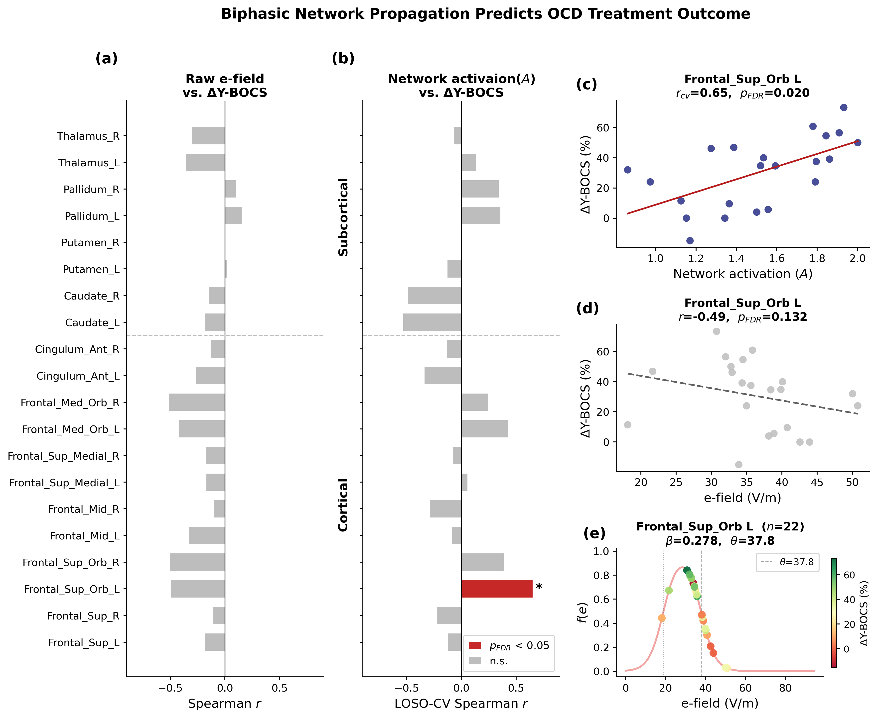
      References
      1. Diedrichsen, J. (2006). A spatially unbiased atlas template of the human cerebellum. NeuroImage, 33(1), 127–138. https://doi.org/10.1016/j.neuroimage.2006.05.056
      2. Ronneberger, O., Fischer, P., Brox, T. (2015). U-Net: Convolutional Networks for Biomedical Image Segmentation. Medical Image Computing and Computer-Assisted Intervention – MICCAI 2015, 9351, 234-241. Springer. https://doi.org/10.1007/978-3-319-24574-4_28
      3. Fonov, V., Evans, A., McKinstry, R., Almli, C., & Collins, D. (2009). Unbiased nonlinear average age-appropriate brain templates from birth to adulthood. NeuroImage, 47(Supplement 1), S102. https://doi.org/10.1016/S1053-8119(09)70884-5

      Acknowledgement
      This research was funded by the Raynor Cerebellum Project. We thank the Brain and Mind Institute at Western University for data acquisition and support. We acknowledge the contributors of the public datasets used in this work: dHCP, BCP, and HCP-YoungAdult. We are grateful to the expert raters for manual mask validation.

      Sunday July 12, 2026 4:20pm - 6:20pm ADT
      Ballroom B2

      4:20pm ADT

      P039: The Self-Organization of Serotonergic Axons at the Surface of a Vertebrate Brain: A Myopic Self-Avoiding FBM Model
      Sunday July 12, 2026 4:20pm - 6:20pm ADT
      Introduction
      Serotonergic axons (fibers) are present in virtually all brain regions of vertebrate animals (from humans to fishes). Within these regions, individual serotonergic fibers typically grow in paths that resemble random walks with memory. At the population level, they produce regionally-specific fiber meshworks that are thought to support the neuroplasticity of local, non-serotonergic neural circuits. We have recently developed a computational framework in which the trajectories of serotonergic fibers are modeled as paths of reflected fractional Brownian motion (rFBM), with simulated fiber densities approximating the biological densities in the forebrains of two phylogenetically distant species, the mouse [1] and the Pacific angelshark [2].


      Methods
      This study focuses on the distribution of serotonergic fibers at the brain surface. Specifically, we investigate the emergence of high-density fiber bands in the dorsal pallium (including the mammalian cerebral cortex) that rFBM cannot capture accurately. We introduce a new animal model, the Pacific electric ray (Tetronarce californica), the forebrain of which has an extremely simple geometry with no ventricles or major fibers in the telencephalon. Its serotonergic fibers are visualized with immunohistochemistry, with comparisons to the mouse and angelshark brains [1,2]. We also introduce a major theoretical extension of rFBM, the reflected myopic self-avoiding FBM, based on a recently developed myopic self-avoiding FBM model [3].


      Results
      In the electric ray telencephalon, serotonergic fibers generally accumulate at higher densities near the pial surface, consistent with findings in other vertebrate species. However, they additionally produce dense bands in the dorsal pallium (homologous to the mammalian cerebral cortex). The emergence of this biologically important feature cannot be explained by simple rFBM but is consistent with our simulations using the reflected myopic self-avoiding FBM, a stochastic process that includes a mean-density interaction between the members of the fiber ensemble. These simulations show that the interaction cuts off the divergence of the density at a reflecting boundary, producing a high-density plateau in the vicinity of the boundary.


      Discussion
      Our results suggest that FBM, with its recently developed theoretical extensions by our group [3,4], can eventually predict the key features of serotonergic fiber densities in any vertebrate brain. This approach relies on the geometry and regional viscoelasticity of the brain and is agnostic to anatomically-defined brain nuclei and their biological function. This research contributes to the understanding of the minimal set of principles that lead to the self-organization of the fundamental brain architecture. In addition, it stimulates the theoretical development of FBM-related stochastic processes, with broad applications in other fields.


      References
      1. Janušonis, S., Haiman, J.H., Metzler, R., Vojta, T. (2023) Predicting the distribution of serotonergic axons [...]. Front. Comput. Neurosci. 17: 1189853. https://doi.org/10.3389/fncom.2023.1189853  
      2. Janušonis, S., Metzler, R., Vojta, T. (2025) The organization of serotonergic fibers in the Pacific angelshark brain [...]. Front. Neurosci. 19: 1602116. https://doi.org/10.3389/fnins.2025.1602116
      3. House, J., Bakhshizada, R., Janušonis, S., Metzler, R., Vojta, T. (2025) Fractional Brownian motion with mean-density interaction [...]. Phys. Rev. E 112: 034119. https://doi.org/10.1103/w5pk-bw5r
      4. Wang, W., Balcerek, M., Burnecki, K., et al. (2023) Memory-multi-fractional Brownian motion with continuous correlation. Phys. Rev. Res. 5: L032025. https://doi.org/10.1103/PhysRevResearch.5.L032025

      Acknowledgement
      This research was funded by an NSF-BMBF CRCNS grant (NSF #2112862 to SJ & TV).

      Sunday July 12, 2026 4:20pm - 6:20pm ADT
      Ballroom B2

      4:20pm ADT

      P040: Dendritic calcium dynamics shape functionally relevant human E-/MEG ~20Hz beta events: a biophysical modeling study
      Sunday July 12, 2026 4:20pm - 6:20pm ADT
      Introduction
      Electroencephalography (EEG) and Magnetoencephalography (MEG) are widely used non-invasive techniques to record electric brain activity from the human brain. While it has long been known that synchronous intracellular currents in dendrites of neocortical pyramidal neurons underlie E/MEG signals, only few theories on the computational role of E/MEG dynamics consider dendritic computations. For instance, high-amplitude transient ~20Hz oscillations known as cortical beta-events, dominate human E/MEG signals and have been repeatedly argued to support perception and motor action by orchestrating spiking activity [1,2]. Yet, the neural generators of lower frequency oscillations and their effect on dendritic activity are underexplored.


      Methods
      Here, we present a biophysically detailed neocortical circuit model that is optimized to study how dendritic processes manifest in human E/MEG signals. Somatic conductances were tuned to reproduce dynamics observed in in vitro experiments from human donors. The active conductances in the pyramidal neuron dendrites were tuned to reproduce non-linear processes associated with intracellular calcium. The layer 5 neurons exhibit calcium plateau potentials in response to high-frequency somatic spiking, coincident somatic and dendritic inputs, and strong feedback inputs, as repeatedly shown in the literature [3,4]. The layer 2/3 pyramidal neurons generate shorter dendritic spikes that have recently been reported in human neurons [5,6].


      Results
      Expanding prior theories on the generation of beta events [7,8], we demonstrate how dendritic calcium spikes, in combination with somatic and dendritic inhibition by GABAergic interneurons, can produce the characteristic waveform shapes at 15-25 Hz observed in human E/MEG data. These findings complement previous reports of dendritic calcium spikes being detectable at the cortical surface in rodents [9], by demonstrating that these spikes are associated with large currents that dominate the E/MEG signal. To test and constrain model predictions, we show preliminary evidence comparing simulated extracellular fields to laminar recordings during homologous beta events in rodents.


      Discussion
      Our modeling work makes important contributions to understanding the role of dendritic calcium dynamics in the multiscale neural generators of functionally relevant human brain oscillations. Across species translation of our results provides a powerful framework to examine the causal influence of dendritic processes and other beta event generating mechanisms in sensory perception and motor action.  The circuit is packaged and distributed within the user-friendly Human Neocortical Neurosolver (HNN) software (https://hnn.brown.edu) designed for multiscale interpretation of human E/MEG signals, making our tools and results available to a broad neuroscience community.


      References
      [1] Shin, H. et al. (2017). elife, 6, e29086.
      [2] Bonaiuto, J. J. et al. (2021). NeuroImage, 242, 118479.
      [3] Larkum, M. E. et al. (1999). Proceedings of the National Academy of Sciences, 96(25), 14600-14604.
      [4] Larkum, M. et al. (1999). Nature, 398(6725), 338-341.
      [5] Gidon, A. et al. (2020). Science, 367(6473), 83-87.
      [6] Gooch, H. M. et al. (2022). Cell reports, 41(3).
      [7] Sherman, M. A. et al. (2016). Proceedings of the National Academy of Sciences, 113(33), E4885-E4894.
      [8] Law, R. G. et al. (2022). Cerebral Cortex, 32(4), 668-688.
      [9] Suzuki, M., & Larkum, M. E. (2017). Nature communications, 8(1), 276.
      Sunday July 12, 2026 4:20pm - 6:20pm ADT
      Ballroom B2

      4:20pm ADT

      P041: Predicting natural video motion from spiking activity across the mouse visual pathway
      Sunday July 12, 2026 4:20pm - 6:20pm ADT
      Introduction
      Visual information from individual photoreceptors represents only simple light intensity information. More complex visual information, such as motion, is discerned by considering the combined responses from several receptors, or over a duration. The exact processes and locations at which encoding steps occur along the visual pathway are unclear. Yet, by aligning response preferences of neurons to the presence of specific visual stimuli, specialised encoding regions may be identified. Using computer vision methods, we demonstrate the ability to extract simple visual components from natural stimuli and, using electrophysiological data in mice, predict neuronal optical flow response preferences across the visual pathway.

      Methods
      Electrophysiological recordings from the public Allen Brain Observatory dataset, comprising responses of 32 mice (Mus musculus) to varied artificial and natural stimuli, were processed to detect spiking action potentials [1]. Dense optical flow analysis was performed to extract motion magnitude and direction by estimating local neighbourhood displacement between frames. Magnitudes were weighted by their cosine direction components to assess correlation between spiking rate and motion magnitude in 8 directions. Moreover, region-specific logistic regression models were trained, using either drifting grating or natural video stimulus-response data, to predict predominant global motion direction for a novel natural video from spiking rates.

      Results
      Subsets of neurons within regions displayed correlation between spiking rate and optical flow magnitude consistently across repeated presentations, but due to motion direction bias within the video, horizontal direction preferences were more represented than vertical ones. Regional regression models were able to predict predominant motion direction, with accuracy varying across regions, and specific direction performance reliant on sufficient training examples. Both models trained using only drifting gratings, or only natural video, displayed high direction prediction accuracy to a novel video. Hence, we identify a subset of visual pathway cells with directional coding preferences to natural video motion consistent with rate-based coding.

      Discussion
      This study was motivated by previous attempts to train models to predict high-resolution pixel images from spiking activity, whereby models were unable to generalise to predict novel stimuli [2]. Our work elucidates possible shortcomings in such an approach that warrant further investigation: mouse spiking activity contains less relevant pixel information than we previously believed, quantified by our analysis, likely due to a specialisation for motion over acuity. This study represents the first to compare regional differences in visual feature prediction, using electrophysiological activity, for a novel natural video. Future work aims to explore more specific feature predictions, including foreground/background motion discrimination.

      References
      1.      Siegle, J. H., Jia, X., Durand, S., Gale, S., Bennett, C., Graddis, N., Heller, G., Ramirez, T. K., Choi, H., Luviano, J. A., Groblewski, P. A., Ahmed, R., Arkhipov, A., Bernard, A., Billeh, Y. N., Brown, D., Caldejon, S., Casal, L., Cho, A., … Koch, C. (2021). Survey of spiking in the mouse visual system reveals functional hierarchy. Nature, 592(7852), 86–92. https://doi.org/10.1038/s41586-020-03171-x
      2.      Chen, Y., Beech, P., Yin, Z., Jia, S., Zhang, J., Yu, Z., & Liu, J. K. (2024). Decoding dynamic visual scenes across the brain hierarchy. PLoS Computational Biology, 20(8), e1012297. https://doi.org/10.1371/journal.pcbi.1012297

      Acknowledgement
      This project utilises the open source Allen brain observatory visual coding neuropixels dataset from the Allen Institute for Brain Science [1].
      Sunday July 12, 2026 4:20pm - 6:20pm ADT
      Ballroom B2

      4:20pm ADT

      P042: Neural Heterogeneity Controls Neural Network Development
      Sunday July 12, 2026 4:20pm - 6:20pm ADT
      Introduction
      We study the role of interaction between synaptic plasticity rules and cellular physiology in producing useful connectivity in neural populations. We leverage two key aspects of biological neural networks: 1) neurons and the synapses connecting them are inherently diverse in their structure and electrophysiological properties, and 2) synapses are highly plastic and subject to activity-dependent changes in strength, which can be mathematically formalized by rules such as spike-timing-dependent plasticity.


      Methods
      We address this question in networks of quadratic integrate-and-fire (QIF) neurons endowed with STDP. We develop a multi-population mean-field model [1] that incorporates spike synchronization, allowing it to reproduce synaptic weight evolution in heterogeneous spiking neural networks — something conventional rate models fail to capture. Mathematically, synaptic evolution is driven by variables that trace past spiking events with time constants determined by the respective learning rule [2].


      Results
      We find that the evolving connectivity patterns are the natural result of an interaction between neural heterogeneity and STDP. The mean-field model captures complex network structure even when it is relatively coarse-grained compared to the network of QIF neurons. As potential applications, we demonstrate that this model can flexibly store associative memory items, and encode memory sequences with repeating items.


      Discussion
      We conclude that the mean-field model can accurately predict synaptic pattern formation in heterogeneous spiking networks. Not only can the model be used for analysis methods such as bifurcation analysis that are not available for discontinuous spiking neuron models, but it should also be applicable for a larger family of synaptic plasticity rules [3].


      References
      [1] Richard Gast, Thomas R. Knösche, and Helmut Schmidt (2021). Mean-field approximations of networks of spiking neurons with short-term synaptic plasticity. Physical Review E, 104(4):044310.


      [2] Morrison, Abigail, Markus Diesmann, and Wulfram Gerstner (2008). Phenomenological models of synaptic plasticity based on spike timing. Biological Cybernetics 98(6): 459-478.


      [3] Pfister, Jean-Pascal, and Wulfram Gerstner (2006). Triplets of spikes in a model of spike timing-dependent plasticity. Journal of Neuroscience 26(38): 9673-9682.

      Acknowledgement
      This work was supported by a Lumina-Quaeruntur fellowship (LQ100302301 awarded to H.S.) founded by the Czech Academy of Sciences, the Czech Science Foundation (No. 25-15412L), and the Brain dynamics project (No. CZ.02.01.01/00/22_008/0004643) funded by the European Regional Development Fund.

      Speakers
      HS

      Helmut Schmidt

      Scientific researcher, Institute of Computer Science, Czech Academy of Sciences
      Sunday July 12, 2026 4:20pm - 6:20pm ADT
      Ballroom B2

      4:20pm ADT

      P043: Modeling Network Effects of Transcranial Magnetic Stimulation on Obsessive Compulsive Disorder
      Sunday July 12, 2026 4:20pm - 6:20pm ADT
      Introduction

      Transcranial magnetic stimulation (TMS) induces electric fields (E-fields) that propagate through white matter pathways, influencing distributed brain networks beyond the stimulation site. Deep TMS using the H7 coil is an FDA-cleared treatment for obsessive-compulsive disorder (OCD), targeting the medial prefrontal cortex (mPFC) and anterior cingulate cortex (ACC), key nodes of the cortico-striato-thalamo-cortical (CSTC) circuit. However, how individual differences in E-field distribution and network propagation contribute to clinical response remains unclear. We hypothesized that a dose-dependent local neuronal response, amplified through structural connectivity, predicts treatment outcome following deep TMS in OCD.


      Methods

      Twenty-two patients with OCD received 6 weeks of TMS (20 Hz, 100% rMT) targeting the mPFC/ACC. Individual E-field distributions were computed and averaged within 90 AAL brain regions. Network activation was modeled as A = (I − αC)-1 r, where C is the structural connectome, α is the network coupling strength, and r = f(e; β, θ) represents the local neuronal response to E-field magnitude e. Four neuronal response models were evaluated: excitatory/inhibitory sigmoid, exponential decay, and biphasic. (α, β, θ) were optimized across CSTC ROIs via leave-one-subject-out cross-validation. Predicted network activation was correlated with percentage improvement on the Yale-Brown Obsessive-Compulsive Scale (Y-BOCS, Spearman).


      Results

      Across four candidate neuronal response models, only the biphasic model yielded a cross-validated prediction that survived FDR correction (Fig. 1). Network activation within the left superior orbital frontal region was positively correlated with percentage Y-BOCS improvement (r = 0.65, p_fdr = 0.012). The optimized parameters (β = 0.189, θ = 37.78, α = 0.879) defined an optimal E-field window of ~20–38 V/m; stimulation beyond this range produces reduced neuronal responses. The strong coupling (α = 0.879) indicates local effects are greatly amplified through structural connectivity. Parameter estimates were highly stable across LOSO folds (interquartile range = 0 for all parameters).


      Discussion

      These findings suggest that individual differences in therapeutic response to deep TMS may depend on both local E-field magnitude and its propagation through long-range structural networks. Furthermore, the left frontal superior orbital region might be an effective TMS target for OCD treatment. The biphasic dose-response suggests an optimal E-field window for therapeutic stimulation. Below this window, stimulation is insufficient to drive meaningful neuronal responses; above it, excessive E-field likely reduces neuronal output, forming an inverted-U dose-response relationship. Together, these results support a network-based framework for the mechanisms of deep TMS in OCD and highlight the importance of individualized E-field optimization.

      Figure 1. Correlations between E-field/network activation and clinical outcome in the CSTC ROIs. (a) Raw E-field showed no significant correlations. (b) The biphasic neuronal response model identified the left superior orbital frontal cortex as significantly associated with percentage Y-BOCS improvement. (c-f) Network activation/raw E-field correlation maps, and fitted biphasic neuronal response fReferences
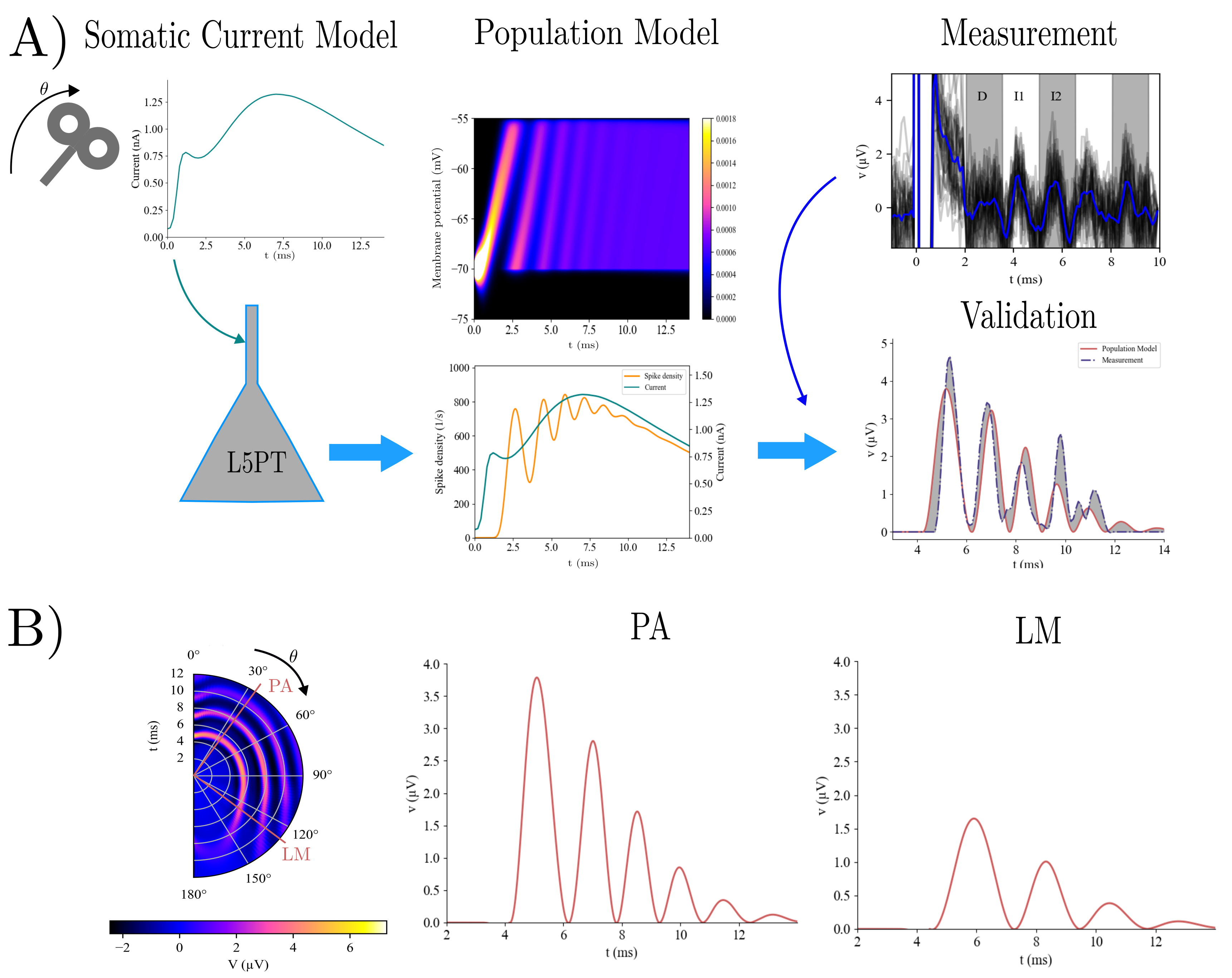
      1. Harel, M., Perini, I., Kämpe, R., Alyagon, U., Shalev, H., Besser, I., ... & Zangen, A. (2022). Repetitive transcranial magnetic stimulation in alcohol dependence: a randomized, double-blind, sham-controlled proof-of-concept trial targeting the medial prefrontal and anterior cingulate cortices. Biological psychiatry, 91(12), 1061-1069.
      2. Burguiere, E., Monteiro, P., Mallet, L., Feng, G., & Graybiel, A. M. (2015). Striatal circuits, habits, and implications for obsessive–compulsive disorder. Current opinion in neurobiology, 30, 59-65.


      Acknowledgement
      No
      Speakers
      HS

      Helmut Schmidt

      Scientific researcher, Institute of Computer Science, Czech Academy of Sciences
      JH

      Jaroslav Hlinka

      Senior researcher, Institute of Computer Science of the Czech Academy of Sciences
      Currently                                I am leading the and also serve as the Head of the Department of Complex Systems and as the Chair of the Council of the of the Czech Academy of Sciences.
      Brief bio After obtaining master degrees in Psychology from Charles University (2005) and in Mathematics from Czech Technical University (2006), I went on the quest of applying mathematics in helping to understand the complex activity of human brain through neuroimaging data analysis... Read More →
      Sunday July 12, 2026 4:20pm - 6:20pm ADT
      Ballroom B2

      4:20pm ADT

      P044: Discovering the dynamics of evoked responses to near-threshold tactile stimuli: A layer-specific neural mass model of the somatosensory microcircuit
      Sunday July 12, 2026 4:20pm - 6:20pm ADT
      Introduction
      The processing of tactile stimuli relies on complex dynamics within cortical microcircuits. Near the perceptual threshold, unperceived stimuli evoke somatosensory responses that differ from perceived ones, a phenomenon extensively studied with EEG [1]. However, the neural mechanisms underlying this transition are poorly understood. Neural mass models are a tool to describe dynamics of cortical circuits and give a mechanistic understanding of their functions. Here, we present such a model to investigate these unknown dynamics.


      Methods
      The model consists of two cortical columns representing the primary and secondary somatosensory cortex, each with granular, supra-, and infra-granular layers. It includes pyramidal neurons and interneuron populations of three different types (somatostatin-, parvalbumin-, and vasoactive-intestinal-peptide-expressing interneurons). Mean firing rate and membrane potential are defined based on the Jansen-Rit model [2]. Connectivity, cell counts and synaptic properties are obtained from animal studies and previous models [3,4].


      Results
      Our approach reveals the characteristics of the somatosensory microcircuit dynamics with respect to model parameters. The model allows for precise predictions of how connectivity pattern and excitation-inhibition balance of each neuronal population shapes its individual functional role in generating tactile evoked responses and in letting input pass to higher cortical areas, reflecting the perception of the tactile stimulus. In combination with feedforward bottom-up input, top-down input from higher areas influences perceptual gating. An observation model transforms firing rates and membrane potential into EEG- and LFP-like signals, allowing future fitting to real recordings to improve interpretability and validity.


      Discussion
      Our model offers a biologically plausible approach to investigate somatosensory perception. It provides hypothetical mechanisms underlying the processing of tactile stimuli and the transition from subliminal to supraliminal responses. By bridging the gap between macroscopic measurements and microscopic neural dynamics, this model enhances our understanding of the mechanisms underlying tactile perception at multiple levels. Future work will fit the model to empirical data from near-threshold detection tasks.


      References
      1. Forschack, N., Nierhaus, T., Müller, M. M., & Villringer, A. (2020). Dissociable neural correlates of stimulation intensity and detection in somatosensation. NeuroImage, 217, 116908.
      2. Jansen, B. H., & Rit, V. G. (1995). Electroencephalogram and visual evoked potential generation in a mathematical model of coupled cortical columns. Biological Cybernetics, 73(4), 357–366.
      3. Isbister, J. B., Ecker, A., Pokorny, ... & Reimann, M. W. (2024). Modeling and Simulation of Neocortical Micro- and Mesocircuitry. Part II: Physiology and Experimentation. bioRxiv.
      4. Jiang, H.-J., Qi, G., Duarte, R., Feldmeyer, D., & Albada, S. J. van. (2023). A Layered Microcircuit Model of Somatosensory Cortex with Three Interneuron Types and Cell-Type-Specific Short-Term Plasticity. bioRxiv.

      Acknowledgement
      This work was supported by the IMPRS programs. We thank the members of our group BrainNets  and the Neurology department for insightful discussions and continuous feedback. We are also grateful to previous interns who contributed to preliminary simulations.
      Sunday July 12, 2026 4:20pm - 6:20pm ADT
      Ballroom B2

      4:20pm ADT

      P045: A biophysical model of coil-orientation dependent Transcranial Magnetic Stimulation (TMS) evoked I-waves in the motor cortex
      Sunday July 12, 2026 4:20pm - 6:20pm ADT
      Introduction
      Transcranial magnetic stimulation (TMS) is a promising non-invasive neuromodulation procedure. The magnetic field generated by the TMS coil induces a short-lasting electric field and elicits firing in targeted cortical neurons. In experiments targeting the human motor cortex, TMS produced repetitive descending cortical volleys known as D- and I-waves representing sustained strong firing activity for about 10ms post stimulation. The underlying biophysical mechanisms remains incompletely understood [1]. We aim to address this gap by leveraging novel modeling approaches.

      Methods
      We propose a novel model of I-wave generation (Fig. 1) A). Our model builds upon a recent electric-field-coupling approach that computes precise somatic current fluctuations of Layer 5 pyramidal tract neurons (L5PT) in the motor cortex [2]. This approach reproduces the sensitivity of an average input current to those neurons to changes in the TMS-coil orientation. The activity of the L5PT neurons is modeled by a Fokker-Planck-based stochastic population model, which allows for the recovery of the membrane potential distribution of the targeted neurons as well as a spike density. From this spike density we further compute a voltage signal that can be compared to epidural recordings of I-waves at the spinal cord.

      Results
      Our model is able to replicate signal characteristics of I-waves [3, 4] within biophysically plausible parameter ranges (Fig. 1) B). It further reproduces key experimental findings, including the sensitivity of I-waves to coil orientation, electric field strength, and synaptic parameters. Finally, we extended the analysis of quantitative I-wave characteristic by peak-to-peak delays and amplitude ratios that may be more tractable for experimental comparison and compared those between our model and existing data.

      Discussion

      Using our method, we were able to reproduce I-wave characteristics in unprecedented detail. The use of a neural population model for computing the I-waves allowed for a sophisticated analysis of the influence of many parametric dependencies that are commonly reserved for computationally inexpensive neural mass models, while still retaining sudden transient effects that are outside the applicability of traditional mean field models. Our comparison to measured data proved this approach to be promising and gives rise to a bottom-up biophysically based parsimonious I-wave model that may enable predictions for changes of motor pathways under various influences, such as plasticity, medication, or pathology.

      Figure 1. A) The somatic current model [3] generates coil orientation-sensitive somatic currents (first column). They are then applied to the L5PT model which computes membrane potential distribution and spike density for it (second column). This is then transformed to a potential and compared to measured I-waves. B) Orientation dependency of I-waves (first column) and potentials for putative parietal-anter
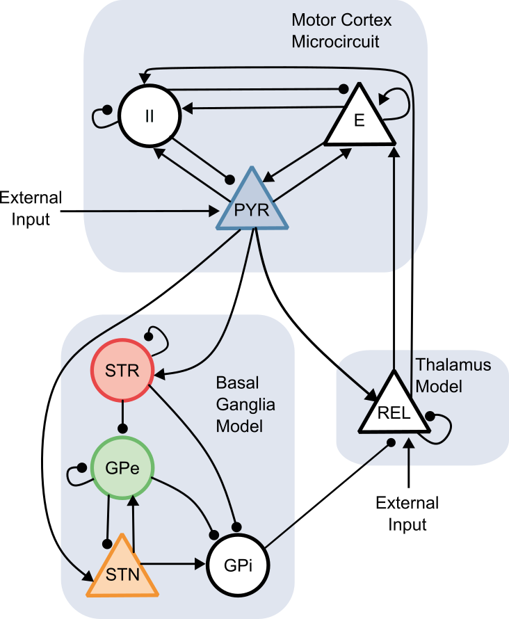
      References
      1. Ziemann, U. (2020). I-waves in motor cortex revisited. Exp. brain research, 238(7), 1601-1610.
      2. Miller, A., Knösche, T. R., & Weise, K. (2025). A coupling model of transcranial magnetic stimulation induced electric fields to neural state variables. bioRxiv, 2025-08. 
      3. Di Lazzaro, V., & Ziemann, U. (2013). The contribution of transcranial magnetic stimulation in the functional evaluation of microcircuits in human motor cortex. Front. in neural circ., 7, 18. 
      4. Di Lazzaro, V., Pilato, F., Oliviero, A., Dileone, M., Saturno, E., Mazzone, P., ... & Rothwell, J. C. (2006). Origin of facilitation of motor-evoked potentials after paired magnetic stimulation: direct recording of epidural activity in conscious humans. Jrnl of neurophys., 96(4), 1765-1771. 


      Acknowledgement
      -
      Sunday July 12, 2026 4:20pm - 6:20pm ADT
      Ballroom B2

      4:20pm ADT

      P046: Phase-Dependent Deep Brain Stimulation to Suppress Pathological Neural Oscillations in Parkinson’s Disease
      Sunday July 12, 2026 4:20pm - 6:20pm ADT
      Introduction
      Parkinson's disease (PD) is characterised by elevated oscillatory activity in the low-beta frequency range (13–20 Hz), a hallmark correlated with hypokinesia and is thought to arise from pathophysiological changes within the basal ganglia thalamocortical (BGTC) network. Phase-dependent deep brain stimulation (pdDBS) is a proposed alternative stimulation strategy to clinically standard high-frequency DBS, delivering pulses at targeted phases of the beta oscillatory cycle to either synchronise or desynchronise oscillatory amplitude. Despite experimental and computational work in the field, existing investigations of pdDBS have yet to examine how patient-specific responses to stimulation shape oscillatory dynamics at the network level.

      Methods
      We employ a BGTC neural mass model [1], to investigate the effects of phase-dependent DBS across 8 populations in the BGTC network (Figure 1). Model parameters are estimated through simulation-based inference using biologically informed priors, fit to reproduce pathological oscillatory activity seen in PD [2]. We use a novel method for modelling STN DBS incorporating orthodromic and antidromic invasion of axonal collaterals [3], describing how stimulation perturbs effective firing rates across the network. Approximate Bayesian Computation is used to optimise DBS activation parameters and stimulation phase for maximal suppression of oscillatory activity.

      Results
      Consistent with existing models, the BGTC model successfully reproduces plausible firing rates and spectra in line with animal models. Applying the optimisation framework across a range of STN DBS activation parameters, initial results show that the optimal stimulation phase for oscillatory suppression depends on the relative activation of fibre pathways during stimulation. Consistent with experimental findings, this suppression is accompanied by increased oscillatory activity at adjacent frequency bands.

      Discussion
      This work offers a framework for understanding patient-specific responses to phase-dependent DBS, showing that optimal stimulation phases are not universal but vary according to how DBS perturbs the BGTC network through different fibre pathways. Beyond its immediate relevance to optimising phase-dependent DBS in PD, the framework generalises to various pulsatile brain stimulation strategies aimed at shifting oscillatory dynamics towards less pathological states. Ongoing work will extend the analysis to network level responses to stimulation and evaluating responses when GPi is used as the stimulation target.

      Figure 1. The BGTC circuit connectivity implemented in the neural mass model includes cortical excitatory (E), inhibitory interneuron (II), and deep pyramidal (DP) populations, as well as the striatum, globus pallidus externus (GPe), globus pallidus internus (GPi), subthalamic nucleus (STN), and thalamic relay nuclei (REL). Shaded regions indicate nodes with spectral data used for model fitting.
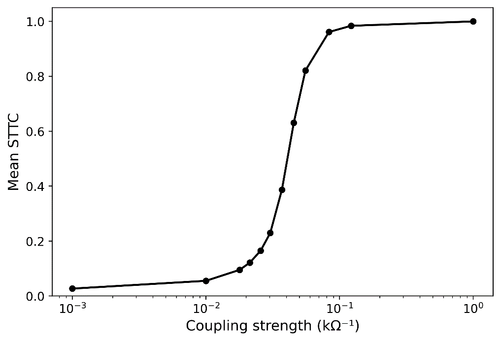
      References
      1.     van Albada, S. J., et al. (2009). Mean-field modeling of the basal ganglia-thalamocortical system. II: Dynamics of parkinsonian oscillations. Journal of Theoretical Biology, 257(4), 664–688. https://doi.org/10.1016/j.jtbi.2008.12.013
      2.     West, T. O., et al. (2022). Stimulating at the right time to recover network states in a model of the cortico-basal ganglia-thalamic circuit. PLOS Computational Biology, 18(3), e1009887. https://doi.org/10.1371/journal.pcbi.1009887
      3.     Crompton, et al. (2025, April 24). A Unified Computational Framework for Implementing Impact of Deep Brain Stimulation in Neural Circuits. [Conference Presentation]. Krembil Research Day, Toronto, Canada

      Acknowledgement
      We acknowledge the financial support of the Branch Out Neurological Foundation and the Max-Planck Center for Neural Science and Technology (P.K) as well as the Natural Sciences and Engineering Council (NSERC) RGPIN-2022-05181 (L.M).
      Sunday July 12, 2026 4:20pm - 6:20pm ADT
      Ballroom B2

      4:20pm ADT

      P047: Feature Extraction of Neuronal Morphology with a Variational Auto-Encoder
      Sunday July 12, 2026 4:20pm - 6:20pm ADT
      Introduction
      A single pyramidal neuron can compute XOR using its dendritic structure [1]. This suggests that neuronal morphology is closely related to computational capability. Since neurons exhibit diverse morphologies [2], individual neurons may possess distinct computational capabilities. To reveal the relationship between neuronal morphology and computational capability, simulation experiments using neuron models with diverse morphologies are effective. However, constructing realistic neuron models that capture the morphologies of neurons is difficult because the essential features that characterize neuronal morphology are poorly understood. This study aims to investigate whether a Variational Auto-Encoder (VAE) is effective for feature extraction.

      Methods
      A toy neuron dataset consisting of single-branch neurons was created to train the VAE. The toy neurons were generated using several morphometric features, including node type (elongation, branch, or terminal), angle, and elongation length. The VAE was trained to map input neuronal morphologies to a low-dimensional latent space and reconstruct them from the space. The space was analyzed using latent traversal [3]. In this method, the VAE was first inputted a toy neuron morphology from the dataset, which was then reconstructed. Next, the value of one variable spanning the space was varied while the others were fixed. We then evaluated which morphometric features the latent variable represented based on changes in the reconstructed morphology.

      Results
      The VAE successfully reconstructed morphologies that resembled the toy neurons. The reconstructed morphologies gradually varied in response to changes in the latent variables. Those changes reflected the morphometric features used to create the toy neurons. When the input toy neuron data were replaced with another neuron, the features represented by each latent variable sometimes differed; however, across all variables in the latent space, all the features were extracted.

      Discussion
      These results suggest that the VAE is a useful approach for extracting morphometric features. The dependence of the extracted features on the input morphology suggests that the VAE may implicitly cluster training data in the latent space and extract cluster-specific morphometric features. Future work is to apply the proposed approach to real neuronal data, such as pyramidal neurons.

      References
      [1] Gidon, A., et al. (2020). Dendritic action potentials and computation in human layer 2/3 cortical neurons. Science. 367:83–87.
      [2] Peng, H., et al. (2021). Morphological diversity of single neurons in molecularly defined cell types. Nature. 598 (7879):174–181.
      [3] Burgess, C. P., et al. (2018) Understanding disentangling in beta-VAE. arXiv preprint arXiv:1804.03599.


      Acknowledgement
      This research was supported by AMED under Grant Number JP25wm0625418h0001.

      Sunday July 12, 2026 4:20pm - 6:20pm ADT
      Ballroom B2

      4:20pm ADT

      P048: Noise-Driven Spiking Dynamics and Synchronization Transitions in an Ensemble of Neuromorphic Oscillators
      Sunday July 12, 2026 4:20pm - 6:20pm ADT
      Introduction
      Some neural systems are assumed to operate near a critical point between order and disorder, where collective dynamics enable efficient information processing and computational capabilities [1,2]. Neuromorphic hardware systems provide an experimental platform for investigating such network dynamics in an engineered system. Networks of coupled oscillators have been proposed as promising systems for studying synchronization, stochastic spiking dynamics, and potential signatures of critical dynamics in neural-like systems [3]. We study the synchronization behavior of a network of stochastic relaxation-type oscillators driven by noise that generate neuron-like spiking in a neuromorphic hardware system with event-based spike timing readout.

      Methods
      We experimentally investigate a network of 36 neuron-inspired oscillators. Each node of the network is implemented as a relaxation-type oscillator based on a programmable unijunction transistor, which implements neuron-like threshold firing dynamics. Stochastic spike generation is induced through externally injected electrical noise, leading to Poisson-like spiking dynamics. The oscillators are coupled via resistive connections in an all-to-all topology with tunable coupling strength. Spike events are recorded using a novel event-based readout system that captures the precise spike times for all oscillators, enabling spike-train based analysis of the resulting network dynamics.

      Results
      To quantify synchronization in the oscillator network, we compute the mean spike time tiling coefficient (STTC) across all oscillator pairs, a spike-train based correlation measure that quantifies temporal spike coincidences while remaining robust to differences in firing rate. While systematically varying the coupling resistances, the mean STTC increases continuously with coupling strength, indicating a gradual emergence of collective synchronization in the oscillator network (Fig. 1). This behavior is consistent with theoretical predictions for synchronization transitions in ensembles of coupled oscillators, where increasing coupling promotes phase locking and collective dynamics across the network.

      Discussion
      Phase transitions are frequently discussed in the context of collective neural dynamics and potential signatures of criticality in the brain [1,2,4]. While the observed behavior is consistent with a continuous synchronization transition, the presence of such a transition alone does not constitute sufficient evidence for criticality. Additional signatures such as scale-free activity statistics or critical scaling of network correlations are required to establish critical dynamics. Our results show that neuromorphic oscillator networks provide a controllable experimental platform for studying collective spike dynamics. Future work will investigate statistical indicators of criticality and the influence of coupling architecture and noise.

      Figure 1. Mean spike time tiling coefficient (STTC) as a function of coupling strength in a network of 36 coupled stochastic relaxation-type oscillators. Increasing coupling drives the system from weakly correlated spiking activity toward global synchronization, indicating a continuous synchronization transition.


      References
      1. Beggs, J. M., & Plenz, D. (2003). Neuronal Avalanches in Neocortical Circuits. Journal of Neuroscience, 23(35), 11167-11177. https://doi.org/10.1523/JNEUROSCI.23-35-11167.2003
      2. Shew, W. L., & Plenz, D. (2012). The Functional Benefits of Criticality in the Cortex. Neuroscientist, 19(1), 88-100. https://doi.org/10.1177/1073858412445487
      3. Feketa, P., Meurer, T., & Kohlstedt, H. (2022). Structural plasticity driven by task performance leads to criticality signatures in neuromorphic oscillator networks. Scientific Reports, 12(1), 15321. https://doi.org/10.1038/s41598-022-19386-z
      4. Chialvo, D. (2010). Emergent complex neural dynamics. Nature Physics, 6(10), 744-750. https://doi.org/10.1038/nphys1803

      Acknowledgement
      -
      Speakers
      avatar for Wilhelm Braun

      Wilhelm Braun

      Junior Research Group Leader, Kiel University (CAU Kiel), Faculty of Engineering, Department of Electrical and Information Engineering
      Early nervous systems, functional neuronal networks, stochastic neural dynamics, animal behavior, reinforcement learning, network reconstruction
      Sunday July 12, 2026 4:20pm - 6:20pm ADT
      Ballroom B2
       
      Share Modal

      Share this link via

      Or copy link

      Filter sessions
      Apply filters to sessions.
      Filtered by Date -