Loading…
Venue: Ballroom B1 clear filter
arrow_back View All Dates
Sunday, July 12
 

9:00am ADT

Announcements
Sunday July 12, 2026 9:00am - 9:10am ADT

Sunday July 12, 2026 9:00am - 9:10am ADT
Ballroom B1

9:10am ADT

Keynote 2: Mac Shine, "The Neurobiological Basis of Consciousness"
Sunday July 12, 2026 9:10am - 10:10am ADT
Understanding the neural basis of consciousness requires mechanistic accounts that span multiple scales of brain organisation. Yet most existing theoretical frameworks operate at the macroscale, offering systems-level predictions without prescribing the cellular and circuit-level mechanisms that implement them. Here I argue that the multiscale architecture of the thalamocortical system offers a principled solution to this problem. Drawing on theoretical and computational work from our group, I show how the core/matrix organisation of the thalamus, in combination with the nonlinear dendritic integration properties of L5B pyramidal neurons, generates the conditions necessary for both the global state of consciousness and the specific contents of experience. A biophysical microcircuit model, extended to a corticothalamic neural mass framework, reproduces key empirical phenomena across multiple perturbation regimes - including anaesthesia, optogenetic manipulation, and pharmacological intervention - and makes predictions at the macroscale that are consistent with leading theoretical accounts.


Sunday July 12, 2026 9:10am - 10:10am ADT
Ballroom B1

10:40am ADT

FO1: The Synapse-Pairing Tradeoff: How Clustering, Bursts, and Dendritic Location Enable Robust Plasticity In-Vivo
Sunday July 12, 2026 10:40am - 11:10am ADT
Dhuruva Priyan Gowri Mariyappan*1,2,3, Nghi V Nguyen2,3,4, Giuseppe Chindemi5, András Ecker6, Sabrina Tazerart2,4, James Isbister7, Darshan Mandge7, Diana E. Mitchell2,4, Michael W Reimann7, Roberto Araya4,2, Eilif B Muller2,3,4
Department of Computer Science and Operations Research, Faculty of Arts and Science, Université de Montréal, Montréal, Canada
Centre de Recherche Azrieli du CHU Sainte-Justine, Montréal, Canada
Mila Quebec AI Institute, Montréal, Canada
Department of Neurosciences, Faculty of Medicine, Université de Montréal, Montréal, Canada
ETH AI Center, Zurich, Switzerland
Cytocast Hungary Kft, Budapest, Hungary
Open Brain Institute, Lausanne, Switzerland
*Email: [email protected]


Introduction
Cortical representations are thought to arise from stable network motifs formed by neuronal assemblies, with synaptic plasticity between pyramidal cells (PCs) playing a central role in their formation, competition, and maintenance. While rules governing such synaptic changes have been well characterized in slice conditions, their implications for learning in awake behaving animals remain an active area of research. Here we use biophysically detailed simulations with calibrated ion channels, background synaptic activity, and calcium-based plasticity rules to investigate mechanisms enabling reliable plasticity in-vivo. We find that spatially clustered activation and burst firing offer robust pathways for LTP under physiological conditions.

Methods
We used biophysically detailed simulations of a large-scale in-silico cortical microcircuit of rat somatosensory cortex with a calcium-based plasticity model capturing LTP and Long-Term Depression (LTD) as a function of integrated postsynaptic calcium. We parameterized voltage-gated Na⁺, K⁺, Ca²⁺, and Bk channels throughout the dendritic tree based on experimental data. To reproduce the high-conductance state of awake cortex, we incorporated stochastic background activity using Dendritic Extra-Excitatory Synapses (DEES) at 1.1 synapses/μm. We investigated clustered plasticity in L2/3 PC and L5-TTPC basal and apical dendrites under both in-vitro and in-vivo-like extracellular calcium concentrations.

Results
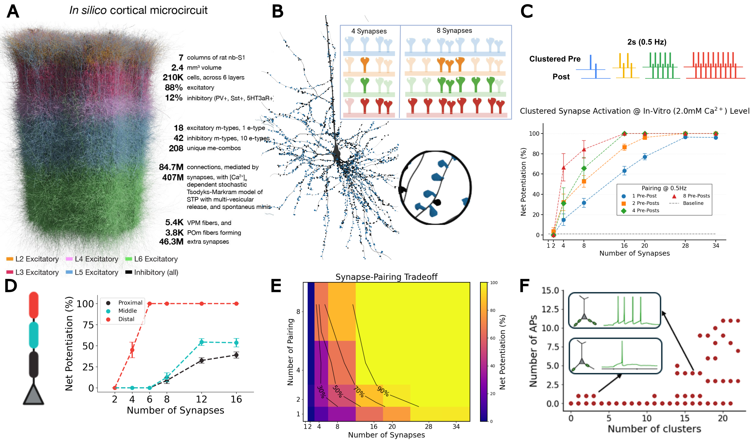
Synchronous activation of ≥11 clustered synapses generates dendritic plateau potentials (≥100 ms) that induce LTP in distal basal dendrites (Fig. 1). We identify a synapse-pairing tradeoff, where dendrites effectively trade the number of co-activated synapses for pairing repetitions: 16-synapse clusters achieve one-shot learning, while 8-synapse clusters require 3+ pairings. This tradeoff exhibits spatial gradients: distal dendrites achieve LTP independent of backpropagating action potentials, while proximal clusters require spike-timing coincidence. When multiple basal clusters coactivate, summated depolarizations trigger somatic bursts; both presynaptic and postsynaptic bursts drive robust LTP with all-or-none threshold dynamics.

Discussion

These findings establish multiple plasticity mechanisms within a single neuron—spatial clustering, location-dependent learning modes, and dual burst pathways—providing testable predictions for how cortical circuits implement flexible, hierarchical learning. Distal dendrites enable unsupervised learning via cluster-based LTP independent of bAPs, while proximal regions implement supervised learning requiring spike-timing coincidence. Apical dendrites receiving top-down signals can generate bursts or couple with somatic spikes via backpropagation-activated calcium (BAC) firing, a substrate for top-down plasticity modulation. These mechanisms reveal how dendrites trade synapse number for pairing repetitions under noisy physiological conditions.

Figure 1. A, In silico cortical microcircuit. B, L5-TTPC with magnified cluster showing plasticity for 4 vs 8 co-active synapses. C, Clustered pre-post pairing (0.5 Hz); net potentiation vs synapse number. D, Spatial learning gradient E, Synapse-pairing tradeoff heatmap. F, Basal cluster coactivation triggers somatic burst.

References
1. Chindemi, G., Abdellah, M., Amsalem, O., Benavides-Piccione, R., Delattre, V., Doron, M., Ecker, A., Jaquier, A. T., King, J., Kumbhar, P., Monney, C., Perin, R., Rössert, C., Tuncel, A. M., Van Geit, W., DeFelipe, J., Graupner, M., Segev, I., Markram, H., & Muller, E. B. (2022). A calcium-based plasticity model for predicting long-term potentiation and depression in the neocortex.
2. Ecker, A., Egas Santander, D., Abdellah, M., Alonso, J. B., Bolaños-Puchet, S., Chindemi, G., Gowri Mariyappan, D. P., Isbister, J. B., Ki
Sunday July 12, 2026 10:40am - 11:10am ADT
Ballroom B1

11:10am ADT

O1: Biologically plausible credit assignment via neuronal frequency multiplexing
Sunday July 12, 2026 11:10am - 11:30am ADT

Li Ji-An1,2,3Marcus K. Benna*3

Neurosciences Graduate Program, University of California San Diego, La Jolla, CA, USA
Department of Psychology, New York University, New York, NY, USA
Department of Neurobiology, University of California San Diego, La Jolla, CA, USA

*Email: [email protected]

Introduction
Backpropagation has been highly successful for training artificial neural networks. However, whether the biological brain implements any variant of backpropagation remains unknown. A key challenge concerns the capability of a single biological neuron to simultaneously encode and transmit feedforward predictions and feedback errors with minimal interference. Here, we propose a neuronal frequency multiplexing framework to address this challenge.

Methods
Each model neuron has multiple compartments that multiplex signals in the frequency domain. One dendrite acts as a low-pass filter, and extracts feedforward prediction signals from the low-frequency, direct-current components of the inputs. Another dendrite acts as a high-pass filter, and extracts feedback error signals from high-frequency, oscillatory components of the inputs. The soma integrates both signals, transmitting them to other neurons through its firing rate, which consists of a slowly varying prediction component and an oscillatory error component.

Results
We demonstrate that this frequency multiplexing algorithm using a simple, local learning rule closely approximates backpropagation in fully connected networks trained on the MNIST dataset and in convolutional networks trained on the CIFAR-10 dataset, achieving comparable performance and similar learning speed as a function of the number of training epochs.

Discussion
Our framework implements backpropagation-like training of functionally feedforward neural networks using continuously running, recurrently connected neuronal populations that simultaneously encode and propagate both prediction and error signals with minimal interference. This represents a new solution to the long-standing problem of biologically plausible credit assignment, suggesting a potential computational role for oscillatory signals in coordinating synaptic plasticity.

Speakers
MK

Marcus K Benna

UC San Diego
Sunday July 12, 2026 11:10am - 11:30am ADT
Ballroom B1

11:30am ADT

O2: A unifying account of rTMS and rTFUS neurostimulation effects based on calcium-dependent synaptic plasticity theory and an equivalent-energy principle
Sunday July 12, 2026 11:30am - 11:50am ADT
John D. Griffiths*1,2,3,4, Kevin Kadak1,3, Yupeng Tian1,5,6

1Krembil Centre for Neuroinformatics, Centre for Addiction and Mental Health, Toronto, Canada
2Department of Psychiatry, University of Toronto, Canada
3Institute of Medical Sciences, University of Toronto, Canada
4Institute of Biomedical Engineering, University of Toronto, Canada
5Dept. Mathematics, University of Toronto, Canada 
6Fields Institute for Mathematical Sciences, Toronto, Canada


*Email: [email protected]

Introduction
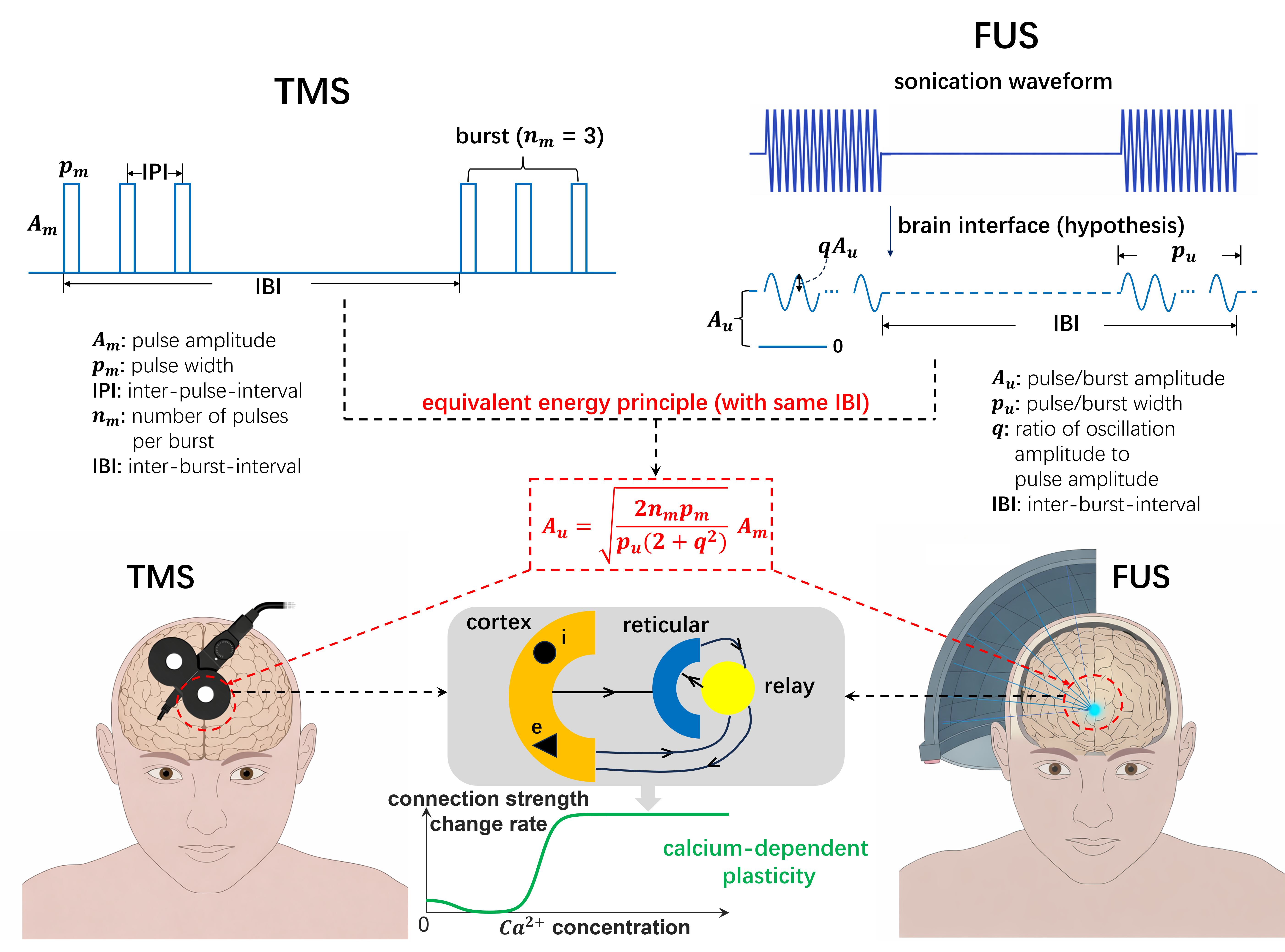
Repetitive transcranial magnetic stimulation (rTMS) and transcranial focused ultrasound stimulation (rTFUS) are noninvasive neuromodulation techniques with established and promising clinical applications, respectively.Though their primary mechanisms of action differ (electromagnetic vs. acoustic),both exert clinically relevant effects through stimulation-induced synaptic plasticity. Despite rich neurophysiological understanding of plasticity, a validated theoretical framework describing noninvasive neurostimulation-induced plasticity remains to be developed. We present a unified mean-field modelling framework for rTMS- and rTFUS-induced plasticity, grounded in calcium-dependent plasticity theory embedded in a corticothalamic circuit[1,2,3].

Methods
We extended the Fung-Robinson calcium-dependent plasticity model [1] by embedding it in a multi-population corticothalamic circuit generating alpha oscillations, implemented in NFTsim. For rTMS validation, a within-subject TMS-EEG experiment (N=21; 5 visits) tested 5 iTBS protocols varying inter-burst frequency and pulses-per-burst, measuring motor-evoked potentials (MEPs) and resting-state EEG alpha power. For rTFUS, a novel equivalent-energy principle (Fig.1) scaled continuous FUS burst amplitudes to deliver equivalent energy to corresponding rTMS waveforms, enabling direct model comparison across modalities. Predictions were compared against published rTFUS motor plasticity data across varying inter-burst frequencies and durations [4,5].

Results
Weaker iTBS protocols (3Hz/3PPB, 5Hz/2PPB) produced paradoxically stronger LTP-like MEP facilitation than standard iTBS (5Hz/3PPB), while the strongest protocol (7Hz/3PPB) robustly sign-flipped to LTD. MEP and resting-state alpha power showed a consistent inverse relationship across all protocols, supporting EEG as a plasticity biomarker outside the motor system. The corticothalamic model reproduced correct MEP directionality in 5/5 protocols and rank-ordering in 4/5, and captured alpha directionality in 4/5.Applying the equivalent-energy principle to rTFUS, the same model replicated published cTB-FUS plasticity results and accounted for the LTD/LTP sign-flip in cTB-TMS (40s vs. 80s) but not cTB-FUS, explained by waveform shape alone [4,5].

Discussion
These findings support a 'less is more' principle: gentler stimulation paradoxically yields stronger plasticity effects, with over-stimulation causing sign-reversal to LTD. The consistent MEP-alpha inverse relationship opens the possibility of using scalp EEG as a protocol-agnostic plasticity readout. The equivalent-energy principle provides a principled bridge between rTMS and rTFUS modelling, enabling the same calcium-dependent corticothalamic framework to account for both modalities without re-parameterization. Together these results establish a foundation for in silico exploration of the largely unmapped rTMS/rTFUS protocol space, with direct implications for optimizing clinical neuromodulation [2,3].

Figure 1. Equivalent energy principle for aligning rTMS and rTFUS plasticity models. rTMS delivers discrete pulse bursts; rTFUS delivers continuous bursts filtered by the skull interface to sub-1kHz sinusoids. The principle constrains parameters so both modalities deliver equal energy at the same carrier frequency, enabling unified mean-field modelling of calcium-dependent plasticity.


References
  1. Fung & Robinson (2014). Neural field theory of synaptic metaplasticity with applications to theta burst stimulation. J Theor Biol, 340, 164–176. https://doi.org/10.1016/j.jtbi.2013.09.021
  2. Kadak K, et al. (2026, submitted). Less is more: gentle protocols induce stronger facilitatory effects than standard iTBS through calcium-dependent metaplasticity.
  3. Tian Y, et al. (2026, submitted). Equivalent energy principle and calcium-dependent plasticity theory unify TMS and FUS effects.
  4. Zeng K, et al. (2024). Motor cortex plasticity by theta burst transcranial ultrasound. Ann Neurol, 91(2), 238–252. https://doi.org/10.1002/ana.26294
  5. Gamboa OL, et al. (2010). Reversal of theta burst after-effect with prolonged stimulation. Exp Brain Res, 204(2), 181–187. https://doi.org/10.1007/s00221-010-2293-4

Acknolwedgments
We acknowledge funding from the Krembil Foundation, Labbatt Foundation, UofT EMHSeed, and Fields Institute for Mathematical Sciences, that supported this work. 
Speakers
Sunday July 12, 2026 11:30am - 11:50am ADT
Ballroom B1

11:50am ADT

O3: Compartmentalized learning through coupled electrochemical adaptation in cortical pyramidal neurons
Sunday July 12, 2026 11:50am - 12:10pm ADT
Beatriz Barros1,2, Raquel Figueiredo1,2Renato Duarte*1, 2


1Center for Neuroscience and Cell Biology (CNC-UC), University of Coimbra, Portugal
2Centre for Innovative Biomedicine and Biotechnology (CiBB), University of Coimbra, Portugal
*Email: [email protected]

Introduction
A single cortical neuron simultaneously expresses Hebbian STDP proximally and cooperative plasticity distally, couples excitatory and inhibitory weight changes, and maintains homeostatic stability across timescales spanning seconds to days. Computational models treat these phenomena separately, yet the underlying molecular cascades are shared, shaped by local dendritic morphology and chemical composition. This convergence means that compartment-specific learning rules, local E/I balance, and multi-timescale homeostasis are not independent, but mechanistically coupled through intracellular dynamics. What emerges computationally from this coupling, and how it reshapes our understanding of single-neuron learning, remains an open question.

Methods
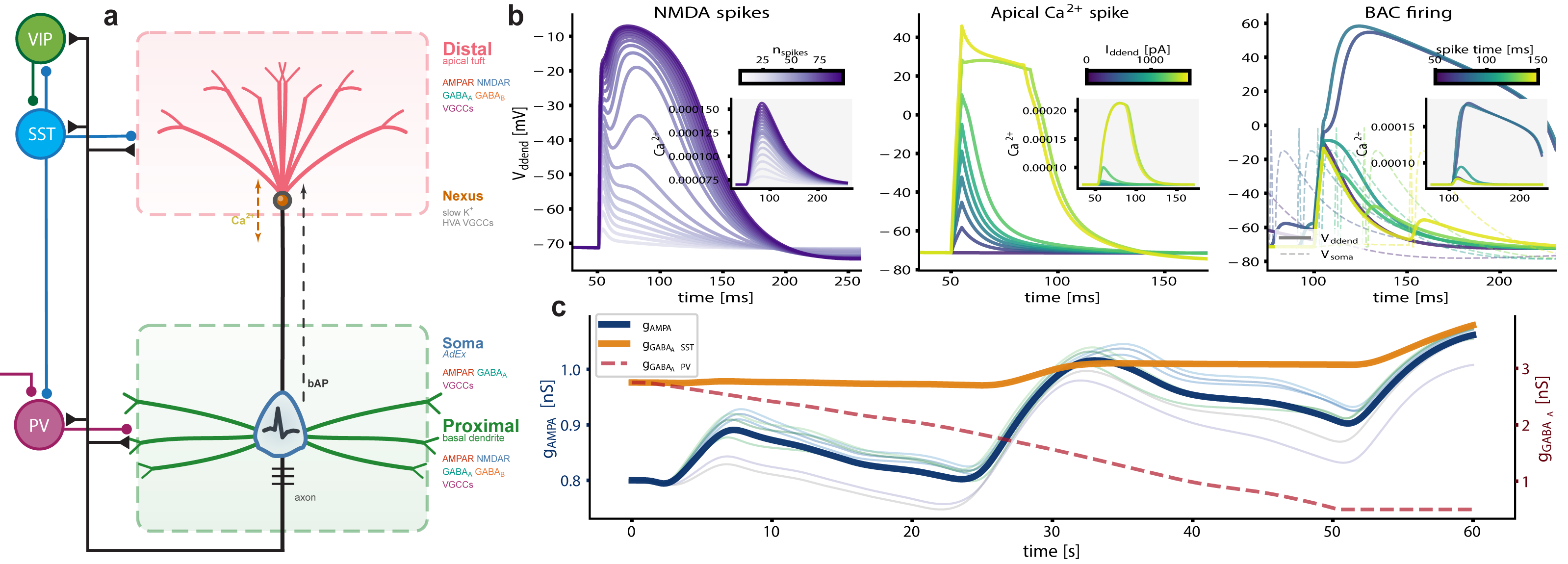
We build on a three-compartment neuron model [2], augmented with active, electrogenic dendritic processes (Fig. 1). Local calcium currents feed a slow dendritic integrator that drives calcium-dependent plasticity [3] at every synapse. Compartment-specific learning emerges naturally: bAP-dominated proximal calcium produces Hebbian STDP, while VGCC/NMDAR-dominated distal calcium yields cooperative plasticity. Inhibitory synapses read the same calcium with inverted thresholds, coupling E/I balance without explicit homeostatic targets. We extend this via stargazin phosphorylation [4], anchoring AMPAR trafficking, Kv7.2-mediated intrinsic excitability, and synaptic scaling in a three-tier cascade spanning seconds to days.

Results
A single plasticity rule, operating on compartment-resolved calcium trace, produces Hebbian STDP proximally (bAP-dominated) and cooperative, timing-independent plasticity distally (VGCC/NMDAR-dominated), matching recent in vivo observations [1]. Shared calcium maintains coupled E/I balance locally, without explicit homeostatic targets, and allows accurate stimulus representation from the response to localized perturbations. The stargazin cascade reproduces multiphasic homeostatic dynamics with intrinsic plasticity preceding synaptic scaling. We show that the apparent diversity of cortical plasticity rules is an emergent phenomenon, a consequence of intracellular dynamics and proceed to investigate its functional consequences.

Discussion
Compartment-specific signaling produces qualitatively different learning rules from the same mechanism, reframing credit assignment in cortical circuits [1] and emphasizing intracellular signaling as a primary locus of learning and memory. Stimulus associations, selectivity, and stability emerge from dendritic biophysics and are co-modulated with shared electrochemical substrates. Beyond the biophysical details, the framework we present here raises broader questions: can local, compartmentalized balance serve as a natural learning objective? And how does coupling different adaptation mechanisms shape information representation and memorization, with intracellular dynamics acting as a stack-like memory?

Figure 1. Augmented tripod neuron with compartment-specific electrogenic events and coupled E/I plasticity. (a) Circuit schematic with compartment-resolved receptors and interneuron targeting. (b) NMDA plateaus, apical Ca²⁺ spikes, and BAC firing with dendritic calcium transients (insets). (c) Shared calcium couples excitatory and inhibitory weight dynamics, actively maintaining E/I balance.References
[1] Wright, W. J., Hedrick, N. G., & Komiyama, T. (2025). Distinct synaptic plasticity rules operate across dendritic compartments in vivo during learning. Science, 388(6744), 322-328.
[2] Quaresima, A., Fitz, H., Duarte, R., van den Broek, D., Hagoort, P., & Petersson, K. M. (2023). The Tripod neuron: a minimal structural reduction of the dendritic tree. The Journal of Physiology, 601(15), 3265-3295.
[3] Graupner, M. & Brunel, N. (2012). Calcium-based plasticity model explains sensitivity of synaptic changes to spike pattern, rate, and dendritic location. PNAS, 109(10), 3991-3996.
[4] Rodrigues, M. V. et al. (2024). Type I TARPs regulate Kv7.2 potassium channels and susceptibility to seizures. bioRxiv, 2024.08.09.607194.

Acknowledgments
This work was supported by national funds through FCT—Foundation for Science and Technology, I.P., under the project HetSyn (2023.13758.PEX).

Speakers
avatar for Renato Duarte

Renato Duarte

Assistant Researcher, Center for Neuroscience and Cell Biology (CNC), University of Coimbra
Sunday July 12, 2026 11:50am - 12:10pm ADT
Ballroom B1

12:10pm ADT

O4: Rewarding Control: Feedback-Phase-Dependent 2Hz Medial Frontal Transcranial Alternating Current Stimulation Shifts the Expected Value of Control
Sunday July 12, 2026 12:10pm - 12:30pm ADT
Authors: Robert Louis Treuting1, Eric Rawls*1,2
Affiliations: 1Department of Psychology, University of North Carolina Wilmington, Wilmington, NC, USA, 2Department of Psychiatry and Behavioral Sciences, University of Minnesota Twin Cities, Minneapolis, MN, USA
*Email: [email protected]

Introduction
Medial frontal delta activity is a plausible control signal because the Reward Positivity (RewP) tracks outcome evaluation and prediction errors, and our prior simultaneous EEG-fMRI work localized key signed prediction-error contributions of the RewP to medial frontal cortex [1,2]. Here we tested whether 2 Hz transcranial alternating current stimulation (tACS) targeting this generator changes motivated cognitive control. We predicted that stimulation would alter latent decision parameters in an Expected Value of Control Stroop rather than merely speeding responses [2,3].

Methods
Seven healthy participants completed a reward×efficacy Stroop with congruent/incongruent targets across sham and active 2 Hz medial frontal tACS sessions. Accuracy was analyzed with binomial generalized linear mixed models and reaction time with Gamma models. We then fit a hierarchical Wiener diffusion model in brms to decompose behavior into drift rate (mu), boundary separation (bs), and nondecision time (ndt). In a second model restricted to active trials, sine and cosine terms indexed the phase of stimulation at the prior trial’s feedback.

Results
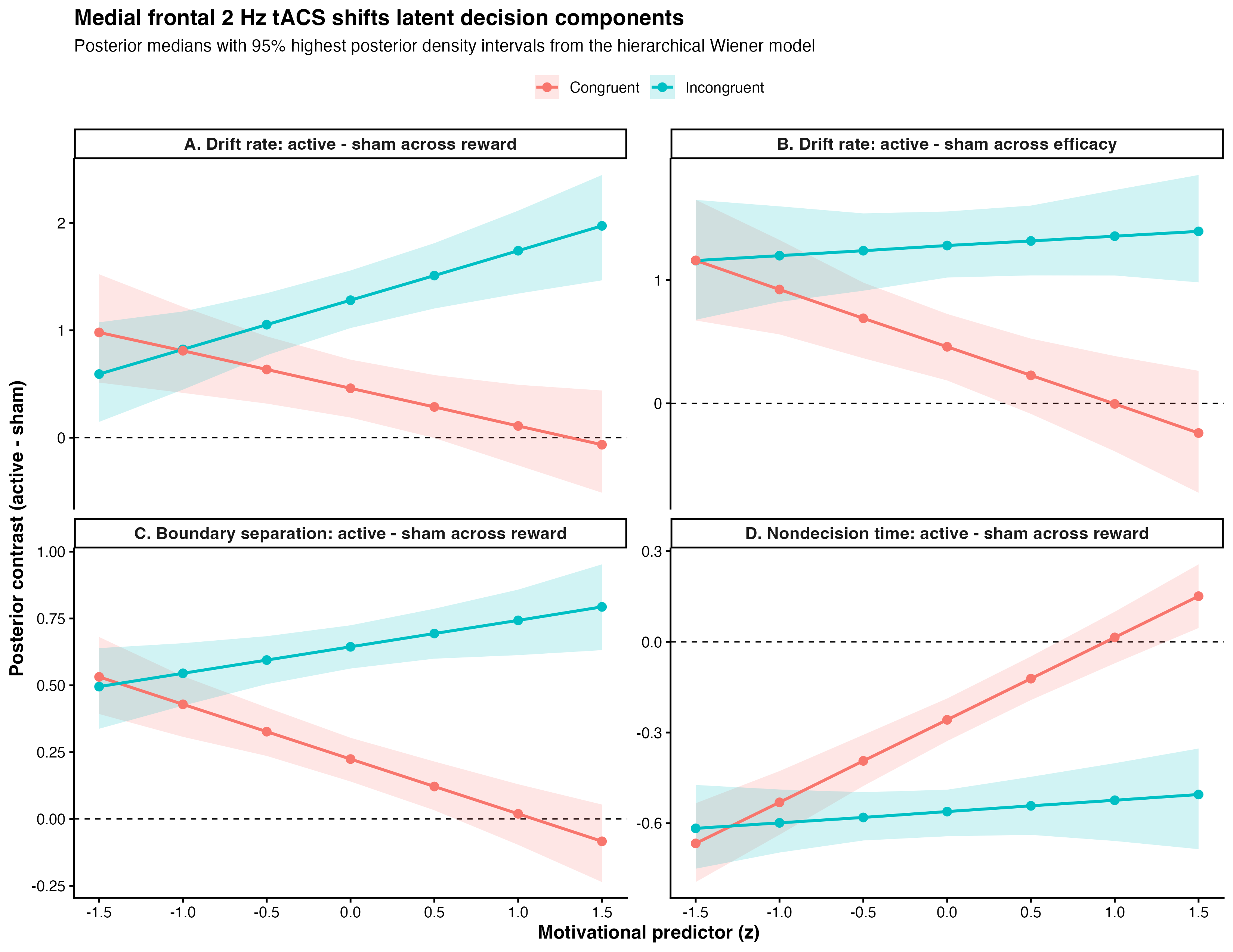
Active stimulation improved accuracy (χ²(1)=8.76, p=0.003) without a main reaction-time benefit, arguing against nonspecific speeding. In the diffusion model, stimulation shifted all three latent components: drift, boundary, and nondecision time. Posterior contrasts showed robust drift benefits throughout incongruent trials and selective benefits in congruent trials when reward or efficacy was low (Fig. 1). In active trials, prior-feedback phase predicted subsequent drift (feed_cos=0.39 [0.19, 0.59]; feed_sin=-0.26 [-0.48, -0.05]), consistent with causal modulation of a RewP-linked medial frontal mechanism for updating expected value of control from recent outcomes [1,2].

Discussion
These results suggest that medial frontal delta stimulation does not simply energize behavior. Instead, it changes how outcome information is converted into the next trial’s control state. Computationally, active 2 Hz tACS improved evidence accumulation while also reshaping caution and peripheral processing.

Figure 1. Active minus sham posterior contrasts from the hierarchical Wiener diffusion model. Top panels show drift-rate changes across reward and efficacy, separated by congruency. Bottom panels show boundary-separation and nondecision-time changes across reward. Points indicate posterior medians; ribbons show 95% highest posterior density intervals.

References
[1] Rawls, E., Demro, C., Teich, C. D., Zhang, J., Wang, A., Heilbronner, S. R., Mueller, B. A., Sponheim, S. R., & MacDonald, A. W., III. (2026). The search for RewP: Dissociating cortical generators of electrophysiological signed and unsigned prediction errors using simultaneous EEG-fMRI [Manuscript in preparation].
[2] Frömer, R., Lin, H., Dean Wolf, C. K., Inzlicht, M., & Shenhav, A. (2021). Expectations of reward and efficacy guide cognitive control allocation. Nature Communications, 12, 1030.
[3] Ratcliff, R., & McKoon, G. (2008). The diffusion decision model. Neural Computation, 20(4), 873–922.

Acknowledgments
We thank the Brain, Data, and Causality Lab and our University of Minnesota collaborators for the prior simultaneous EEG-fMRI work that motivated this study, and for foundational discussion of RewP source modeling and expected value of control.

Speakers
Sunday July 12, 2026 12:10pm - 12:30pm ADT
Ballroom B1

2:00pm ADT

FO2: Norepinephrine Restores Cortical Dynamics and Enables Machine Learning–Based Severity Mapping in a Multiscale Model of Parkinson’s Disease
Sunday July 12, 2026 2:00pm - 2:30pm ADT
Jeeyune Jung*1,3,   Adam Newton1,3,  Donald Doherty1,3,  Hong-Yuan Chu2,3,  Samuel Neymotin4 , William Lytton1,3
1. Department of Physiology and Pharmacology, SUNY Downstate Health Sciences University, Brooklyn, NY, USA 
2. Department of Pharmacology and Physiology, Georgetown University Medical Center, Washington, DC, USA 
3. Aligning Science Across Parkinson’s (ASAP) Collaborative Research Network, Chevy Chase, MD, USA
Center for Biomedical Imaging & Neuromodulation, Nathan Kline Institute, Orangeburg, NY, USA

*Email: [email protected]


Introduction
Parkinson’s disease (PD) involves not only basal ganglia dopamine loss but also cortical dysfunction, including excessive beta synchronization, abnormal beta–gamma coupling, altered bursting, and impaired corticospinal recruitment. Early locus coeruleus degeneration may reduce cortical norepinephrine (NE), disrupting cell-type-specific gain control in pyramidal tract (PT) and intratelencephalic (IT) neurons. We tested whether experimentally constrained NE modulation restores cortical excitability and network dynamics in an advanced MitoPark motor cortex model and whether NE-sensitive cortical biomarkers support severity mapping and prediction of dopamine-therapy response.


Methods
Whole-cell patch-clamp recordings from Layer 5 PT and IT neurons quantified NE (10 µM)-induced changes in firing–current relationships. Conductance-based single-cell models were fit to baseline and NE responses and embedded in a biophysically detailed laminar M1 network model in NEURON/NetPyNE. Parkinsonian simulations incorporated reduced PT5B intrinsic excitability and reduced thalamocortical drive, with disease-stage-dependent NE scaling from PK/PD modeling. From the resulting simulations, we extracted cortical biomarkers including PT5B firing, IT5B beta-synchronized bursting, IT/PT imbalance, beta power, beta-burst duration, beta–high gamma phase-amplitude coupling, and avalanche slope, which were then used for severity mapping. 

Results
NE exerted opposite intrinsic effects across Layer 5 pyramidal subtypes: PT firing increased, whereas IT repetitive firing decreased. In the Parkinsonian network, NE-dependent conductance changes partially rescued pathological dynamics: PT5B firing increased by ~60–70%, IT5B bursting declined, the IT/PT activity ratio shifted toward control-like values, and pathological beta–high gamma phase-amplitude coupling decreased by ~40%. Across the biomarker set, NE shifted cortical dynamics toward the control regime. In the machine-learning framework, greater deviation from control tracked greater disease severity and predicted progressively shorter and weaker levodopa benefit as NE support declined.
Discussion
These findings identify NE-sensitive intrinsic gain control as a mechanistic bridge between single-cell excitability and pathological cortical state in PD. Loss of noradrenergic modulation may directly contribute to corticospinal under-recruitment, hypersynchronous beta activity, and broader cortical biomarker abnormalities, whereas restoring NE-dependent PT/IT balance may complement dopamine-based therapy. This multiscale framework links cellular mechanisms, network dysfunction, severity mapping, and predicted treatment response, positioning noradrenergic modulation as a promising strategy for advanced PD.

References
1. Dura-Bernal, S., et al. (2023). Multiscale model of primary motor cortex circuits predicts in vivo cell-type-specific, behavioral state-dependent dynamics. Cell Reports.
2. Chu, H. Y., et al. (2024). Dysfunction of motor cortices in Parkinson’s disease. Cerebral Cortex.
3. Doherty, D. W., et al. (2025). Enhanced beta power emerges from simulated parkinsonian primary motor cortex. npj Parkinson’s Disease.

Acknowledgments
This work was supported by the Aligning Science Across Parkinson’s (ASAP) Collaborative Research Network. We thank colleagues for providing experimental data used to constrain the model. Computational resources were provided by SUNY Downstate Health Sciences University.


Sunday July 12, 2026 2:00pm - 2:30pm ADT
Ballroom B1

2:30pm ADT

O5: Impact of EAAT2 Dysfunction on AMPA/NMDA-Mediated Excitability in Neuronal Activity
Sunday July 12, 2026 2:30pm - 2:50pm ADT
Hannah van Susteren1,*, Guillaume Girier2,*, Michel J.A.M. van Putten3,4 , Jaroslav Hlinka2, Helmut Schmidt2, Hil G.E. Meijer1


1 Department of Applied Mathematics, University of Twente, Enschede, the Netherlands
2 Institute of Computer Science, Czech Academy of Sciences, Prague, Czech Republic
3 Department of Neurology and Clinical Neurophysiology, University of Twente, Enschede, the Netherlands
4 Medisch Spectrum Twente, Enschede, the Netherlands
* These authors contributed equally to this work.


Email: [email protected]


Introduction
Epilepsy is among the most prevalent neurological disorders. The astrocytic excitatory amino acid transporter (EAAT2) plays a key role in regulating excitability, by controlling extracellular glutamate levels and glutamate receptor activation [1,2]. Reduced EAAT2 expression has been reported in several epilepsy patients [1,3]. However, the impact of neuron-astrocyte interactions on hyperexcitability on single cell level is underexplored. We developed a biophysical model of a presynaptic neuron and astrocyte to explore the relation between astrocytic EAAT2-mediated glutamate clearance, presynaptic glutamate receptors and bursting activity.

Methods
We build on our previous work [4,5], where we consider a presynaptic neuron and an astrocyte in a finite extracellular space (ECS). This model describes sodium, potassium, chloride dynamics as well as calcium-dependent exocytosis and glutamate-glutamine (GG) recycling. For this study, we add a potassium bath with diffusion to the ECS to induce neuronal bursting (Fig. 1A). Additionally, we implement the presynaptic glutamate receptors AMPA and NMDA, which are important in regulating hyperexcitability. Lastly, we study the impact of the antiseizure drugs perampanel and memantine by simulating the effect of these AMPA and NMDA receptor antagonists.

Results
We induce neuronal bursting by increasing extracellular potassium in the bath. We first examine how AMPA and NMDA permeabilities affect burst frequency (Fig. 1C), where frequency refers to spike frequency during the last burst or during tonic firing, to fit the NMDA/AMPA ratio to experimental data [6]. Higher permeabilities increase neuronal firing and intracellular calcium, triggering a feedback loop that enhances neuronal glutamate release. Reducing EAAT permeability raises burst frequency and induces tonic firing (Fig. 1B). Finally, AMPA and NMDA antagonists, perampanel and memantine [7], reduce firing despite elevated extracellular glutamate, with perampanel showing a more significant reduction in firing frequency (Fig. 1D). 

Discussion
Our results show that reduced EAAT expression, as observed in several epilepsy patients, results in increased extracellular glutamate and overstimulation of excitatory glutamate receptors. Furthermore, we show that the AMPA and NMDA receptor permeabilities affect burst frequency. Receptor antagonists such as perampanel and memantine are able to reduce firing. In conclusion, our detailed neuron–astrocyte model provides insight into the effects of reduced EAAT expression and receptor antagonists on hyperexcitability.
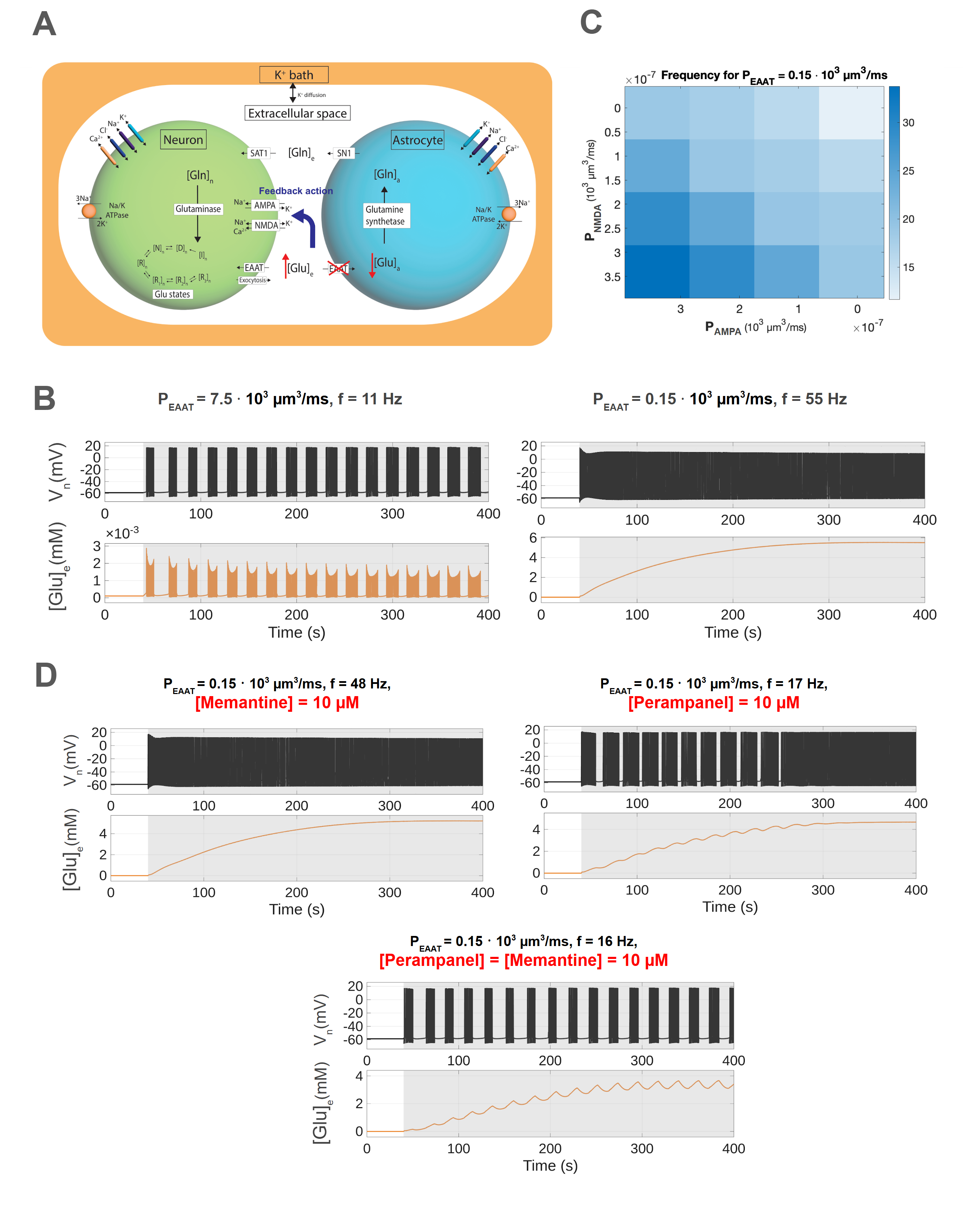
Figure 1. A: Three-compartment model illustrating the GG-cycle during EAAT2 knockout. B: The membrane potential, spike frequency f and ECS glutamate at different EAAT2 permeabilities. C: Spike frequency within bursts as a function of NMDA and AMPA receptor permeability.  D: Neuronal activity at fixed EAAT2 permeability (PEAAT=0.15 * 103  µm3/ms) under antagonist conditions.

References
[1] Green, J. L., dos Santos, W. F., & Fontana, A. C. K. (2021). Biochemical Pharmacology, 10.1016/j.bcp.2021.114786
[2] Scimemi, A., Tian, H., & Diamond, J. S. (2009). The Journal of Neuroscience, 10.1523/JNEUROSCI.4845-09.2009
[3] Barker-Haliski, M., & White, H. (2015). Cold Spring Harbor perspectives in medicine, 10.1101/cshperspect.a022863
[4] van Susteren, H., Rose, C. R., van Putten, M. J., & Meijer, H. G. (2025). bioRxiv, 10.1101/2025.11.10.687543
[5] Kalia, M., et al. (2021).  PLOS Computational Biology, 10.1371/journal.pcbi.1009019
[6] Watt, A. J., Sjöström, P. J., Häusser, M., Nelson, S. B., & Turrigiano, G. G. (2004).  Nature neuroscience, 10.1038/nn1220
[7] Chen, T.-S., Huang, T.-H., Lai, M.-C., & Huang, C.-W. (2023).  Biomedicines, 10.3390/biomedicines11030783

Acknowledgments
HVS, HGEM, MJAMVP funded from the DFG, FOR2795 ‘Synapses under stress’ to CRR (Prof. Dr. Christine R. Rose) (Ro2327/13-2 and 14-2).
GG, HS, and JH were supported by the ERDF-Project Brain dynamics, No. CZ.02.01.01/00/22\_008/0004643, a Lumina-Quaeruntur fellowship (LQ100302301), and the long-term strategic development financing of the Institute of Computer Science (RVO:67985807).

Speakers
HV

Hannah van Susteren

PhD student, University of Twente
Sunday July 12, 2026 2:30pm - 2:50pm ADT
Ballroom B1

2:50pm ADT

O6: Multiscale modeling of neural markers for adaptive deep brain stimulation in Parkinson’s Disease
Sunday July 12, 2026 2:50pm - 3:10pm ADT

Alberto Mazzoni1
,2,*, Federico Fattorini1,2, Nicolò Meneghetti1,2


1The Biorobotics Institute, Scuola Superiore Sant’Anna, Pisa, Italy
2Department of Excellence for Robotics and AI, Scuola Superiore Sant’Anna, Pisa, Italy


*Email: [email protected]


Introduction
Parkinson’s disease (PD) is a common neurodegenerative disorder causing severe impairments. Drug-resistant patients are treated with Deep Brain Stimulation (DBS) of the basal ganglia (BG), with current efforts focused on adaptive DBS. The most relevant biomarkers for adaptive DBS is the power in the beta ([12, 30] Hz) and gamma ([30, 100] Hz) range, yet the mechanisms underlying these rhythms remain unclear. Computational models can provide mechanistic insights into pathophysiology and test new stimulation treatments. Using spiking and morphological models, we show how beta and gamma resonances emerge from BG interactions, how DBS reshapes these dynamics, and how they are reflected in subthalamic nucleus local field potentials (LFPs).

Methods
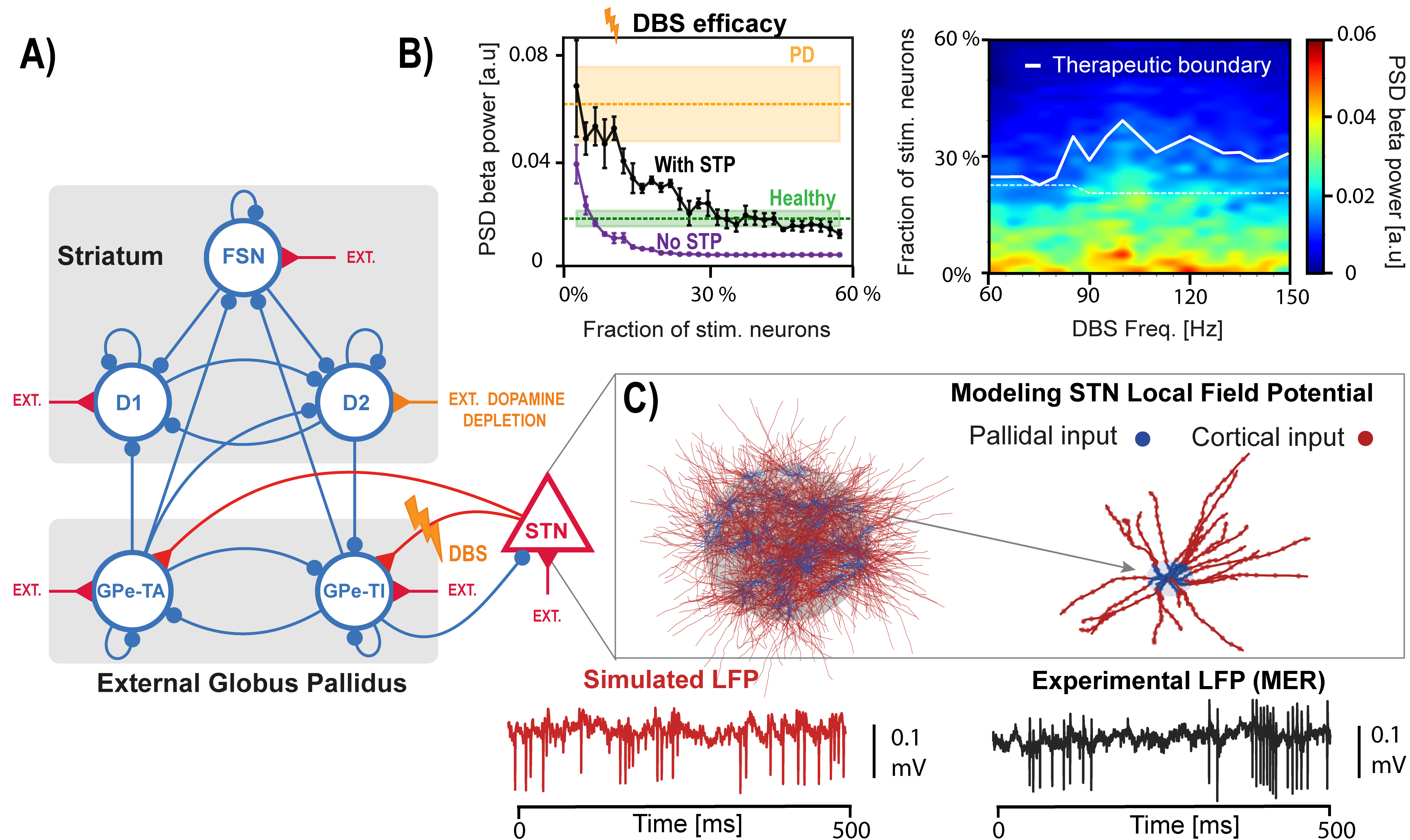
We implemented a spiking model of the basal ganglia with six neuronal populations: three within the striatum, two within the external globus pallidus, and the subthalamic nucleus (STN) (Fig. 1A) [1]. Dopamine depletion was simulated by modulating striatal inputs. We dissected the network mechanisms underlying beta and gamma resonances and we simulated STN DBS, considering short-term synaptic plasticity (Fig. 1B) [2]. To simulate the signals recorded by DBS electrodes we developed a population of morphological STN neurons model and computed LFPs associated with network activity through volume conduction theory (Fig. 1C).

Results
We show how beta oscillations arise from two independent loops in the BG model that strongly synchronize when dopamine is depleted [1]. STN DBS disrupts these oscillations, although without synaptic plasticity it requires unrealistically low stimulation levels (Fig 1B left). The model also supports the hypothesis that gamma-range stimulation can be as effective as the clinical standard of ~130 Hz used in PD (Fig. 1B right) [2]. Gamma oscillations emerge through recurrent inhibition in pallidal and striatal populations. Different from cortical ones, STN LFPs are largely noise-dominated due to weak correlations and symmetric neuronal morphology, becoming informative only when strong beta synchronization is present.

Discussion
We characterized in a spiking model of BG beta and gamma rhythmogenesis and their alteration due to PD-related dopamine depletion and DBS. Moreover, we investigated the origin of STN LFPs driving adaptive DBS, by integrating spiking and morphological modeling. Overall, we provide a multiscale framework for better understanding Parkinsonian dynamics and DBS mechanisms, showing how network modeling can clarify treatment mechanisms and guide improved stimulation strategies.

Figure 1. A) Spiking network model of the basal ganglia.  B) Left: efficacy of STN DBS as a function of the fraction of stimulated neurons, with and without short-term plasticity (STP). Right: effect of stimulation frequencies on beta spectral power.  C) Top: STN population and morphological neuron model receiving cortical and pallidal inputs. Bottom: simulated and recorded local field potentials (LFPs).


References

1. Ortone, A., Vergani, A. A., Ahmadipour, M., Mannella, R., & Mazzoni, A. (2023). Dopamine depletion leads to pathological synchronization of distinct basal ganglia loops in the beta band. PLOS Computational Biology, 19(4), 1–31. https://doi.org/10.1371/journal.pcbi.1010645
2. Ahmadipour, M., Fattorini, F., Meneghetti, N., & Mazzoni, A. (2026). In silico model of basal ganglia deep brain stimulation in Parkinson’s disease captures range of effective parameters for pathological beta power suppression. PLOS Computational Biology, 22(2), e1013280. https://doi.org/10.1371/journal.pcbi.1013280
Acknowledgments
This work was supported by the Italian Ministry of University and Research, under the complementary actions to the NRRP ‘Fit4MedRob - Fit for Medical Robotics’ Grant (# PNC0000007).


Speakers
Sunday July 12, 2026 2:50pm - 3:10pm ADT
Ballroom B1

3:10pm ADT

O7: Decreased alpha/theta temporal ExSEnt of left prefrontal cortex: a robust biomarker of dementia
Sunday July 12, 2026 3:10pm - 3:30pm ADT
Sara Kamali*1, Fabiano Baroni1, Pablo Varona1

1Department of Computer Engineering, Autonomous University of Madrid, Madrid, Spain

*Email: [email protected]

Introduction
Dementia involves progressive cognitive decline, with Alzheimer’s disease (AD) and frontotemporal dementia (FTD) as major subtypes. Electroencephalography (EEG) provides a noninvasive and accessible measure of brain dynamics and able to capture pathological slowing, often reflected by increased theta-to-alpha power ratio (TAR) [1]. Reduced signal complexity has also been reported in dementia. We tested whether Extrema-Segmented Entropy (ExSEnt), which separates temporal and amplitude irregularity, improves discrimination and interpretability beyond conventional electroencephalography biomarkers [2].

Methods
We analyzed resting-state EEG from 88 subjects, including 36 with AD, 23 with FTD, and 29 controls [3]. After preprocessing and independent component analysis, we classified brain sources into four clusters: right and left prefrontal cortices (R/LPFC) and right and left visual associations (R/LVA). Then, we computed a set of classical spectral and complexity features. We also measured the ExSEnt metrics, which quantifies the entropy of extrema-based segment durations, amplitudes, and their pair. To find the stable features for dementia detection, we performed stability selection with elastic-net logistic regression and nested leave-one-subject-out validation [4].

Results
ExSEnt improved discrimination mainly in the LPFC, where balanced accuracy increased from 71.5% to 82.6%. In that region, ExSEnt features showed high selection stability and replaced conventional measures as dominant predictors in 3 out of 4 brain areas. The most informative variables were temporal and joint temporal-amplitude entropy measures in alpha and theta bands, with negative coefficients indicating reduced irregularity and reduced dynamical variability in dementia. Other regions showed weaker or inconsistent gains, suggesting a localized effect rather than a global one. 

Discussion
ExSEnt provides a compact and interpretable measure for EEG-based dementia classification. The results suggest that dementia is associated not only with spectral slowing but also with reduced diversity of extrema timing and temporal-amplitude variability in LPFC dynamics. Because ExSEnt is explainable by design, when combined with stability selection and sparse linear classification, it yields robust biomarkers with direct physiological interpretation. These findings support ExSEnt as a promising candidate for explainable dementia screening and motivate future validation against cognitive severity measures.

References
[1] Partanen, J. V., Soininen, H., & Riekkinen, P. J. (1986). Does an ACTH derivative (Org 2766) prevent deterioration of EEG in Alzheimer's disease?. Electroencephalography and clinical neurophysiology, 63(6), 547-551.
[2] Kamali, S., Baroni, F., & Varona, P. (2025). ExSEnt: Extrema-Segmented Entropy Analysis of Time Series. arXiv preprint arXiv:2509.07751.
[3] Miltiadous, A., Tzimourta, K. D., Afrantou, T., Ioannidis, P., Grigoriadis, N., Tsalikakis, D. G., ... & Tzallas, A. T. (2023). A dataset of scalp EEG recordings of Alzheimer’s disease, frontotemporal dementia and healthy subjects from routine EEG. Data, 8(6), 95.
[4] Meinshausen, N., & Bühlmann, P. (2010). Stability selection. Journal of the Royal Statistical Society Series B: Statistical Methodology, 72(4), 417-473.

      Acknowledgments
      Funded by PID2024-155923NB-I00 and CPP2023-010818.
      Speakers
      Sunday July 12, 2026 3:10pm - 3:30pm ADT
      Ballroom B1

      3:30pm ADT

      O8: Frequency-dependent modulation of cortical traveling waves during general anesthesia
      Sunday July 12, 2026 3:30pm - 3:50pm ADT
      Duan Li*, Anthony G. Hudetz

      Center for Consciousness Science, Department of Anesthesiology, University of Michigan, Ann Arbor, MI

      *Email: [email protected]

      Introduction
      General anesthetics profoundly reshape neuronal activity and functional interactions, yet how they alter the spatiotemporal organization of the cortex is incompletely understood. Traveling waves provide a framework for examining coordinated neural activity across the cortex, but their modulation by anesthesia remains underexplored. Recently we showed that cortical dynamics under anesthesia exhibit spontaneous transitions among discrete states, including a paradoxical state characterized by low delta power and high complexity in deep anesthesia[1]. To investigate how traveling waves vary across cortical states, we recorded hemispheric ECoG at multiple anesthetic depths and analyzed wave dynamics as a function of cortical state and frequency.

      Methods
      ECoG was recorded from the right hemisphere with chronically implanted 32-site flexible polymer arrays (4×8 grid) at four desflurane concentrations (6-0%) for 1 h each. Cortical states were identified using PCA of power spectrograms followed by density-based clustering across concentrations. Within each state, traveling wave episodes were detected based on stabile spatial phase gradient patterns [2] between consecutive time points [3] in the delta, theta, and gamma bands. Wave occurrence and pattern richness were quantified, the latter defined as the entropy of SVD eigenvalues of the episode similarity matrix. Plane waves were identified and further classified as feedforward (posterior-to-anterior) or feedback (anterior-to-posterior).

      Results
      Seven states were identified. S1-S5 broadly tracked anesthetic depth but were not tied to a specific level. S6 corresponded to burst suppression, and S7 indicated a paradoxical state mostly during deep anesthesia. In S1, theta waves were mainly feedback-directed, whereas gamma were feedforward-directed. As anesthesia deepened, delta waves became more frequent but showed reduced pattern diversity. In contrast, the occurrence and diversity of theta and gamma patterns were largely preserved. Although gamma feedforward dominance persisted, theta feedback dominance was suppressed during deep anesthesia. In S7, reductions in delta complexity and theta organization were only partially reversed, despite reduced delta power and wake-like complexity.

      Discussion
      General anesthesia differentially modulates cortical traveling waves across frequency bands. Loss of theta feedback dominance paired with preserved gamma feedforward propagation suggests disrupted top-down integration despite maintained bottom-up information flow. Reduced diversity of delta waves during deep anesthesia suggests a shift toward more stereotyped, low-information dynamics. Importantly, traveling waves do not fully recover in the paradoxical state suggesting that spectrally activated deep anesthetic states lack coordinated spatiotemporal organization necessary for conscious processing. These findings suggest that traveling wave dynamics provide complementary insight into brain state dynamics beyond spectral power or complexity.

      References
      1. Li, D., & Hudetz, A. G. (2025). Dynamic electrocortical states and paradoxical complexity during desflurane anesthesia. bioRxiv. https://doi.org/10.1101/2025.10.13.682019
      2. Muller, L., Piantoni, G., Koller, D., Cash, S. S., Halgren, E., & Sejnowski, T. J. (2016). Rotating waves during human sleep spindles organize global patterns of activity that repeat precisely through the night. eLife, 5, e17267. https://doi.org/10.7554/eLife.17267
      3. Das, A., Zabeh, E., Ermentrout, B., & Jacobs, J. (2024). Planar, spiral, and concentric traveling waves distinguish cognitive states in human memory. bioRxiv. https://doi.org/10.1101/2024.01.26.577456

      Acknowledgments
      Research was supported by the National Institute of General Medical Sciences of the National Institutes of Health under award number R01-GM056398 and the Center for Consciousness Science, Department of Anesthesiology, University of Michigan Medical School, Ann Arbor, Michigan, USA. The authors express their gratitude to Dr. Shiyong Wang for his assistance in performing the experiments.

      Speakers
      DL

      Duan Li

      Associate Research Scientist, University of MIchigan
      Sunday July 12, 2026 3:30pm - 3:50pm ADT
      Ballroom B1
       
      Share Modal

      Share this link via

      Or copy link

      Filter sessions
      Apply filters to sessions.
      Filtered by Date -