Loading…
Venue: Ballroom B1 clear filter
Saturday, July 11
 

5:00pm ADT

Welcome and Announcememts
Saturday July 11, 2026 5:00pm - 5:20pm ADT

Saturday July 11, 2026 5:00pm - 5:20pm ADT
Ballroom B1

5:20pm ADT

Keynote 1: Bratislav Misic
Saturday July 11, 2026 5:20pm - 6:20pm ADT

Saturday July 11, 2026 5:20pm - 6:20pm ADT
Ballroom B1
 
Sunday, July 12
 

9:00am ADT

Announcements
Sunday July 12, 2026 9:00am - 9:10am ADT

Sunday July 12, 2026 9:00am - 9:10am ADT
Ballroom B1

9:10am ADT

Keynote 2: Mac Shine, "The Neurobiological Basis of Consciousness"
Sunday July 12, 2026 9:10am - 10:10am ADT
Understanding the neural basis of consciousness requires mechanistic accounts that span multiple scales of brain organisation. Yet most existing theoretical frameworks operate at the macroscale, offering systems-level predictions without prescribing the cellular and circuit-level mechanisms that implement them. Here I argue that the multiscale architecture of the thalamocortical system offers a principled solution to this problem. Drawing on theoretical and computational work from our group, I show how the core/matrix organisation of the thalamus, in combination with the nonlinear dendritic integration properties of L5B pyramidal neurons, generates the conditions necessary for both the global state of consciousness and the specific contents of experience. A biophysical microcircuit model, extended to a corticothalamic neural mass framework, reproduces key empirical phenomena across multiple perturbation regimes - including anaesthesia, optogenetic manipulation, and pharmacological intervention - and makes predictions at the macroscale that are consistent with leading theoretical accounts.


Sunday July 12, 2026 9:10am - 10:10am ADT
Ballroom B1

10:40am ADT

FO1: The Synapse-Pairing Tradeoff: How Clustering, Bursts, and Dendritic Location Enable Robust Plasticity In-Vivo
Sunday July 12, 2026 10:40am - 11:10am ADT
Dhuruva Priyan Gowri Mariyappan*1,2,3, Nghi V Nguyen2,3,4, Giuseppe Chindemi5, András Ecker6, Sabrina Tazerart2,4, James Isbister7, Darshan Mandge7, Diana E. Mitchell2,4, Michael W Reimann7, Roberto Araya4,2, Eilif B Muller2,3,4
Department of Computer Science and Operations Research, Faculty of Arts and Science, Université de Montréal, Montréal, Canada
Centre de Recherche Azrieli du CHU Sainte-Justine, Montréal, Canada
Mila Quebec AI Institute, Montréal, Canada
Department of Neurosciences, Faculty of Medicine, Université de Montréal, Montréal, Canada
ETH AI Center, Zurich, Switzerland
Cytocast Hungary Kft, Budapest, Hungary
Open Brain Institute, Lausanne, Switzerland
*Email: [email protected]


Introduction
Cortical representations are thought to arise from stable network motifs formed by neuronal assemblies, with synaptic plasticity between pyramidal cells (PCs) playing a central role in their formation, competition, and maintenance. While rules governing such synaptic changes have been well characterized in slice conditions, their implications for learning in awake behaving animals remain an active area of research. Here we use biophysically detailed simulations with calibrated ion channels, background synaptic activity, and calcium-based plasticity rules to investigate mechanisms enabling reliable plasticity in-vivo. We find that spatially clustered activation and burst firing offer robust pathways for LTP under physiological conditions.

Methods
We used biophysically detailed simulations of a large-scale in-silico cortical microcircuit of rat somatosensory cortex with a calcium-based plasticity model capturing LTP and Long-Term Depression (LTD) as a function of integrated postsynaptic calcium. We parameterized voltage-gated Na⁺, K⁺, Ca²⁺, and Bk channels throughout the dendritic tree based on experimental data. To reproduce the high-conductance state of awake cortex, we incorporated stochastic background activity using Dendritic Extra-Excitatory Synapses (DEES) at 1.1 synapses/μm. We investigated clustered plasticity in L2/3 PC and L5-TTPC basal and apical dendrites under both in-vitro and in-vivo-like extracellular calcium concentrations.

Results
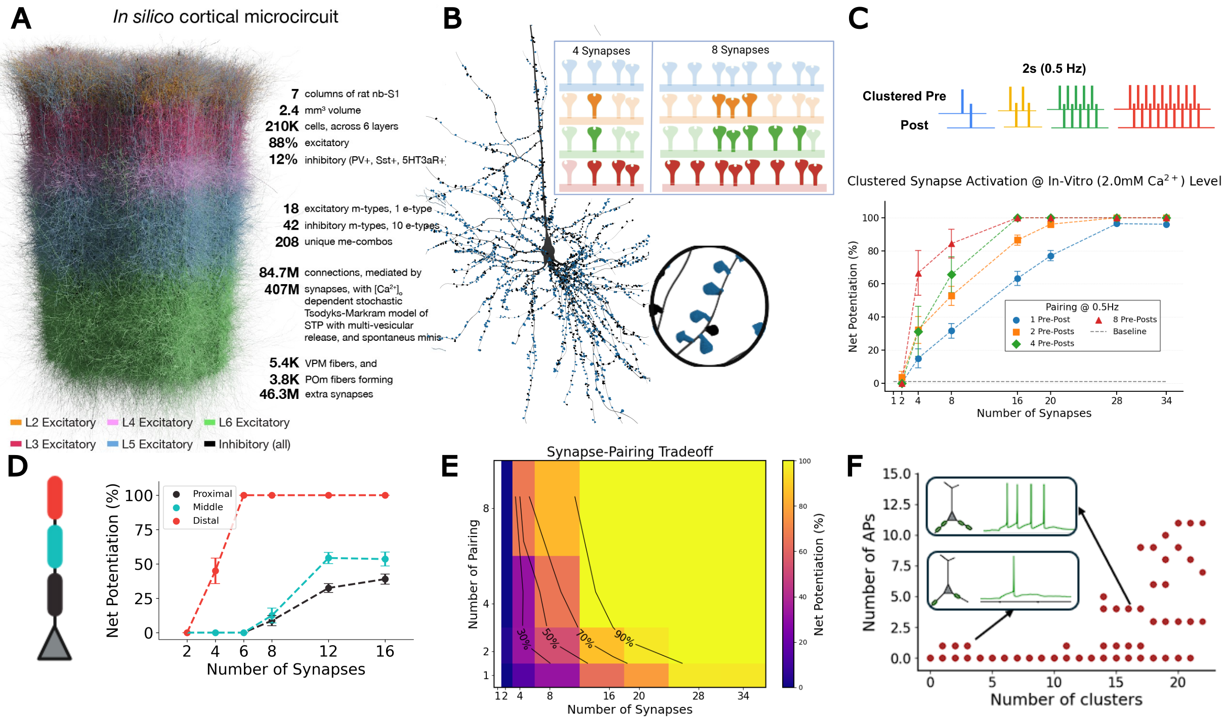
Synchronous activation of ≥11 clustered synapses generates dendritic plateau potentials (≥100 ms) that induce LTP in distal basal dendrites (Fig. 1). We identify a synapse-pairing tradeoff, where dendrites effectively trade the number of co-activated synapses for pairing repetitions: 16-synapse clusters achieve one-shot learning, while 8-synapse clusters require 3+ pairings. This tradeoff exhibits spatial gradients: distal dendrites achieve LTP independent of backpropagating action potentials, while proximal clusters require spike-timing coincidence. When multiple basal clusters coactivate, summated depolarizations trigger somatic bursts; both presynaptic and postsynaptic bursts drive robust LTP with all-or-none threshold dynamics.

Discussion

These findings establish multiple plasticity mechanisms within a single neuron—spatial clustering, location-dependent learning modes, and dual burst pathways—providing testable predictions for how cortical circuits implement flexible, hierarchical learning. Distal dendrites enable unsupervised learning via cluster-based LTP independent of bAPs, while proximal regions implement supervised learning requiring spike-timing coincidence. Apical dendrites receiving top-down signals can generate bursts or couple with somatic spikes via backpropagation-activated calcium (BAC) firing, a substrate for top-down plasticity modulation. These mechanisms reveal how dendrites trade synapse number for pairing repetitions under noisy physiological conditions.

Figure 1. A, In silico cortical microcircuit. B, L5-TTPC with magnified cluster showing plasticity for 4 vs 8 co-active synapses. C, Clustered pre-post pairing (0.5 Hz); net potentiation vs synapse number. D, Spatial learning gradient E, Synapse-pairing tradeoff heatmap. F, Basal cluster coactivation triggers somatic burst.

References
1. Chindemi, G., Abdellah, M., Amsalem, O., Benavides-Piccione, R., Delattre, V., Doron, M., Ecker, A., Jaquier, A. T., King, J., Kumbhar, P., Monney, C., Perin, R., Rössert, C., Tuncel, A. M., Van Geit, W., DeFelipe, J., Graupner, M., Segev, I., Markram, H., & Muller, E. B. (2022). A calcium-based plasticity model for predicting long-term potentiation and depression in the neocortex.
2. Ecker, A., Egas Santander, D., Abdellah, M., Alonso, J. B., Bolaños-Puchet, S., Chindemi, G., Gowri Mariyappan, D. P., Isbister, J. B., Ki
Sunday July 12, 2026 10:40am - 11:10am ADT
Ballroom B1

11:10am ADT

O1: Biologically plausible credit assignment via neuronal frequency multiplexing
Sunday July 12, 2026 11:10am - 11:30am ADT

Li Ji-An1,2,3Marcus K. Benna*3

Neurosciences Graduate Program, University of California San Diego, La Jolla, CA, USA
Department of Psychology, New York University, New York, NY, USA
Department of Neurobiology, University of California San Diego, La Jolla, CA, USA

*Email: [email protected]

Introduction
Backpropagation has been highly successful for training artificial neural networks. However, whether the biological brain implements any variant of backpropagation remains unknown. A key challenge concerns the capability of a single biological neuron to simultaneously encode and transmit feedforward predictions and feedback errors with minimal interference. Here, we propose a neuronal frequency multiplexing framework to address this challenge.

Methods
Each model neuron has multiple compartments that multiplex signals in the frequency domain. One dendrite acts as a low-pass filter, and extracts feedforward prediction signals from the low-frequency, direct-current components of the inputs. Another dendrite acts as a high-pass filter, and extracts feedback error signals from high-frequency, oscillatory components of the inputs. The soma integrates both signals, transmitting them to other neurons through its firing rate, which consists of a slowly varying prediction component and an oscillatory error component.

Results
We demonstrate that this frequency multiplexing algorithm using a simple, local learning rule closely approximates backpropagation in fully connected networks trained on the MNIST dataset and in convolutional networks trained on the CIFAR-10 dataset, achieving comparable performance and similar learning speed as a function of the number of training epochs.

Discussion
Our framework implements backpropagation-like training of functionally feedforward neural networks using continuously running, recurrently connected neuronal populations that simultaneously encode and propagate both prediction and error signals with minimal interference. This represents a new solution to the long-standing problem of biologically plausible credit assignment, suggesting a potential computational role for oscillatory signals in coordinating synaptic plasticity.

Speakers
MK

Marcus K Benna

UC San Diego
Sunday July 12, 2026 11:10am - 11:30am ADT
Ballroom B1

11:30am ADT

O2: A unifying account of rTMS and rTFUS neurostimulation effects based on calcium-dependent synaptic plasticity theory and an equivalent-energy principle
Sunday July 12, 2026 11:30am - 11:50am ADT
John D. Griffiths*1,2,3,4, Kevin Kadak1,3, Yupeng Tian1,5,6

1Krembil Centre for Neuroinformatics, Centre for Addiction and Mental Health, Toronto, Canada
2Department of Psychiatry, University of Toronto, Canada
3Institute of Medical Sciences, University of Toronto, Canada
4Institute of Biomedical Engineering, University of Toronto, Canada
5Dept. Mathematics, University of Toronto, Canada 
6Fields Institute for Mathematical Sciences, Toronto, Canada


*Email: [email protected]

Introduction
Repetitive transcranial magnetic stimulation (rTMS) and transcranial focused ultrasound stimulation (rTFUS) are noninvasive neuromodulation techniques with established and promising clinical applications, respectively.Though their primary mechanisms of action differ (electromagnetic vs. acoustic),both exert clinically relevant effects through stimulation-induced synaptic plasticity. Despite rich neurophysiological understanding of plasticity, a validated theoretical framework describing noninvasive neurostimulation-induced plasticity remains to be developed. We present a unified mean-field modelling framework for rTMS- and rTFUS-induced plasticity, grounded in calcium-dependent plasticity theory embedded in a corticothalamic circuit[1,2,3].

Methods
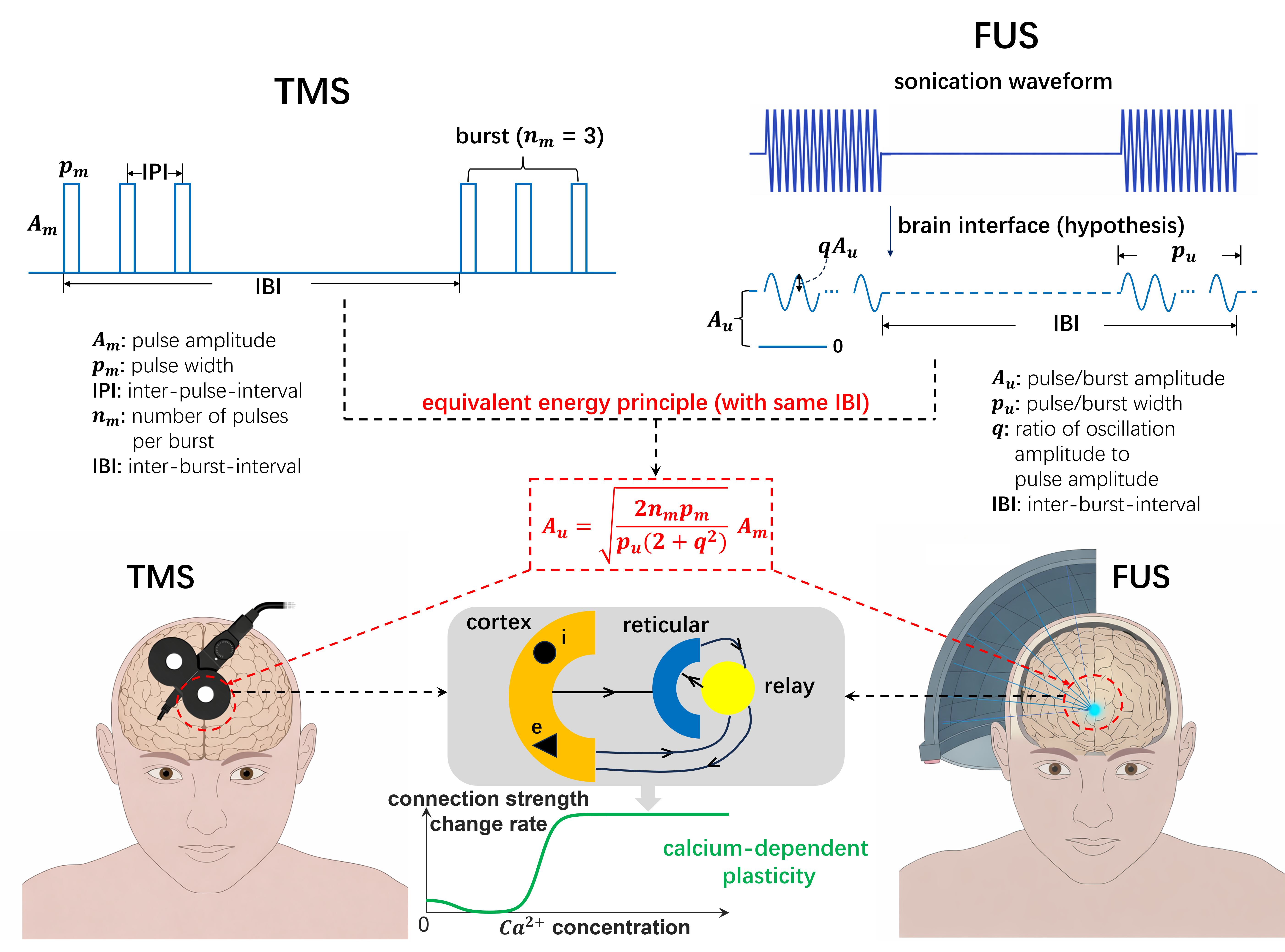
We extended the Fung-Robinson calcium-dependent plasticity model [1] by embedding it in a multi-population corticothalamic circuit generating alpha oscillations, implemented in NFTsim. For rTMS validation, a within-subject TMS-EEG experiment (N=21; 5 visits) tested 5 iTBS protocols varying inter-burst frequency and pulses-per-burst, measuring motor-evoked potentials (MEPs) and resting-state EEG alpha power. For rTFUS, a novel equivalent-energy principle (Fig.1) scaled continuous FUS burst amplitudes to deliver equivalent energy to corresponding rTMS waveforms, enabling direct model comparison across modalities. Predictions were compared against published rTFUS motor plasticity data across varying inter-burst frequencies and durations [4,5].

Results
Weaker iTBS protocols (3Hz/3PPB, 5Hz/2PPB) produced paradoxically stronger LTP-like MEP facilitation than standard iTBS (5Hz/3PPB), while the strongest protocol (7Hz/3PPB) robustly sign-flipped to LTD. MEP and resting-state alpha power showed a consistent inverse relationship across all protocols, supporting EEG as a plasticity biomarker outside the motor system. The corticothalamic model reproduced correct MEP directionality in 5/5 protocols and rank-ordering in 4/5, and captured alpha directionality in 4/5.Applying the equivalent-energy principle to rTFUS, the same model replicated published cTB-FUS plasticity results and accounted for the LTD/LTP sign-flip in cTB-TMS (40s vs. 80s) but not cTB-FUS, explained by waveform shape alone [4,5].

Discussion
These findings support a 'less is more' principle: gentler stimulation paradoxically yields stronger plasticity effects, with over-stimulation causing sign-reversal to LTD. The consistent MEP-alpha inverse relationship opens the possibility of using scalp EEG as a protocol-agnostic plasticity readout. The equivalent-energy principle provides a principled bridge between rTMS and rTFUS modelling, enabling the same calcium-dependent corticothalamic framework to account for both modalities without re-parameterization. Together these results establish a foundation for in silico exploration of the largely unmapped rTMS/rTFUS protocol space, with direct implications for optimizing clinical neuromodulation [2,3].

Figure 1. Equivalent energy principle for aligning rTMS and rTFUS plasticity models. rTMS delivers discrete pulse bursts; rTFUS delivers continuous bursts filtered by the skull interface to sub-1kHz sinusoids. The principle constrains parameters so both modalities deliver equal energy at the same carrier frequency, enabling unified mean-field modelling of calcium-dependent plasticity.


References
  1. Fung & Robinson (2014). Neural field theory of synaptic metaplasticity with applications to theta burst stimulation. J Theor Biol, 340, 164–176. https://doi.org/10.1016/j.jtbi.2013.09.021
  2. Kadak K, et al. (2026, submitted). Less is more: gentle protocols induce stronger facilitatory effects than standard iTBS through calcium-dependent metaplasticity.
  3. Tian Y, et al. (2026, submitted). Equivalent energy principle and calcium-dependent plasticity theory unify TMS and FUS effects.
  4. Zeng K, et al. (2024). Motor cortex plasticity by theta burst transcranial ultrasound. Ann Neurol, 91(2), 238–252. https://doi.org/10.1002/ana.26294
  5. Gamboa OL, et al. (2010). Reversal of theta burst after-effect with prolonged stimulation. Exp Brain Res, 204(2), 181–187. https://doi.org/10.1007/s00221-010-2293-4

Acknolwedgments
We acknowledge funding from the Krembil Foundation, Labbatt Foundation, UofT EMHSeed, and Fields Institute for Mathematical Sciences, that supported this work. 
Speakers
Sunday July 12, 2026 11:30am - 11:50am ADT
Ballroom B1

11:50am ADT

O3: Compartmentalized learning through coupled electrochemical adaptation in cortical pyramidal neurons
Sunday July 12, 2026 11:50am - 12:10pm ADT
Beatriz Barros1,2, Raquel Figueiredo1,2Renato Duarte*1, 2


1Center for Neuroscience and Cell Biology (CNC-UC), University of Coimbra, Portugal
2Centre for Innovative Biomedicine and Biotechnology (CiBB), University of Coimbra, Portugal
*Email: [email protected]

Introduction
A single cortical neuron simultaneously expresses Hebbian STDP proximally and cooperative plasticity distally, couples excitatory and inhibitory weight changes, and maintains homeostatic stability across timescales spanning seconds to days. Computational models treat these phenomena separately, yet the underlying molecular cascades are shared, shaped by local dendritic morphology and chemical composition. This convergence means that compartment-specific learning rules, local E/I balance, and multi-timescale homeostasis are not independent, but mechanistically coupled through intracellular dynamics. What emerges computationally from this coupling, and how it reshapes our understanding of single-neuron learning, remains an open question.

Methods
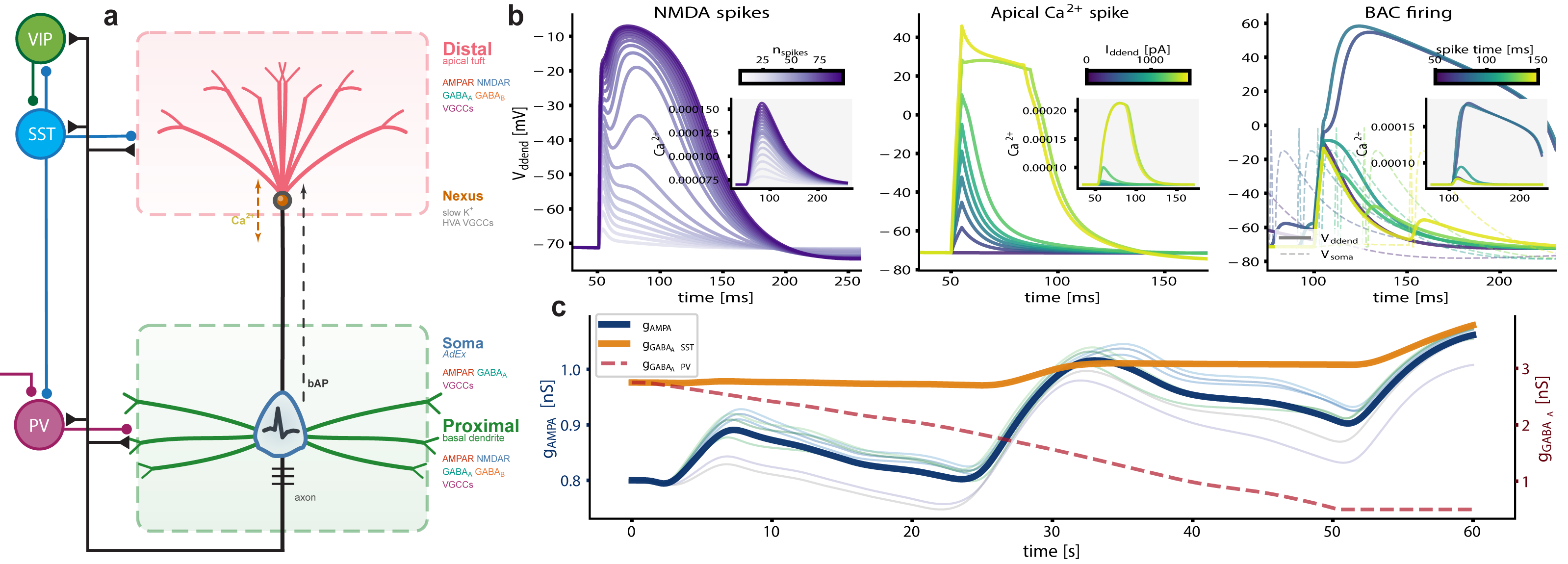
We build on a three-compartment neuron model [2], augmented with active, electrogenic dendritic processes (Fig. 1). Local calcium currents feed a slow dendritic integrator that drives calcium-dependent plasticity [3] at every synapse. Compartment-specific learning emerges naturally: bAP-dominated proximal calcium produces Hebbian STDP, while VGCC/NMDAR-dominated distal calcium yields cooperative plasticity. Inhibitory synapses read the same calcium with inverted thresholds, coupling E/I balance without explicit homeostatic targets. We extend this via stargazin phosphorylation [4], anchoring AMPAR trafficking, Kv7.2-mediated intrinsic excitability, and synaptic scaling in a three-tier cascade spanning seconds to days.

Results
A single plasticity rule, operating on compartment-resolved calcium trace, produces Hebbian STDP proximally (bAP-dominated) and cooperative, timing-independent plasticity distally (VGCC/NMDAR-dominated), matching recent in vivo observations [1]. Shared calcium maintains coupled E/I balance locally, without explicit homeostatic targets, and allows accurate stimulus representation from the response to localized perturbations. The stargazin cascade reproduces multiphasic homeostatic dynamics with intrinsic plasticity preceding synaptic scaling. We show that the apparent diversity of cortical plasticity rules is an emergent phenomenon, a consequence of intracellular dynamics and proceed to investigate its functional consequences.

Discussion
Compartment-specific signaling produces qualitatively different learning rules from the same mechanism, reframing credit assignment in cortical circuits [1] and emphasizing intracellular signaling as a primary locus of learning and memory. Stimulus associations, selectivity, and stability emerge from dendritic biophysics and are co-modulated with shared electrochemical substrates. Beyond the biophysical details, the framework we present here raises broader questions: can local, compartmentalized balance serve as a natural learning objective? And how does coupling different adaptation mechanisms shape information representation and memorization, with intracellular dynamics acting as a stack-like memory?

Figure 1. Augmented tripod neuron with compartment-specific electrogenic events and coupled E/I plasticity. (a) Circuit schematic with compartment-resolved receptors and interneuron targeting. (b) NMDA plateaus, apical Ca²⁺ spikes, and BAC firing with dendritic calcium transients (insets). (c) Shared calcium couples excitatory and inhibitory weight dynamics, actively maintaining E/I balance.References
[1] Wright, W. J., Hedrick, N. G., & Komiyama, T. (2025). Distinct synaptic plasticity rules operate across dendritic compartments in vivo during learning. Science, 388(6744), 322-328.
[2] Quaresima, A., Fitz, H., Duarte, R., van den Broek, D., Hagoort, P., & Petersson, K. M. (2023). The Tripod neuron: a minimal structural reduction of the dendritic tree. The Journal of Physiology, 601(15), 3265-3295.
[3] Graupner, M. & Brunel, N. (2012). Calcium-based plasticity model explains sensitivity of synaptic changes to spike pattern, rate, and dendritic location. PNAS, 109(10), 3991-3996.
[4] Rodrigues, M. V. et al. (2024). Type I TARPs regulate Kv7.2 potassium channels and susceptibility to seizures. bioRxiv, 2024.08.09.607194.

Acknowledgments
This work was supported by national funds through FCT—Foundation for Science and Technology, I.P., under the project HetSyn (2023.13758.PEX).

Speakers
avatar for Renato Duarte

Renato Duarte

Assistant Researcher, Center for Neuroscience and Cell Biology (CNC), University of Coimbra
Sunday July 12, 2026 11:50am - 12:10pm ADT
Ballroom B1

12:10pm ADT

O4: Rewarding Control: Feedback-Phase-Dependent 2Hz Medial Frontal Transcranial Alternating Current Stimulation Shifts the Expected Value of Control
Sunday July 12, 2026 12:10pm - 12:30pm ADT
Authors: Robert Louis Treuting1, Eric Rawls*1,2
Affiliations: 1Department of Psychology, University of North Carolina Wilmington, Wilmington, NC, USA, 2Department of Psychiatry and Behavioral Sciences, University of Minnesota Twin Cities, Minneapolis, MN, USA
*Email: [email protected]

Introduction
Medial frontal delta activity is a plausible control signal because the Reward Positivity (RewP) tracks outcome evaluation and prediction errors, and our prior simultaneous EEG-fMRI work localized key signed prediction-error contributions of the RewP to medial frontal cortex [1,2]. Here we tested whether 2 Hz transcranial alternating current stimulation (tACS) targeting this generator changes motivated cognitive control. We predicted that stimulation would alter latent decision parameters in an Expected Value of Control Stroop rather than merely speeding responses [2,3].

Methods
Seven healthy participants completed a reward×efficacy Stroop with congruent/incongruent targets across sham and active 2 Hz medial frontal tACS sessions. Accuracy was analyzed with binomial generalized linear mixed models and reaction time with Gamma models. We then fit a hierarchical Wiener diffusion model in brms to decompose behavior into drift rate (mu), boundary separation (bs), and nondecision time (ndt). In a second model restricted to active trials, sine and cosine terms indexed the phase of stimulation at the prior trial’s feedback.

Results
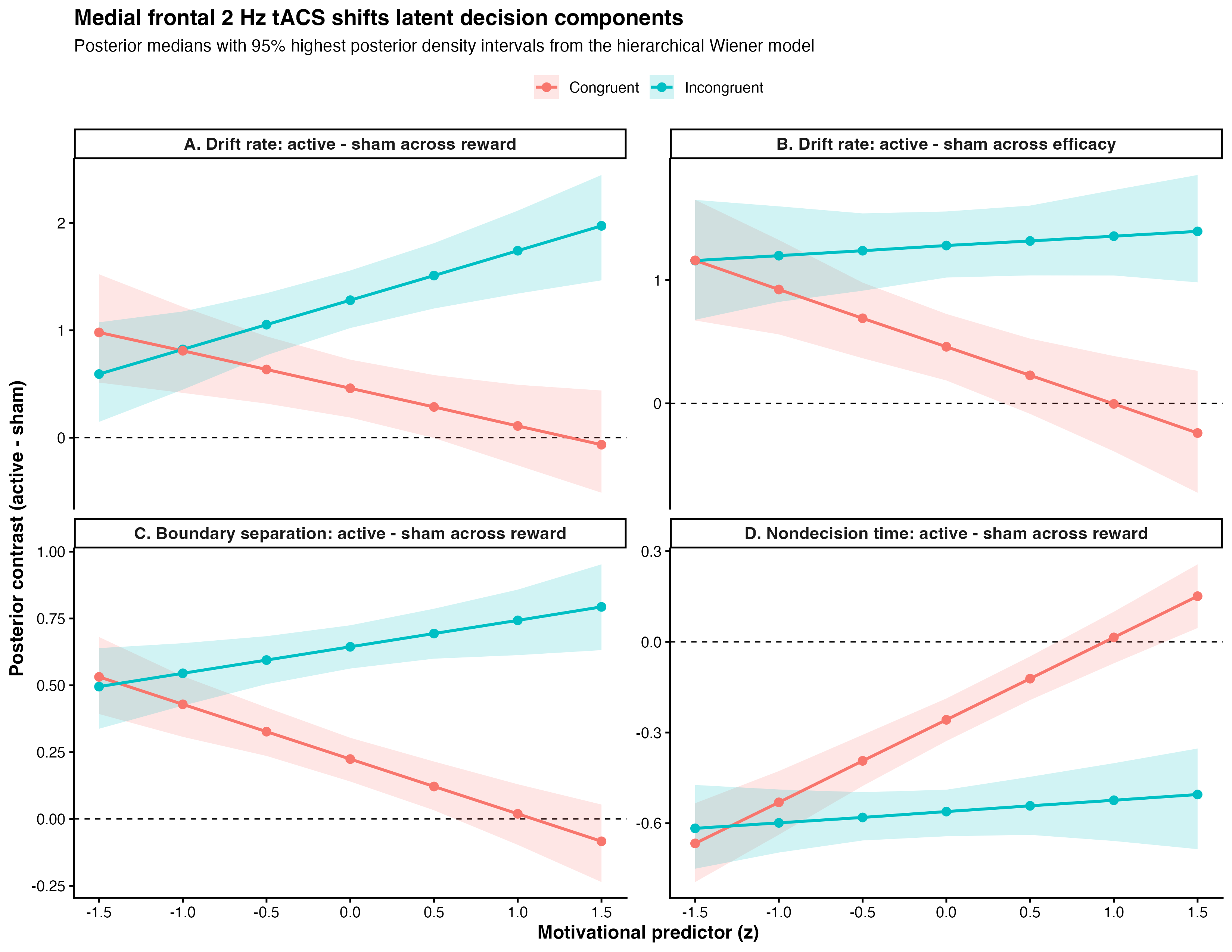
Active stimulation improved accuracy (χ²(1)=8.76, p=0.003) without a main reaction-time benefit, arguing against nonspecific speeding. In the diffusion model, stimulation shifted all three latent components: drift, boundary, and nondecision time. Posterior contrasts showed robust drift benefits throughout incongruent trials and selective benefits in congruent trials when reward or efficacy was low (Fig. 1). In active trials, prior-feedback phase predicted subsequent drift (feed_cos=0.39 [0.19, 0.59]; feed_sin=-0.26 [-0.48, -0.05]), consistent with causal modulation of a RewP-linked medial frontal mechanism for updating expected value of control from recent outcomes [1,2].

Discussion
These results suggest that medial frontal delta stimulation does not simply energize behavior. Instead, it changes how outcome information is converted into the next trial’s control state. Computationally, active 2 Hz tACS improved evidence accumulation while also reshaping caution and peripheral processing.

Figure 1. Active minus sham posterior contrasts from the hierarchical Wiener diffusion model. Top panels show drift-rate changes across reward and efficacy, separated by congruency. Bottom panels show boundary-separation and nondecision-time changes across reward. Points indicate posterior medians; ribbons show 95% highest posterior density intervals.

References
[1] Rawls, E., Demro, C., Teich, C. D., Zhang, J., Wang, A., Heilbronner, S. R., Mueller, B. A., Sponheim, S. R., & MacDonald, A. W., III. (2026). The search for RewP: Dissociating cortical generators of electrophysiological signed and unsigned prediction errors using simultaneous EEG-fMRI [Manuscript in preparation].
[2] Frömer, R., Lin, H., Dean Wolf, C. K., Inzlicht, M., & Shenhav, A. (2021). Expectations of reward and efficacy guide cognitive control allocation. Nature Communications, 12, 1030.
[3] Ratcliff, R., & McKoon, G. (2008). The diffusion decision model. Neural Computation, 20(4), 873–922.

Acknowledgments
We thank the Brain, Data, and Causality Lab and our University of Minnesota collaborators for the prior simultaneous EEG-fMRI work that motivated this study, and for foundational discussion of RewP source modeling and expected value of control.

Speakers
Sunday July 12, 2026 12:10pm - 12:30pm ADT
Ballroom B1

2:00pm ADT

FO2: Norepinephrine Restores Cortical Dynamics and Enables Machine Learning–Based Severity Mapping in a Multiscale Model of Parkinson’s Disease
Sunday July 12, 2026 2:00pm - 2:30pm ADT
Jeeyune Jung*1,3,   Adam Newton1,3,  Donald Doherty1,3,  Hong-Yuan Chu2,3,  Samuel Neymotin4 , William Lytton1,3
1. Department of Physiology and Pharmacology, SUNY Downstate Health Sciences University, Brooklyn, NY, USA 
2. Department of Pharmacology and Physiology, Georgetown University Medical Center, Washington, DC, USA 
3. Aligning Science Across Parkinson’s (ASAP) Collaborative Research Network, Chevy Chase, MD, USA
Center for Biomedical Imaging & Neuromodulation, Nathan Kline Institute, Orangeburg, NY, USA

*Email: [email protected]


Introduction
Parkinson’s disease (PD) involves not only basal ganglia dopamine loss but also cortical dysfunction, including excessive beta synchronization, abnormal beta–gamma coupling, altered bursting, and impaired corticospinal recruitment. Early locus coeruleus degeneration may reduce cortical norepinephrine (NE), disrupting cell-type-specific gain control in pyramidal tract (PT) and intratelencephalic (IT) neurons. We tested whether experimentally constrained NE modulation restores cortical excitability and network dynamics in an advanced MitoPark motor cortex model and whether NE-sensitive cortical biomarkers support severity mapping and prediction of dopamine-therapy response.


Methods
Whole-cell patch-clamp recordings from Layer 5 PT and IT neurons quantified NE (10 µM)-induced changes in firing–current relationships. Conductance-based single-cell models were fit to baseline and NE responses and embedded in a biophysically detailed laminar M1 network model in NEURON/NetPyNE. Parkinsonian simulations incorporated reduced PT5B intrinsic excitability and reduced thalamocortical drive, with disease-stage-dependent NE scaling from PK/PD modeling. From the resulting simulations, we extracted cortical biomarkers including PT5B firing, IT5B beta-synchronized bursting, IT/PT imbalance, beta power, beta-burst duration, beta–high gamma phase-amplitude coupling, and avalanche slope, which were then used for severity mapping. 

Results
NE exerted opposite intrinsic effects across Layer 5 pyramidal subtypes: PT firing increased, whereas IT repetitive firing decreased. In the Parkinsonian network, NE-dependent conductance changes partially rescued pathological dynamics: PT5B firing increased by ~60–70%, IT5B bursting declined, the IT/PT activity ratio shifted toward control-like values, and pathological beta–high gamma phase-amplitude coupling decreased by ~40%. Across the biomarker set, NE shifted cortical dynamics toward the control regime. In the machine-learning framework, greater deviation from control tracked greater disease severity and predicted progressively shorter and weaker levodopa benefit as NE support declined.
Discussion
These findings identify NE-sensitive intrinsic gain control as a mechanistic bridge between single-cell excitability and pathological cortical state in PD. Loss of noradrenergic modulation may directly contribute to corticospinal under-recruitment, hypersynchronous beta activity, and broader cortical biomarker abnormalities, whereas restoring NE-dependent PT/IT balance may complement dopamine-based therapy. This multiscale framework links cellular mechanisms, network dysfunction, severity mapping, and predicted treatment response, positioning noradrenergic modulation as a promising strategy for advanced PD.

References
1. Dura-Bernal, S., et al. (2023). Multiscale model of primary motor cortex circuits predicts in vivo cell-type-specific, behavioral state-dependent dynamics. Cell Reports.
2. Chu, H. Y., et al. (2024). Dysfunction of motor cortices in Parkinson’s disease. Cerebral Cortex.
3. Doherty, D. W., et al. (2025). Enhanced beta power emerges from simulated parkinsonian primary motor cortex. npj Parkinson’s Disease.

Acknowledgments
This work was supported by the Aligning Science Across Parkinson’s (ASAP) Collaborative Research Network. We thank colleagues for providing experimental data used to constrain the model. Computational resources were provided by SUNY Downstate Health Sciences University.


Sunday July 12, 2026 2:00pm - 2:30pm ADT
Ballroom B1

2:30pm ADT

O5: Impact of EAAT2 Dysfunction on AMPA/NMDA-Mediated Excitability in Neuronal Activity
Sunday July 12, 2026 2:30pm - 2:50pm ADT
Hannah van Susteren1,*, Guillaume Girier2,*, Michel J.A.M. van Putten3,4 , Jaroslav Hlinka2, Helmut Schmidt2, Hil G.E. Meijer1


1 Department of Applied Mathematics, University of Twente, Enschede, the Netherlands
2 Institute of Computer Science, Czech Academy of Sciences, Prague, Czech Republic
3 Department of Neurology and Clinical Neurophysiology, University of Twente, Enschede, the Netherlands
4 Medisch Spectrum Twente, Enschede, the Netherlands
* These authors contributed equally to this work.


Email: [email protected]


Introduction
Epilepsy is among the most prevalent neurological disorders. The astrocytic excitatory amino acid transporter (EAAT2) plays a key role in regulating excitability, by controlling extracellular glutamate levels and glutamate receptor activation [1,2]. Reduced EAAT2 expression has been reported in several epilepsy patients [1,3]. However, the impact of neuron-astrocyte interactions on hyperexcitability on single cell level is underexplored. We developed a biophysical model of a presynaptic neuron and astrocyte to explore the relation between astrocytic EAAT2-mediated glutamate clearance, presynaptic glutamate receptors and bursting activity.

Methods
We build on our previous work [4,5], where we consider a presynaptic neuron and an astrocyte in a finite extracellular space (ECS). This model describes sodium, potassium, chloride dynamics as well as calcium-dependent exocytosis and glutamate-glutamine (GG) recycling. For this study, we add a potassium bath with diffusion to the ECS to induce neuronal bursting (Fig. 1A). Additionally, we implement the presynaptic glutamate receptors AMPA and NMDA, which are important in regulating hyperexcitability. Lastly, we study the impact of the antiseizure drugs perampanel and memantine by simulating the effect of these AMPA and NMDA receptor antagonists.

Results
We induce neuronal bursting by increasing extracellular potassium in the bath. We first examine how AMPA and NMDA permeabilities affect burst frequency (Fig. 1C), where frequency refers to spike frequency during the last burst or during tonic firing, to fit the NMDA/AMPA ratio to experimental data [6]. Higher permeabilities increase neuronal firing and intracellular calcium, triggering a feedback loop that enhances neuronal glutamate release. Reducing EAAT permeability raises burst frequency and induces tonic firing (Fig. 1B). Finally, AMPA and NMDA antagonists, perampanel and memantine [7], reduce firing despite elevated extracellular glutamate, with perampanel showing a more significant reduction in firing frequency (Fig. 1D). 

Discussion
Our results show that reduced EAAT expression, as observed in several epilepsy patients, results in increased extracellular glutamate and overstimulation of excitatory glutamate receptors. Furthermore, we show that the AMPA and NMDA receptor permeabilities affect burst frequency. Receptor antagonists such as perampanel and memantine are able to reduce firing. In conclusion, our detailed neuron–astrocyte model provides insight into the effects of reduced EAAT expression and receptor antagonists on hyperexcitability.
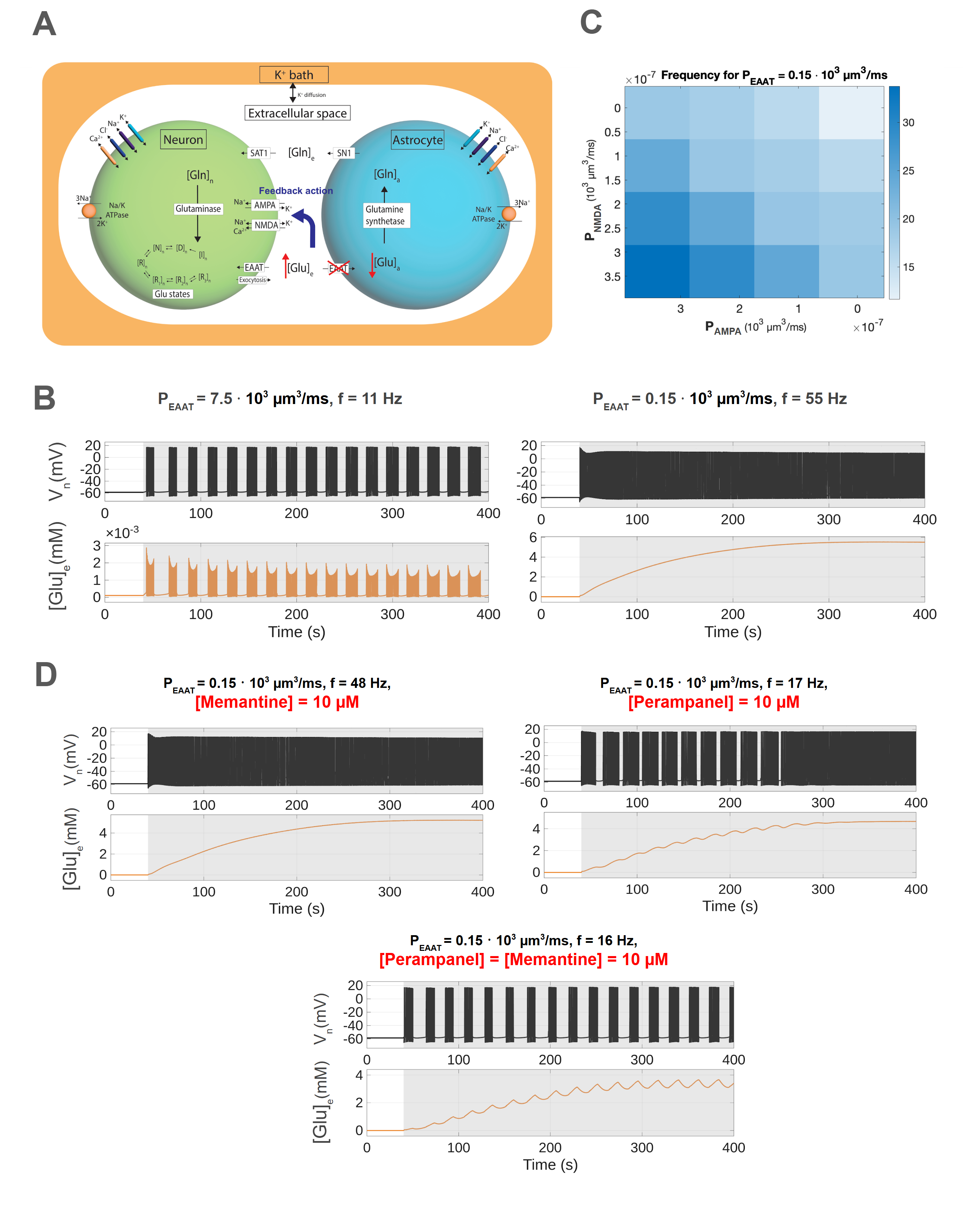
Figure 1. A: Three-compartment model illustrating the GG-cycle during EAAT2 knockout. B: The membrane potential, spike frequency f and ECS glutamate at different EAAT2 permeabilities. C: Spike frequency within bursts as a function of NMDA and AMPA receptor permeability.  D: Neuronal activity at fixed EAAT2 permeability (PEAAT=0.15 * 103  µm3/ms) under antagonist conditions.

References
[1] Green, J. L., dos Santos, W. F., & Fontana, A. C. K. (2021). Biochemical Pharmacology, 10.1016/j.bcp.2021.114786
[2] Scimemi, A., Tian, H., & Diamond, J. S. (2009). The Journal of Neuroscience, 10.1523/JNEUROSCI.4845-09.2009
[3] Barker-Haliski, M., & White, H. (2015). Cold Spring Harbor perspectives in medicine, 10.1101/cshperspect.a022863
[4] van Susteren, H., Rose, C. R., van Putten, M. J., & Meijer, H. G. (2025). bioRxiv, 10.1101/2025.11.10.687543
[5] Kalia, M., et al. (2021).  PLOS Computational Biology, 10.1371/journal.pcbi.1009019
[6] Watt, A. J., Sjöström, P. J., Häusser, M., Nelson, S. B., & Turrigiano, G. G. (2004).  Nature neuroscience, 10.1038/nn1220
[7] Chen, T.-S., Huang, T.-H., Lai, M.-C., & Huang, C.-W. (2023).  Biomedicines, 10.3390/biomedicines11030783

Acknowledgments
HVS, HGEM, MJAMVP funded from the DFG, FOR2795 ‘Synapses under stress’ to CRR (Prof. Dr. Christine R. Rose) (Ro2327/13-2 and 14-2).
GG, HS, and JH were supported by the ERDF-Project Brain dynamics, No. CZ.02.01.01/00/22\_008/0004643, a Lumina-Quaeruntur fellowship (LQ100302301), and the long-term strategic development financing of the Institute of Computer Science (RVO:67985807).

Speakers
HV

Hannah van Susteren

PhD student, University of Twente
Sunday July 12, 2026 2:30pm - 2:50pm ADT
Ballroom B1

2:50pm ADT

O6: Multiscale modeling of neural markers for adaptive deep brain stimulation in Parkinson’s Disease
Sunday July 12, 2026 2:50pm - 3:10pm ADT

Alberto Mazzoni1
,2,*, Federico Fattorini1,2, Nicolò Meneghetti1,2


1The Biorobotics Institute, Scuola Superiore Sant’Anna, Pisa, Italy
2Department of Excellence for Robotics and AI, Scuola Superiore Sant’Anna, Pisa, Italy


*Email: [email protected]


Introduction
Parkinson’s disease (PD) is a common neurodegenerative disorder causing severe impairments. Drug-resistant patients are treated with Deep Brain Stimulation (DBS) of the basal ganglia (BG), with current efforts focused on adaptive DBS. The most relevant biomarkers for adaptive DBS is the power in the beta ([12, 30] Hz) and gamma ([30, 100] Hz) range, yet the mechanisms underlying these rhythms remain unclear. Computational models can provide mechanistic insights into pathophysiology and test new stimulation treatments. Using spiking and morphological models, we show how beta and gamma resonances emerge from BG interactions, how DBS reshapes these dynamics, and how they are reflected in subthalamic nucleus local field potentials (LFPs).

Methods
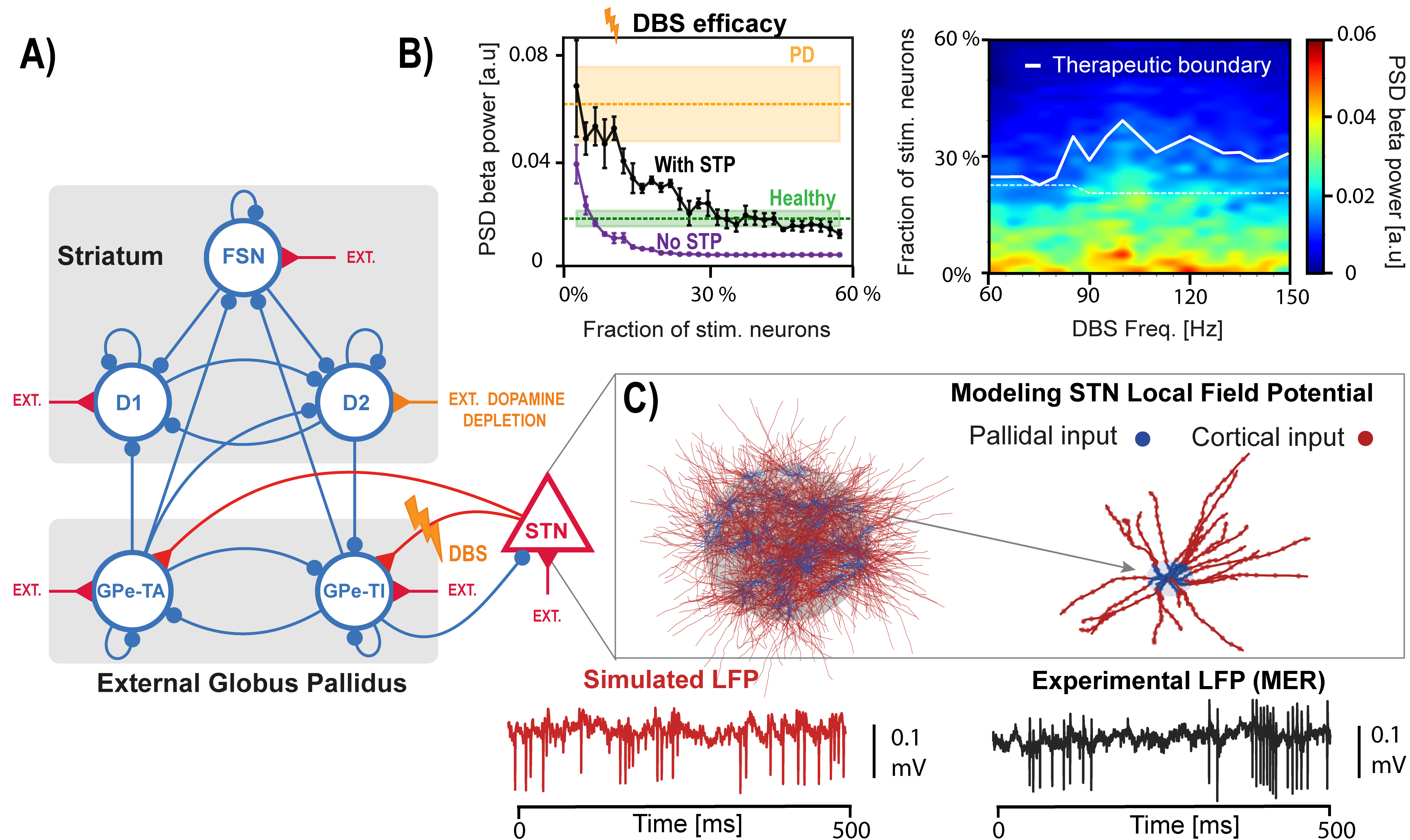
We implemented a spiking model of the basal ganglia with six neuronal populations: three within the striatum, two within the external globus pallidus, and the subthalamic nucleus (STN) (Fig. 1A) [1]. Dopamine depletion was simulated by modulating striatal inputs. We dissected the network mechanisms underlying beta and gamma resonances and we simulated STN DBS, considering short-term synaptic plasticity (Fig. 1B) [2]. To simulate the signals recorded by DBS electrodes we developed a population of morphological STN neurons model and computed LFPs associated with network activity through volume conduction theory (Fig. 1C).

Results
We show how beta oscillations arise from two independent loops in the BG model that strongly synchronize when dopamine is depleted [1]. STN DBS disrupts these oscillations, although without synaptic plasticity it requires unrealistically low stimulation levels (Fig 1B left). The model also supports the hypothesis that gamma-range stimulation can be as effective as the clinical standard of ~130 Hz used in PD (Fig. 1B right) [2]. Gamma oscillations emerge through recurrent inhibition in pallidal and striatal populations. Different from cortical ones, STN LFPs are largely noise-dominated due to weak correlations and symmetric neuronal morphology, becoming informative only when strong beta synchronization is present.

Discussion
We characterized in a spiking model of BG beta and gamma rhythmogenesis and their alteration due to PD-related dopamine depletion and DBS. Moreover, we investigated the origin of STN LFPs driving adaptive DBS, by integrating spiking and morphological modeling. Overall, we provide a multiscale framework for better understanding Parkinsonian dynamics and DBS mechanisms, showing how network modeling can clarify treatment mechanisms and guide improved stimulation strategies.

Figure 1. A) Spiking network model of the basal ganglia.  B) Left: efficacy of STN DBS as a function of the fraction of stimulated neurons, with and without short-term plasticity (STP). Right: effect of stimulation frequencies on beta spectral power.  C) Top: STN population and morphological neuron model receiving cortical and pallidal inputs. Bottom: simulated and recorded local field potentials (LFPs).
References

1. Ortone, A., Vergani, A. A., Ahmadipour, M., Mannella, R., & Mazzoni, A. (2023). Dopamine depletion leads to pathological synchronization of distinct basal ganglia loops in the beta band. PLOS Computational Biology, 19(4), 1–31. https://doi.org/10.1371/journal.pcbi.1010645
2. Ahmadipour, M., Fattorini, F., Meneghetti, N., & Mazzoni, A. (2026). In silico model of basal ganglia deep brain stimulation in Parkinson’s disease captures range of effective parameters for pathological beta power suppression. PLOS Computational Biology, 22(2), e1013280. https://doi.org/10.1371/journal.pcbi.1013280
Acknowledgments
This work was supported by the Italian Ministry of University and Research, under the complementary actions to the NRRP ‘Fit4MedRob - Fit for Medical Robotics’ Grant (# PNC0000007).


Speakers
Sunday July 12, 2026 2:50pm - 3:10pm ADT
Ballroom B1

3:10pm ADT

O7: Decreased alpha/theta temporal ExSEnt of left prefrontal cortex: a robust biomarker of dementia
Sunday July 12, 2026 3:10pm - 3:30pm ADT
Sara Kamali*1, Fabiano Baroni1, Pablo Varona1

1Department of Computer Engineering, Autonomous University of Madrid, Madrid, Spain

*Email: [email protected]

Introduction
Dementia involves progressive cognitive decline, with Alzheimer’s disease (AD) and frontotemporal dementia (FTD) as major subtypes. Electroencephalography (EEG) provides a noninvasive and accessible measure of brain dynamics and able to capture pathological slowing, often reflected by increased theta-to-alpha power ratio (TAR) [1]. Reduced signal complexity has also been reported in dementia. We tested whether Extrema-Segmented Entropy (ExSEnt), which separates temporal and amplitude irregularity, improves discrimination and interpretability beyond conventional electroencephalography biomarkers [2].

Methods
We analyzed resting-state EEG from 88 subjects, including 36 with AD, 23 with FTD, and 29 controls [3]. After preprocessing and independent component analysis, we classified brain sources into four clusters: right and left prefrontal cortices (R/LPFC) and right and left visual associations (R/LVA). Then, we computed a set of classical spectral and complexity features. We also measured the ExSEnt metrics, which quantifies the entropy of extrema-based segment durations, amplitudes, and their pair. To find the stable features for dementia detection, we performed stability selection with elastic-net logistic regression and nested leave-one-subject-out validation [4].

Results
ExSEnt improved discrimination mainly in the LPFC, where balanced accuracy increased from 71.5% to 82.6%. In that region, ExSEnt features showed high selection stability and replaced conventional measures as dominant predictors in 3 out of 4 brain areas. The most informative variables were temporal and joint temporal-amplitude entropy measures in alpha and theta bands, with negative coefficients indicating reduced irregularity and reduced dynamical variability in dementia. Other regions showed weaker or inconsistent gains, suggesting a localized effect rather than a global one. 

Discussion
ExSEnt provides a compact and interpretable measure for EEG-based dementia classification. The results suggest that dementia is associated not only with spectral slowing but also with reduced diversity of extrema timing and temporal-amplitude variability in LPFC dynamics. Because ExSEnt is explainable by design, when combined with stability selection and sparse linear classification, it yields robust biomarkers with direct physiological interpretation. These findings support ExSEnt as a promising candidate for explainable dementia screening and motivate future validation against cognitive severity measures.

References
[1] Partanen, J. V., Soininen, H., & Riekkinen, P. J. (1986). Does an ACTH derivative (Org 2766) prevent deterioration of EEG in Alzheimer's disease?. Electroencephalography and clinical neurophysiology, 63(6), 547-551.
[2] Kamali, S., Baroni, F., & Varona, P. (2025). ExSEnt: Extrema-Segmented Entropy Analysis of Time Series. arXiv preprint arXiv:2509.07751.
[3] Miltiadous, A., Tzimourta, K. D., Afrantou, T., Ioannidis, P., Grigoriadis, N., Tsalikakis, D. G., ... & Tzallas, A. T. (2023). A dataset of scalp EEG recordings of Alzheimer’s disease, frontotemporal dementia and healthy subjects from routine EEG. Data, 8(6), 95.
[4] Meinshausen, N., & Bühlmann, P. (2010). Stability selection. Journal of the Royal Statistical Society Series B: Statistical Methodology, 72(4), 417-473.

      Acknowledgments
      Funded by PID2024-155923NB-I00 and CPP2023-010818.
      Speakers
      Sunday July 12, 2026 3:10pm - 3:30pm ADT
      Ballroom B1

      3:30pm ADT

      O8: Frequency-dependent modulation of cortical traveling waves during general anesthesia
      Sunday July 12, 2026 3:30pm - 3:50pm ADT
      Duan Li*, Anthony G. Hudetz

      Center for Consciousness Science, Department of Anesthesiology, University of Michigan, Ann Arbor, MI

      *Email: [email protected]

      Introduction
      General anesthetics profoundly reshape neuronal activity and functional interactions, yet how they alter the spatiotemporal organization of the cortex is incompletely understood. Traveling waves provide a framework for examining coordinated neural activity across the cortex, but their modulation by anesthesia remains underexplored. Recently we showed that cortical dynamics under anesthesia exhibit spontaneous transitions among discrete states, including a paradoxical state characterized by low delta power and high complexity in deep anesthesia[1]. To investigate how traveling waves vary across cortical states, we recorded hemispheric ECoG at multiple anesthetic depths and analyzed wave dynamics as a function of cortical state and frequency.

      Methods
      ECoG was recorded from the right hemisphere with chronically implanted 32-site flexible polymer arrays (4×8 grid) at four desflurane concentrations (6-0%) for 1 h each. Cortical states were identified using PCA of power spectrograms followed by density-based clustering across concentrations. Within each state, traveling wave episodes were detected based on stabile spatial phase gradient patterns [2] between consecutive time points [3] in the delta, theta, and gamma bands. Wave occurrence and pattern richness were quantified, the latter defined as the entropy of SVD eigenvalues of the episode similarity matrix. Plane waves were identified and further classified as feedforward (posterior-to-anterior) or feedback (anterior-to-posterior).

      Results
      Seven states were identified. S1-S5 broadly tracked anesthetic depth but were not tied to a specific level. S6 corresponded to burst suppression, and S7 indicated a paradoxical state mostly during deep anesthesia. In S1, theta waves were mainly feedback-directed, whereas gamma were feedforward-directed. As anesthesia deepened, delta waves became more frequent but showed reduced pattern diversity. In contrast, the occurrence and diversity of theta and gamma patterns were largely preserved. Although gamma feedforward dominance persisted, theta feedback dominance was suppressed during deep anesthesia. In S7, reductions in delta complexity and theta organization were only partially reversed, despite reduced delta power and wake-like complexity.

      Discussion
      General anesthesia differentially modulates cortical traveling waves across frequency bands. Loss of theta feedback dominance paired with preserved gamma feedforward propagation suggests disrupted top-down integration despite maintained bottom-up information flow. Reduced diversity of delta waves during deep anesthesia suggests a shift toward more stereotyped, low-information dynamics. Importantly, traveling waves do not fully recover in the paradoxical state suggesting that spectrally activated deep anesthetic states lack coordinated spatiotemporal organization necessary for conscious processing. These findings suggest that traveling wave dynamics provide complementary insight into brain state dynamics beyond spectral power or complexity.

      References
      1. Li, D., & Hudetz, A. G. (2025). Dynamic electrocortical states and paradoxical complexity during desflurane anesthesia. bioRxiv. https://doi.org/10.1101/2025.10.13.682019
      2. Muller, L., Piantoni, G., Koller, D., Cash, S. S., Halgren, E., & Sejnowski, T. J. (2016). Rotating waves during human sleep spindles organize global patterns of activity that repeat precisely through the night. eLife, 5, e17267. https://doi.org/10.7554/eLife.17267
      3. Das, A., Zabeh, E., Ermentrout, B., & Jacobs, J. (2024). Planar, spiral, and concentric traveling waves distinguish cognitive states in human memory. bioRxiv. https://doi.org/10.1101/2024.01.26.577456

      Acknowledgments
      Research was supported by the National Institute of General Medical Sciences of the National Institutes of Health under award number R01-GM056398 and the Center for Consciousness Science, Department of Anesthesiology, University of Michigan Medical School, Ann Arbor, Michigan, USA. The authors express their gratitude to Dr. Shiyong Wang for his assistance in performing the experiments.

      Speakers
      DL

      Duan Li

      Associate Research Scientist, University of MIchigan
      Sunday July 12, 2026 3:30pm - 3:50pm ADT
      Ballroom B1
       
      Monday, July 13
       

      9:00am ADT

      Announcements
      Monday July 13, 2026 9:00am - 9:10am ADT

      Monday July 13, 2026 9:00am - 9:10am ADT
      Ballroom B1

      9:10am ADT

      Keynote 3: Blake Richards, "Exponentiated gradients support effective learning in biologically relevant scenarios"
      Monday July 13, 2026 9:10am - 10:10am ADT
      Computational neuroscience relies on gradient descent (GD) for training artificial neural network (ANN) models of the brain. The advantage of GD is that it is effective at learning difficult tasks. However, it produces ANNs that are a poor phenomenological fit to biology, making them less relevant as models of the brain. Specifically, it violates Dale’s law, by allowing synapses to change from excitatory to inhibitory, and leads to synaptic weights that are not log-normally distributed, contradicting experimental data. Here, starting from first principles of optimization theory, I will present an alternative learning algorithm, exponentiated gradient (EG), that respects Dale’s Law and produces log-normal weights, without losing the power of learning with gradients. We show that in biologically relevant settings EG outperforms GD, including learning from sparsely relevant signals and dealing with synaptic pruning. Altogether, our results show that EG is a superior learning algorithm for modelling the brain with ANNs.


      Monday July 13, 2026 9:10am - 10:10am ADT
      Ballroom B1

      10:40am ADT

      FO3: Gene Gradients Reveal Directed Structural Connectivity Across Species
      Monday July 13, 2026 10:40am - 11:10am ADT
      Benjamin S. Sipes*1, Ashish Raj1

      1Department of Radiology and Biomedical Imaging, University of California, San Francisco, San Francisco, CA, United States

      *Email: [email protected]

      Introduction
      Diffusion MRI (dMRI) tractography estimates the brain's white matter structural connectivity (SC) in vivo, but it cannot resolve the directionality of white matter pathways. Yet, much recent work has shown that genes and gene co-expression maps relate to SC across species [1-4]. Here we test whether gene co-expression gradients can infer connection directionality from undirected structural connectivity using the brain’s structure–function relationship.

      Methods
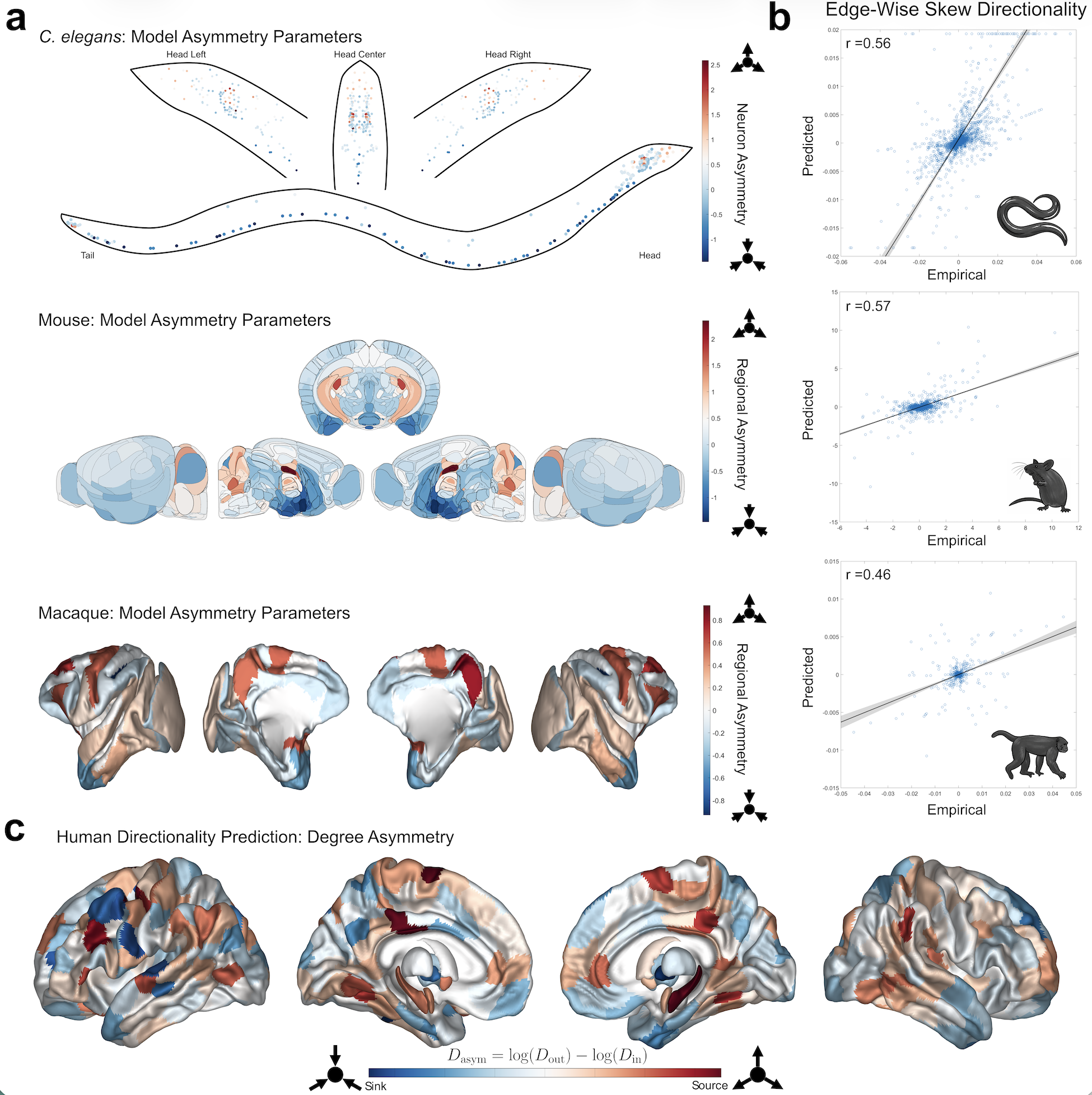
      We introduce asymmetry to SC (C) via a similarity transform with a node-level gauge parameterized by genetic gradients: C̃=ACA^-1, where A=diag(e^{Ga}), with G=[g_1,...,g_k] genetic gradient vectors and a=[a_1,...,a_k]^T gradient weights. We learn gradient weights by fitting a higher order network diffusion (HONeD) model of the SC graph Laplacian, ℒ=I-C̃D_{in}^-1, f(ℒ)=-κI-βℒ+ξℒ^2, to the residual of the Lyapunov equation, f(ℒ)^TΣ+Σf(ℒ)+I [5,6], with stationary covariance (Σ) estimated from functional neuroimaging. We compared our model's performance to ground truth directionality in three species: C. elegans, mouse, and macaque [7-10]. We then ran our model on 770 HCP subjects [11,12]. Public datasets supplied gene expression [13-17].

      Results
      Model-predicted directionality significantly correlated with ground-truth directed edges in all three species. Our model predicted neuron-to-neuron synaptic directionality in C. elegans (r=0.56, p<10^-253) and tracer-based directionality in mouse (r=0.57, p<10^-37) and macaque (r=0.46, p<10^-44) (Fig.1a-b). The optimal numbers of genetic gradients was also different in each species (C. elegans: k=3; Mouse: k=5; Macaque: k=1). We found that humans had optimal test-retest reliability when using k=5 genetic gradients (ICC=0.46). Human predicted degree asymmetry suggests that the hippocampus and posterior cingulate are network sources while temporal poles are network sinks (Fig.1c).

      Discussion
      Although white matter pathways exhibit directionality, estimating their orientation has largely been restricted to tracer-based experiments and a small number of specialized imaging methods. Our results suggest that gene gradients combined with structure–function modeling provide a biologically grounded framework for inferring directed structural connectivity across species, supporting the idea that molecular gradients may encode directional biases in large-scale brain networks. Estimating human SC directionality is valuable not only for basic neuroscience, but also for evaluating circuit-level models of brain function and for studying diseases such as Alzheimer’s, Parkinson’s, and ALS that may propagate along structural pathways [18].

      Figure 1. (a) Model-estimated directionality parameters (e^{Ga}) for the three non-human species: C. elegans (top), Mouse (middle), Macaque (bottom). In the C. elegans plot, each dot represents a single neuron. (b) Scatter plots comparing empirical to predicted skew edges with Pearson correlations listed at the top left (all p<10^{-37}). (c) Predicted human overall degree asymmetry for 414 brain regions.
      Speakers
      avatar for Benjamin Snow Sipes

      Benjamin Snow Sipes

      Graduate Student Researcher, University of California, San Francisco
      My research develops computational approaches for understanding how brain structure shapes neural function. I use graph signal processing, spectral graph theory, and multimodal neuroimaging—including fMRI, diffusion MRI, and MEG—to study structure–function coupling, network... Read More →
      Monday July 13, 2026 10:40am - 11:10am ADT
      Ballroom B1

      11:10am ADT

      O9: Low-Dimensional Communication Subspaces Reveal Distributed Information Across Neural Areas
      Monday July 13, 2026 11:10am - 11:30am ADT
      Farzad Karimi*1,2, Javier G. Orlandi1,2

      1Department of Physics and Astronomy, University of Calgary, Calgary, Canada
      2 Hotchkiss Brain Institute, University of Calgary, AB, Canada

      *Email: [email protected]

      Introduction
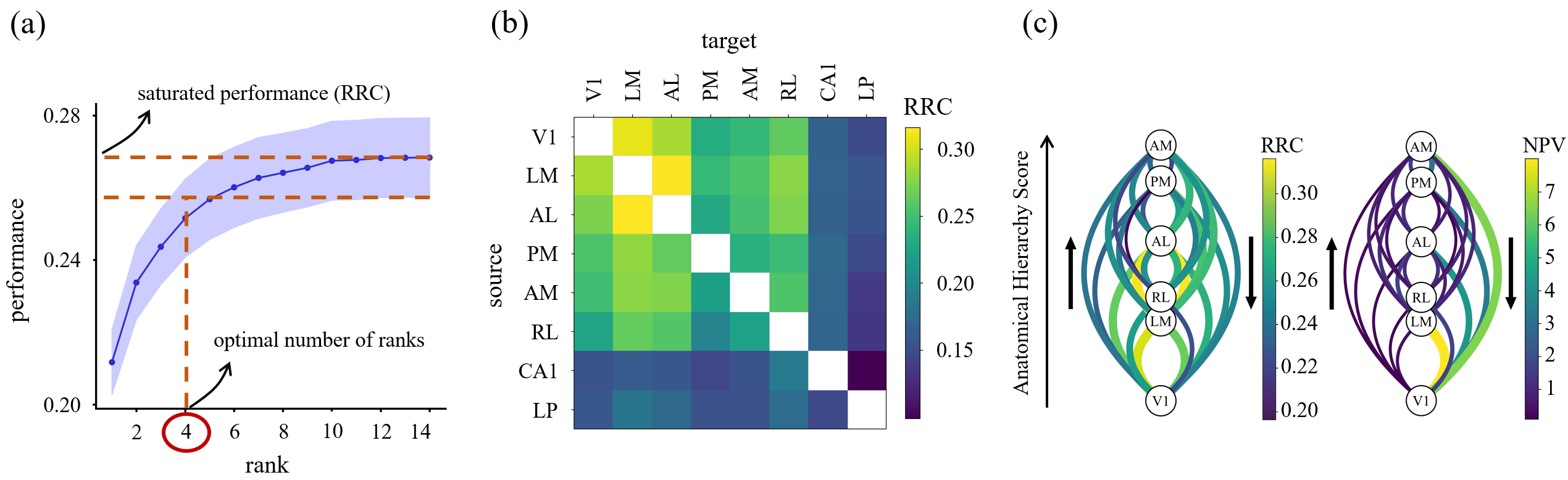
      Recent technological advances allowing us to simultaneously record across thousands of neurons have revealed the presence of distributed representations across the brain [1]. However, the network processes and information pathways that create these distributed representations are still poorly understood. To identify these distributed representations, we measured shared information across brain areas, by introducing a new directed connectivity measure, Reduced Rank Connectivity (RRC). RRC is defined through communication subspaces between neural areas, and by comparing these subspaces we can measure the extent of distributed signals across the brain.

      Methods
      We analyzed Neuropixels recordings from the Allen Institute from 54 mice performing a go/no-go visual change detection task, focusing on six visual cortical areas (V1, LM, AL, RL, AM, PM), as well as the thalamus (LP) and hippocampus (CA1), across two sessions: active behavior and passive replay [2]. To estimate shared information, we applied Reduced Rank Regression (RRR) [3], which predicts target activity from a low-dimensional subspace of a source population. We define the total predictable target activity as a new connectivity measure, called RRC, and distances between subspaces quantify the similarity of shared information across neural areas.

      Results
      We applied RRR to cortical and subcortical areas to analyze information flow across all area pairs combinations. We showed that model performance, defined as the squared correlation between predicted and test data, saturated with only a few predictive dimensions. These results identify low-dimensional communication subspaces between neural areas (Fig. 1a). We observed consistent shared information across the visual cortex, while predictability was lower for subcortical areas (Fig. 1b). Connectivity computed using RRR differed significantly from structural connectivity [4] (Fig. 1c). Our results also show that RRC is modulated by the animal’s engagement with the task (active vs. passive).

      Discussion
      Using RRR on multi-area cortical recordings, we identified robust shared information across visual areas during a discrimination task. RRR performance provides a connectivity measure that captures predictive subspaces rather than coarse averages. The results suggest the presence of low-dimensional communication subspaces between neural areas. Cortical areas can be more easily predicted by their own activity than subcortical areas through these communication subspaces during visual processing. RRC differed from structural connectivity and was modulated by behavioral state.

      Figure 1. Low-dimensional communication subspaces define RRC. (a) Prediction performance vs. rank; saturation defines optimal number of ranks and RRC. (b) Average RRC across animals; cortical areas are more predictable than subc
      Speakers
      avatar for Farzad Karimi

      Farzad Karimi

      PhD student
      Monday July 13, 2026 11:10am - 11:30am ADT
      Ballroom B1

      11:30am ADT

      O10: A mathematical language for large-scale spike recordings from hundreds to thousands of neurons
      Monday July 13, 2026 11:30am - 11:50am ADT
      Alexandra Busch*,1,2,3, Roberto Budzinski2,4, Lyle Muller1,2,3
      1 Department of Mathematics, Western University, London ON, Canada
      2 Fields Lab for Network Computation, Fields Lab, Toronto ON, Canada
      3 Western Institute for Neuroscience, Western University, London ON, Canada
      4  Department of Neuroscience, University of Lethbridge, Lethbridge AB, Canada
      Email: [email protected]

      Introduction
      Recent technological advances now allow simultaneously recording the activity of thousands of neurons while animals engage in cognitive tasks. These datasets can offer an unprecedented window into how the brain computes in real time, but they also challenge existing analytical frameworks. There has been increasing interest in the possibility that coordinated patterns of spikes, such as sequences, may contribute to neural computation [1-3]. However, in contrast to the many methods available for analyzing firing rates, mathematical tools capable of systematically probing spike-time structure at the scale of these next-generation datasets remain limited.

      Methods
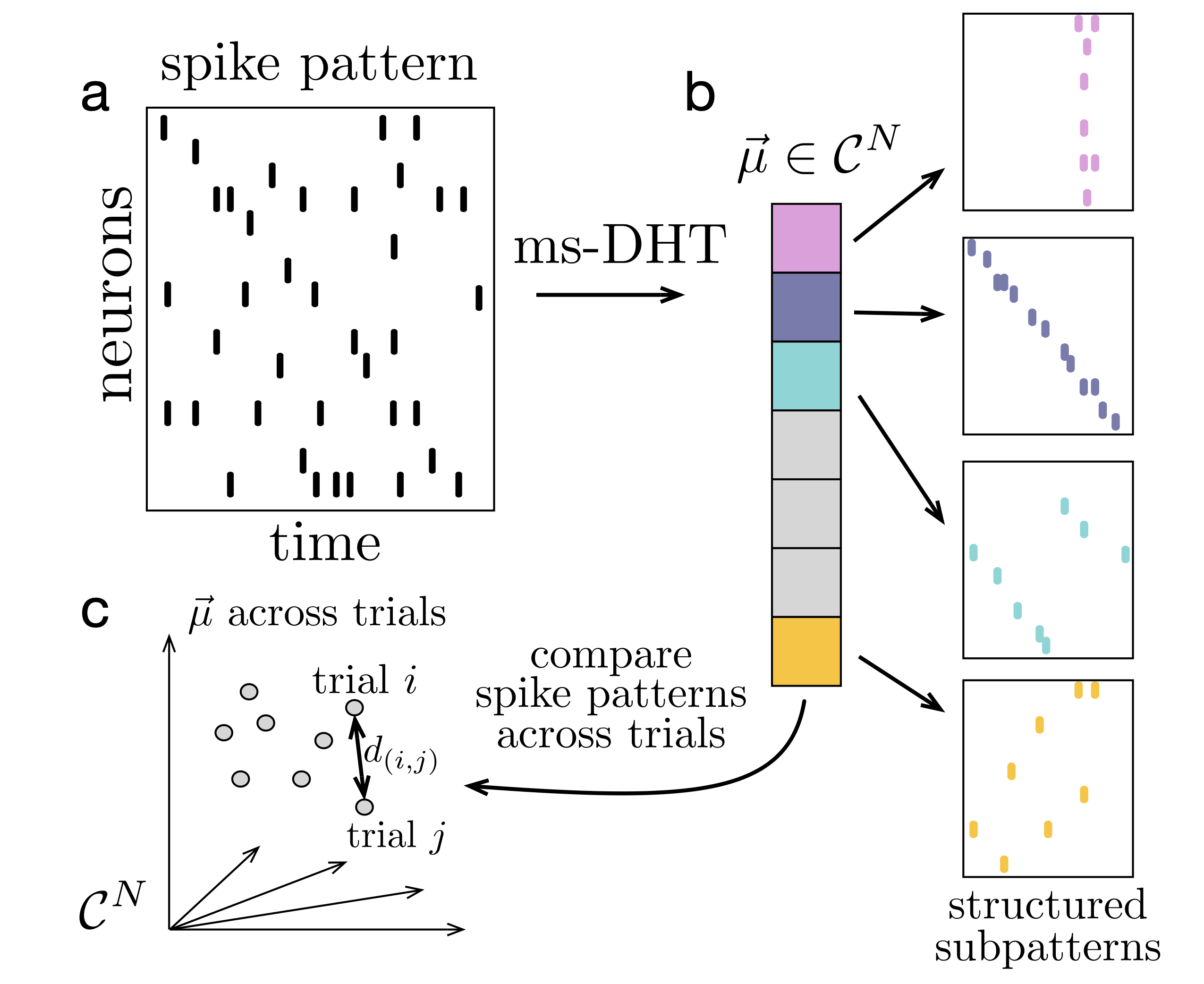
      We introduce a decomposition operator for population spike patterns, termed the multi-sample Discrete Helix Transform (ms-DHT). We derive a generalized inner product that allows the ms-DHT to operate directly on patterns of discrete spikes across thousands of neurons without smoothing. The ms-DHT decomposes these spike patterns into a fixed, interpretable basis, mapping each input pattern to a unique vector that captures the occurrence and timing of every spike (Fig.1). In this representation, distances between spike patterns reduces to the Euclidean distance between their ms-DHT outputs. This distance is invariant to neuron ordering and allows detecting repeating structure ranging from simple spike sequences to complex population motifs.

      Results
      We demonstrate several applications of the ms-DHT to large-scale datasets. Notably, in dual Utah array recordings from the prefrontal cortex of a macaque monkey performing a virtual reality working memory task, the ms-DHT reveals structured spike motifs that predict specific behavioural errors on single trials - before they occur. Further, applications to spiking network simulations with 10,000 neurons demonstrate that the transform operates effectively at the scale of next-generation neural recordings.

      Discussion
      The ms-DHT provides a flexible framework for analyzing large-scale spike patterns. By decomposing spiking activity onto a fixed, interpretable basis using a generalized inner product, the ms-DHT produces unique descriptions of population activity even when neurons emit variable numbers of spikes—a setting that has posed a central challenge for analytical approaches. The resulting representation supports multiple analyses, including clustering and decoding of full spike patterns, detecting repeating substructure through specific helix contributions, and sliding-window analyses that trace the temporal evolution of spike patterns across long recordings.

      FIgure 1. Decomposing spike patterns. The ms-DHT maps a spike pattern (a) to a unique complex-valued vector (b). Each component encodes the strength (amplitude) and timing (phase) of a basis sub-pattern. (c) Distances between spike patterns reduce to Euclidean distances between ms-DHT outputs, which are invariant to neuron order, ensuring behaviourally relevant clusters do not depend on neuron order.
      References
      [1] Xie, W., Wittig, J. H., Chapeton, J. I., El-Kalliny, M., Jackson, S. N., Inati, S. K., & Zaghloul, K. A. (2024). Neuronal sequences in population bursts encode information in human cortex. Nature, 635(8040), 935–942. https://doi.org/10.1038/s41586-024-08075-8
      [2] Chettih, S. N., Mackevicius, E. L., Hale, S., & Aronov, D. (2024). Barcoding of episodic memories in the hippocampus of a food-caching bird. Cell, 187(8), 1922–1935.e20. https://doi.org/10.1016/j.cell.2024.02.032
      [3] Busch, A., Roussy, M., Martinez-Trujillo, J. C., et al. (2024). Neuronal activation sequences in lateral prefrontal cortex encode visuospatial working memory during virtual navigation. Nature Communications, 15, 4471. https://doi.org/10.1038/s41467-024-48664-9

      Acknowledgments
      This work was supported by NSERC, CFREF, NIH, Neuronex NSF, and Canada Research Chairs Program. A.B. gratefully acknowledges a BrainsCAN studentship and NSERC CGS-D. 


      Speakers
      AB

      Alexandra Busch

      PhD Candidate, Western University
      Monday July 13, 2026 11:30am - 11:50am ADT
      Ballroom B1

      11:50am ADT

      O11: SEM to Simulation: Bringing Ultrastructural Detail to Multiscale Modeling
      Monday July 13, 2026 11:50am - 12:10pm ADT
      Cecilia Romaro*1, Matei Coldea2, William W. Lytton3,4, and Robert A. McDougal1,5,6,7,8
      1 Department of Biostatistics, Yale School of Public Health, New Haven, CT, United States
      2 Yale College, Yale University, New Haven, CT, United States
      3 Department of Physiology and Pharmacology & Neurology, SUNY Downstate Health Sciences University, Brooklyn, New York
      4 Department of Neurology, Kings County Hospital Center, Brooklyn, New York
      5 Department of Biomedical Informatics and Data Science, Yale School of Medicine, New Haven, CT, United States
      6 Program in Computational Biology and Bioinformatics, Yale University, New Haven, CT, United States
      7 Wu Tsai Institute, Yale University, New Haven, CT, United States
      8 Interdepartmental Neuroscience Program, Yale University, New Haven, CT, United States
      * Email: [email protected]

      Introduction
      Just as neuron morphology influences spiking behavior and thus network interactions, so too does the 3D placement of spines affect interaction between spines [1] and thus cellular behavior. However fine spine details are not visible under the optical microscopy used for reconstructing neuron morphology and full-cell scanning electron microscopy (SEM) images are generally not feasible due to size constraints. To address these challenges, we developed a tool for the NEURON simulator [2] for importing and editing an SEM reconstruction of a portion of a dendrite, selecting spines, rotating them, and inserting them into a full-cell reconstruction for simulation, using our experimental support for reaction-diffusion multigridding in NEURON.

      Methods
      SEM images may be segmented to identify each spine using standard segmentation software then exported to a TIFF stack. We estimate key electrical properties: approximately equivalent length, diameter, volume, and surface area. Our tool loads the image stack and identifies the voxels forming each spine-dendrite boundary so that we can preserve the connection location after transformations. PySide6 is used to provide a graphical interface allowing spines to be selected and manipulated into position; this can also be done programmatically. An algorithm adds/removes voxels to connect the spine cleanly. Transformed spines can be exported to text files for easy editing, enabling iterative refinement.

      Results
      We present our graphical tool, examples of relevant data sets, and simulation results. The graphical tool allows visualization of both the loaded SEM data and the placed spines after transformations. The simulations leverage our previous work, allowing a synaptic source (e.g., of IP3) to be placed at a precise 3D location within a spine. We validate the multigrid simulation by comparing to a single unified 3D simulation and contrast it to simplified geometry approximations, illustrating their similarities and differences. In particular, our tool allows toggling between the two representations.

      Discussion
      Support for imported spine morphologies brings NEURON a step closer to capturing the intricacies of the human brain. The same tool described here can also directly be used for incorporating SEM data of a dendrite as well. It is not feasible to simulate full cells and networks at this level of detail, nor is that necessarily desirable -- simpler models are often more useful for insights -- but our approach allows us to explore localized behavior in detail in a multiscale context with full cell and network simulations. This tool can give us insight on which details model when and allow us to explore detailed biological questions of synaptic plasticity or the role of morphological changes in disease.

      References

      1. Huertas, M. A., Newton, A. J., McDougal, R. A., Sacktor, T. C., & Shouval, H. Z. (2022). Conditions for synaptic specificity during the maintenance phase of synaptic plasticity. Eneuro, 9(3). https://doi.org/10.1523/ENEURO.0064-22.2022

      2. Hines, M. L., & Carnevale, N. T. (1997). The NEURON simulation environment. Neural computation, 9(6), 1179-1209. https://doi.org/10.1162/neco.1997.9.6.1179

      Acknowledgments
      This research was funded by the National Institute of Mental Health, National Institutes of Health, grant number R01 MH086638. The content is solely the responsibility of the authors and does not necessarily represent the official views of the National Institutes of Health.
      Speakers
      Monday July 13, 2026 11:50am - 12:10pm ADT
      Ballroom B1

      12:10pm ADT

      O12: A new platform technology to explore and leverage the computational properties of biological neural cultures
      Monday July 13, 2026 12:10pm - 12:30pm ADT
      Brett J. Kagan*1, David Hogan1, Andrew Doherty1,  Boon Kien Khoo1,  Johnson Zhou1,  Richard Salib1,  James Stewart1,  Kiaran Lawson1,  Alon Loeffler1,
      1Cortical Labs, Melbourne, Australia
      2 The University of Melbourne, Department of Biochemistry and Pharmacology, Parkville, Melbourne, 3000, Australia

      *Email:[email protected]


      Introduction
      Neural cultures are increasingly explored to understand the computational properties of neural systems due to the controllability and modifiability of these systems. However, BNNs can only be explored reliably as reliable information-processing systems if inputs are delivered in a temporally and structurally consistent manner. In practice, this requires stimulation with precisely controlled structure, microsecond-scale timing, multi-channel synchronization, and the ability to observe and respond to neural activity in real-time. Existing approaches depend on either depend on low-level hardware mechanisms, imposing prohibitive complexity for rapid iteration, or they sacrifice temporal and structural control, undermining consistency.

      Methods
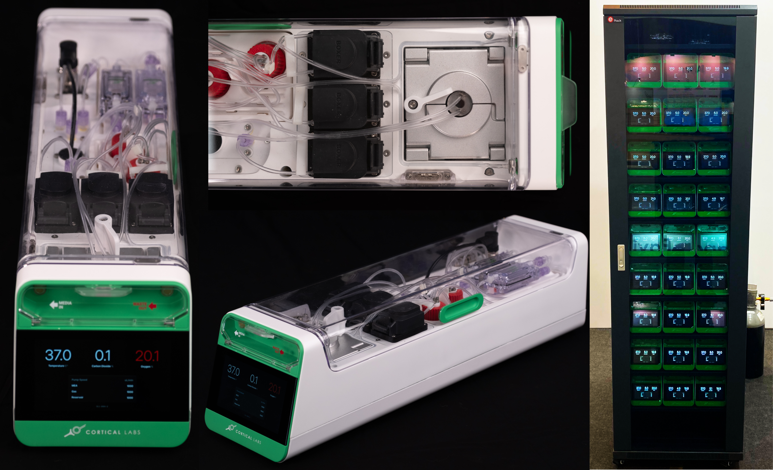
      To resolve this problem. We developed a bespoke but scalable system (the CL1)1 that coupled with a easy to use Application Programming Interface ( CL API)2 to  enables real-time, sub-millisecond closed-loop interactions with neural cultures. The system itself provides real-time closed-loop electrophysiology with integrated life support. For the API design approach, the CL API provides users with precise stimulation semantics, transactional admission, deterministic ordering, and explicit synchronization guarantees. This contract is presented through a declarative Python interface, enabling non-expert programmers to express complex stimulation and closed-loop behavior without managing low-level scheduling or hardware details.

      Results
      The result is a scalable device for interacting with in-vitro neural cell cultures via electrophysiology in a closed-loop real-time environment coupled with an integrated life-support system. The devices are server rack stackable, generating up to 6TB of neural activity data per server rack per day, allowing detailed analysis of electrophysiological data, where each unit can run its own embodied environment. This allows an unparalleled investigation of nearly fully controllable neural systems to explore their dynamics in depth. The flexibility of the Cl1 means that information processing and computation in neural cultures can be explored in many ways, including as reservoir computing, in robotics4, or via games such "Pong"5 or “Doom”.

      Discussion
      The CL1 system coupled with the CL API offers a scalable system for exploring computational dynamics of biological neural networks. Aside from being possible to set up in traditional cell culture laboratories, these systems can be accessed remotely via the cloud where the cell culture methods are managed either by a dedicated company or by partner laboratory groups. This provides a tool for computational neuroscientists, who might otherwise not be able to access these neural cultures, to explore research questions at scale, with precision, and with rapid iteration loops. It is proposed that this availability will allow computational neuroscientists to be able to explore the dynamics of biological neural systems in way never possible before.

      Figure 1. The CL-1 device is scalable desktop device compatible with standard server racks that allows real-time closed-loop interactions with neural cells via an MEA reader. The CL-1 has onboard hardware that interprets simple code via a Python API to allow rapid code development and experimental iterations coupled with a closed-loop perfusion circuit to automatically adjusts gas levels and temperature to
      References
      1) Kagan, B. J. (2025). The CL1 as a platform technology to l
      Speakers
      Monday July 13, 2026 12:10pm - 12:30pm ADT
      Ballroom B1

      2:00pm ADT

      FO4: Selective routing of spatial information in dentate granule cells emerges through disparate combinations of synaptic and intrinsic plasticity
      Monday July 13, 2026 2:00pm - 2:30pm ADT

      Sanjna Kumari*1 and Rishikesh Narayanan1
      1 Cellular Neurophysiology Laboratory, Molecular Biophysics Unit, Indian Institute of Science, Bengaluru 560012, India
      *Email: [email protected]

      Introduction
      Granule cells (GCs) in the dentate gyrus (DG) receive grid-like spatial inputs and contextual inputs from the entorhinal cortex, both broadly tuned to multiple spatial locations. Despite this, GCs elicit sparse spatial firing that is confined to single place fields, thus playing a central role in selective routing of spatial information to the hippocampal circuit. The mechanisms behind the transformation of broadly tuned afferent inputs into sparse and location-specific outputs remains unclear. In this study, we ask if there are physiologically relevant plasticity mechanisms that can mediate selective routing of spatial information towards place-cell emergence and spatial remapping, especially when inhibitory synapses are absent.

      Methods
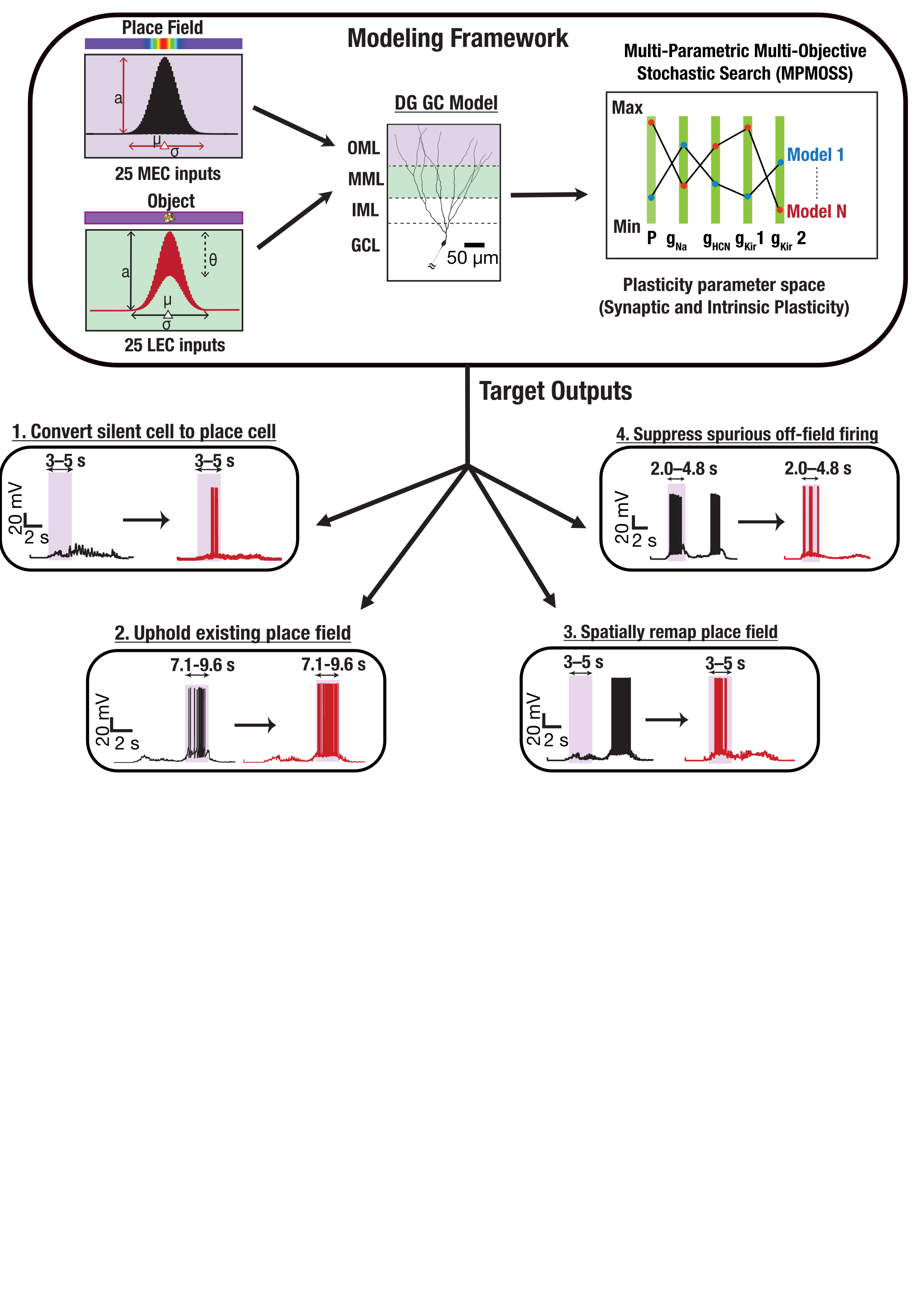
      We employed morphologically and biophysically realistic models of DG GCs (Kumari & Narayanan, 2024), receiving grid-like and contextual spatial inputs from the entorhinal cortex. We employed a stochastic search paradigm in the plasticity space involving fold-changes in excitatory synaptic strengths, persistent sodium (NaP), hyperpolarization-activated cyclic nucleotide-gated (HCN), and inward rectifier potassium (Kir) conductances. We validated plasticity combinations that achieved one of four functional targets relevant to DG spatial tuning: conversion of silent neurons to place cells, uphold existing place field firing, spatial remapping to a new location, and suppression of spurious place fields to obtain a single place field (Fig 1).

      Results
      While excitatory synaptic plasticity alone was insufficient to generate valid spatial tuning, conjunctive synaptic and intrinsic plasticity yielded several valid plasticity combinations for all 4 targets (Valid/Total models for 4 targets: 243/142,000, 325/10,000, 139/5,000, 224/50,000). These valid plasticity combinations manifested pronounced heterogeneity across all fold-changes, unveiling plasticity degeneracy where disparate plasticity combinations yielded similar spatial tuning outcomes. Dimensionality reduction analyses revealed low-dimensional structures in intrinsic measurement and parameter spaces of valid models. In contrast, the plasticity space did not manifest strong constraints on plasticity across different components.

      Discussion
      While inhibitory synaptic inputs have been studied as mechanisms for sculpting spatial tuning, we show that selective routing of information and suppression of off-field firing can be achieved through intrinsic plasticity. Among intrinsic components, we predict the axonal initial segment Kir conductance as the strongest determinant of spatial selectivity. We demonstrate that disparate combinations of concomitant plasticity in excitatory synaptic and intrinsic conductances can mediate the emergence, refinement, and remapping of place fields. We show that co-dependent plasticity in different neuronal components can enable robust yet flexible spatial representations despite heterogeneities in neuronal composition and plasticity mechanisms.

      FIgure 1. Medial and lateral entorhinal cortex inputs impinge on a DG granule cell. Disparate combinations of synaptic and intrinsic plasticity (NaP, HCN, Kir channels) achieved one of four targets: convert silent cell to place cell, uphold existing place field, remap, or suppress spurious firing. Our results show that robust and flexible spatial tuning is achievable through plasticity degeneracy.References
      Kumari, S., & Narayanan, R. (2024). Ion-channel degeneracy and heterogeneities in the emergence of signature physiological characteristics of dentate gyrus granule cells. J Neurophysiol, 132(3), 991-1013. https://doi.org/10.1152/jn.00071.2024

      Speakers
      Monday July 13, 2026 2:00pm - 2:30pm ADT
      Ballroom B1

      2:30pm ADT

      O13: A Developmental Ring Attractor Model for the Head Direction System
      Monday July 13, 2026 2:30pm - 2:50pm ADT
      Shujia Liu*1, 2, Bailu Si1, Michael Herrmann2

      1School of Systems Science, Beijing Normal University, Beijing, China
      2 School of Informatics, The University of Edinburgh, Edinburgh, UK

      *Email: [email protected]

      Introduction
      Most ring attractor models hard-code and phase-biased translation kernels to obtain a stable activity profile (bump) and velocity-driven shifts [1]. This bypasses a key developmental question: Can these stabilizing and translation kernels self-organize, without any pre-set ring topology, from activity statistics under staged multimodal constraints? The Lateral Mammillary Nucleus--Dorsal Tegmental Nucleus (LMN--DTN) loop implicated in head direction system also lacks developmental constraints. Motivated by synfire chain theory [2] we build a plasticity enabled LMN--DTN model and propose: Spontaneous traveling wave statistics plus staged vestibular/visual constraints can drive the emergence of a ring attractor and path integration.

      Methods
      We constructed a rate-based LMN--DTN circuit model with 400 neurons in LMN and two populations of 400 direction--velocity conjunctive cells in DTN. LMN follows leaky integrator dynamics with plastic recurrent excitatory connectivity and a fixed long-range inhibitory kernel. DTN to LMN feedback consists of plastic phase-biased weights gated by the angular velocity input. Training proceeds in functional stages: We first obtain stable traveling wave statistics without external velocity or vision, then update connectivity via STDP-like and structural plasticity, and subsequently introduce long-range inhibition and a visual teacher for representational stabilization and gain

      Results
      With random sparse connectivity, no external velocity input, and no hand-designed ring topology templates, LMN networks spontaneously produce a stable unidirectional traveling wave under the joint action of dominant refractory-like neuronal dynamics and global inhibition, exhibiting consistent phase progression (Fig. 1A). STDP-like and structural plasticity then consolidate the temporal correlations into locally enhanced recurrent excitation (Fig. 1B); long-range inhibition transforms the traveling wave regime into a phase-selectable single bump state. Visual relearning markedly improves short-term angle tracking, yet cumulative drift persists during pure path integration after removing visual information (Fig. 1C).

      Discussion
      Our results indicate that stabilizing and translation kernels of ring attractors need not be hard-coded: intrinsic recurrent dynamics can provide a directional temporal scaffold, which activity-dependent plasticity, staged inhibition, and multimodal constraints shape into a stable bump representation and a learnable translation kernel. Although residual drift remains after visual removal, it is structured rather than arbitrary, suggesting that the model captures much of the required computation while revealing imperfections in the learned kernel. This makes the framework useful both as a proof of principle for de
      Speakers
      Monday July 13, 2026 2:30pm - 2:50pm ADT
      Ballroom B1

      2:50pm ADT

      O14: How synchronization, excitability, and variability shape CPG rhythmic bursting sequences across different time scales
      Monday July 13, 2026 2:50pm - 3:10pm ADT
      Pablo Sanchez-Martin*1, Alicia Garrido-Peña1, Irene Elices1, Carlos Garcia-Saura1, Rafael Levi1, Francisco B. Rodriguez1, Pablo Varona1 

      1Grupo de Neurocomputación Biológica (GNB), Department of Computer Engineering, Universidad Autónoma de Madrid, Madrid, Spain

      *Email: [email protected]

      Introduction
      Rhythmic sequential activity is present in many nervous systems. Neural circuits that generate this activity usually involve intrinsic neuronal variability and different synapse types [1]. Sequential rhythms often require coordination at different time scales to adapt to specific conditions, or to adjust speed and timing to meet functional needs. Previous studies in computational models have assessed how synchronization and excitability can modulate cycle-by-cycle sequential dynamical invariants [2,3]. In this study, we analyzed the interplay among neural synchronization, excitability, and variability to understand how they are related to the sequentiality timing in CPG rhythms.

      Methods
      We acquired long recordings of pyloric CPG neurons of  Carcinus maenas  and extracted the spike timings from intracellular and extracellular time series followed by calculation of all sequence intervals between the PD neurons and the LP. We used metrics of synchronization between the electrically coupled PDs (Victor-Purpura distance, Euclidean distance), excitability for all three neurons (Spike Density Function -SDF-, average ISIs), and interval variability. We identified dynamical invariants in the form of relationships between specific intervals and the instantaneous period. To find relationships between these metrics, we performed analysis at three time scales: whole experiment, segments inside experiments, and cycle-by-cycle analysis.

      Results
      We observed a high level of variability for synchronization, excitability, and the intervals in this system. Ranking each experiment for all metrics revealed a relationship between the variability in the period, the neurons’ SDF, and the strength of the dynamical invariant relationship. Segmenting the data, we found that, in addition to these relationships, synchronization in the PD neurons is related to their excitability. We found non-linear relationships between the excitability of all neurons and their period variability and dynamical invariants. Excitability changes in any neuron were related to the other neurons' excitability at each cycle, although other relationships present at larger time scales were not preserved cycle-by-cycle.

      Discussion
      It is still unclear how robustness and flexibility can be autonomously balanced in neural sequences. Previous works have found evidence that suggests that connectivity asymmetry, i.e., the presence of both slow and fast synapses, could be responsible for the emergence of coordination rules such as sequential dynamical invariants [2, 3]. The LPPDdelay interval and instantaneous period are related cycle-by-cycle, as well as the excitability of all neurons among them. In an intermediate scale, the excitability is non-linearly related to synchronization, variability and strength of the dynamical invariants. In a larger time scale, excitability, variability, and the strength of dynamical invariants are all related, but not synchronization.

      References
      [1] Selverston, A. I., Rabinovich, M. I., Abarbanel, H. D., Elson, R., Szücs, A., Pinto, R. D., ... & Varona, P. (2000). Reliable circuits from irregular neurons: a dynamical approach to understanding central pattern generators. Journal of Physiology-Paris, 94(5-6), 357-374. 
      [2] Berbel, B., Latorre, R., & Varona, P. (2025). Theoretical bases for the relation between excitability, variability and synchronization in sequential neural dynamics. Neurocomputing, 645, 130218. 
      [3] Elices, I., Levi, R., Arroyo, D., Rodriguez, F. B., & Varona, P. (2019). Robust dynamical invariants in sequential neural activity. Scientific Reports, 9(1), 9048. 

      Acknowledgments
      Research funded by grants PID2024-155923NB-I00, PID2023-149669NB-I00 and CPP2023-010818 (MCIN/AEI and ERDF- "A way of making Europe"). 


      Speakers
      PS

      Pablo Sanchez-Marti­n

      PhD Student, Autonomous University of Madrid
      Monday July 13, 2026 2:50pm - 3:10pm ADT
      Ballroom B1

      3:10pm ADT

      O15: Exact mathematical description of computation with transient spatiotemporal dynamics in recurrent neural networks
      Monday July 13, 2026 3:10pm - 3:30pm ADT
      Roberto Budzinski1,2,#, Alexandra Busch2,3,4, Luisa Liboni2,5, Ján Mináč2,3, Lyle Muller2,3,4
      1 Department of Neuroscience, University of Lethbridge, Lethbridge AB, Canada
      2 Fields Lab for Network Computation, Fields Lab, Toronto ON, Canada
      3 Department of Mathematics, Western University, London ON, Canada
      4 Western Institute for Neuroscience, Western University, London ON, Canada
      5 King's University College at Western University, London ON, Canada
      # [email protected]

      Introduction
      Networks throughout physics and biology use spatiotemporal dynamics for computation [1]. In neural systems, waves of neural activity have recently been shown to shape spiking responses, gate perception, and influence behaviour [2]. However, it remains unclear how network connectivity gives rise to neural dynamics and how these dynamics support computation. To address this question, we introduce a new type of recurrent neural network that admits an exact mathematical solution [3,4], enabling us to directly relate network structure to emergent dynamics and the computations those dynamics perform.

      Methods
      We introduce a nonlinear recurrent neural network in which each unit is modeled as a complex-valued oscillator. This complex-valued recurrent neural network (cv-RNN) admits a closed-form solution given by an exact propagator. Importantly, this framework introduces a unified matrix representation of the system that encodes the network's connectivity, including connection strengths and delays, and the input. The exact mathematical solution allows us to control the network dynamics, down to the fine-scale pattern of connectivity, allowing us to use the spatiotemporal patterns that emerge for dynamics-based computation in a wide range of tasks [3,4].

      Results
      We find the cv-RNN can perform a wide range of tasks, including working memory, logic operations, sequence processing, and computer vision, while remaining precise and interpretable mathematically [3,4]. The analytical framework reveals the mechanisms underlying each computation. By exploiting traveling-wave dynamics, the network performs image segmentation and generalizes across different datasets using the same recurrent weights [4]. Further, we create a bio-hybrid version of our cv-RNN, where we leverage patch-clamping techniques to link biological neurons to the recurrent layer, where these neurons can decode the network’s spatiotemporal dynamics and implement computations [3].

      Discussion
      These results demonstrate that structured spatiotemporal dynamics can serve as a powerful computational substrate in recurrent neural networks. The exact solution links connectivity, input, and emergent dynamics within a unified operator framework. This approach provides a principled way to understand how neural circuits may compute through traveling waves and network dynamics. More broadly, it establishes a general framework for connecting network structure, emergent dynamics, and computation, offering new tools for interpreting biological neural activity and for designing transparent dynamical models in artificial intelligence.

      References
      [1] Ermentrout et al. (2001), "Traveling electrical waves in cortex: insights from phase dynamics and speculation on a computational role”, Neuron 29, 33.
      [2] Muller et al. (2018), “Cortical travelling waves: mechanisms and computational principles”, Nature Reviews Neuroscience 19.
      [3] Budzinski et al. (2024) “An exact mathematical description of computation with transient spatiotemporal dynamics in a complex-valued neural network, Communications Physics 7.
      [4] Liboni et al. (2025), “Image segmentation with traveling waves in an exactly solvable recurrent neural network”, Proceedings of the National Academy of Sciences 122.
      Acknowledgments
      This work was supported by NSERC, CFREF, NIH, Neuronex NSF, and Canada Research Chairs Program.


      Speakers
      avatar for Roberto Budzinski

      Roberto Budzinski

      University of Lethbridge
      Monday July 13, 2026 3:10pm - 3:30pm ADT
      Ballroom B1

      3:30pm ADT

      O16: The role of cell types in critical neural activity
      Monday July 13, 2026 3:30pm - 3:50pm ADT
      Adrián Ponce-Alvarez*1,,2,3 and Germán Sumbre4
      1 Departament de Matemàtiques, Universitat Politècnica de Catalunya, 08028 Barcelona, Spain.
      2 Institut de Matemàtiques de la UPC - Barcelona Tech (IMTech), Barcelona, Spain.
      3 Centre de Recerca Matemàtica, Barcelona, Spain.
      4 Institut de Biologie de l’ENS (IBENS), Département de biologie, École normale supérieure, CNRS, INSERM, Université PSL, 75005 Paris, France


      *Email : [email protected]

      Introduction
      Neuronal activity shows statistics consistent with a critical point, a regime that maximize information capacity. Yet, the role of different cell types remains largely unexplored. Models [1] and in vitro studies [2] suggest that excitation–inhibition (E/I) balance is key for self-organized criticality, but how E and I dynamics interact during in vivo critical activity is unclear. Similarly, glial cells such as radial astrocytes (RAs) regulate neuronal function [3], but their role in criticality is unknown. Here, we studied how E/I neuronal activity and astrocyte calcium dynamics contribute to criticality by combining transgenic zebrafish with cell-type-specific calcium indicators, a stochastic network, and model inference.

      Methods
      Spontaneous neuronal activity in the optic tectum (OT) of 10 zebrafish larvae was recorded using light-sheet microscopy. A double-transgenic line expressing GCaMP6f in all neurons and Vglut in glutamatergic neurons identified of E and I cells. Two-photon calcium imaging was performed in 7 larvae expressing GCaMP6f in neurons and RCaMP1b in RAs [3]. OT activity was recorded during spontaneous activity and after mild electrical stimulation, which triggered synchronized Ca²⁺ transients in RAs.
      E and I activity was modelled using a stochastic network displaying critical avalanches at a E/I phase transition [1]. The maximum entropy principle mapped neuronal activity onto statistical models [4], quantifying criticality and detecting deviations.

      Results
      Our results show that neuronal avalanches approached criticality when E and I activity were balanced. Notably, the model accurately captured the observed avalanche statistics and their sensitivity to E/I fluctuations around a critical point defined by balanced excitatory and inhibitory synaptic strengths, where balanced amplification drives network avalanches. Furthermore, we found that RA synchronization shifted tectal neuronal activity away from its spontaneous critical state toward a more ordered regime, with a reduced repertoire of network states and diminished susceptibility to external inputs. These findings demonstrate that glial activity can actively regulate the state of neuronal ensembles, including their proximity to criticality.

      Discussion
      Extensive research highlights the benefits of E/I balance and critical dynamics. Balanced networks enhance amplification, selectivity, and stability, while critical dynamics optimize information processing. Here, we show that neuronal avalanche statistics and their dependence on spontaneous E/I fluctuations in the zebrafish OT match a model reaching criticality at balanced E and I couplings. Moreover, RA synchronization in the OT reshapes collective neuronal activity, consistent with a shift from spontaneous critical dynamics to a more ordered subcritical regime. Our findings show that radial astrocyte activity can shift the state of neuronal ensembles and modulate their proximity to criticality.

      References
      1.     Benayoun, M., et al. (2010). Avalanches in a Stochastic Model of Spiking Neurons. PLoS Comput Biol, 6(7), e1000846. https://doi.org/10.1371/journal.pcbi.1000846
      2.     Shew, W.L., et al. (2011). Information capacity and transmission are maximized in balanced cortical networks with neuronal avalanches. J Neurosci., 31(1), 55-63. https://doi.org/10.1523/JNEUROSCI.4637-10.2011
      3.     Uribe-Arias, A., et al. (2023). Radial astrocyte synchronization modulates the visual system during behavioral-state transitions. Neuron 111, (24), 4040-4057.e6. https://doi.org/10.1016/j.neuron.2023.09.022
      4.     Tkačik, G., et al. (2014). Searching for Collective Behavior in a Large Network of Sensory Neurons. PLoS Comput Biol, 10(1), e1003408. https://doi.org/10.1371/journal.pcbi.1003408

      Acknowledgments
      This study was supported by the Project PID2022-137708NB-I00 funded by MICIU/AEI /10.13039/501100011033 and FEDER, UE. A. Ponce-Alvarez was supported by a Ramón y Cajal fellowship (RYC2020-029117-I) funded by MICIU/AEI/10.13039/501100011033 and “ESF Investing in your future”. G. Sumbre was supported by ERC CoG 726280.


      Speakers
      avatar for Adrián Ponce-Alvarez

      Adrián Ponce-Alvarez

      postdoc, Polytechnic University of Catalonia
      Monday July 13, 2026 3:30pm - 3:50pm ADT
      Ballroom B1
       
      Tuesday, July 14
       

      2:10pm ADT

      Keynote 4
      Tuesday July 14, 2026 2:10pm - 3:20pm ADT

      Tuesday July 14, 2026 2:10pm - 3:20pm ADT
      Ballroom B1

      4:00pm ADT

      Member's meeting
      Tuesday July 14, 2026 4:00pm - 5:00pm ADT

      Tuesday July 14, 2026 4:00pm - 5:00pm ADT
      Ballroom B1
       
      Share Modal

      Share this link via

      Or copy link

      Filter sessions
      Apply filters to sessions.