Loading…
Type: Oral Session Featured clear filter
Sunday, July 12
 

10:40am ADT

FO1: The Synapse-Pairing Tradeoff: How Clustering, Bursts, and Dendritic Location Enable Robust Plasticity In-Vivo
Sunday July 12, 2026 10:40am - 11:10am ADT
Dhuruva Priyan Gowri Mariyappan*1,2,3, Nghi V Nguyen2,3,4, Giuseppe Chindemi5, András Ecker6, Sabrina Tazerart2,4, James Isbister7, Darshan Mandge7, Diana E. Mitchell2,4, Michael W Reimann7, Roberto Araya4,2, Eilif B Muller2,3,4
Department of Computer Science and Operations Research, Faculty of Arts and Science, Université de Montréal, Montréal, Canada
Centre de Recherche Azrieli du CHU Sainte-Justine, Montréal, Canada
Mila Quebec AI Institute, Montréal, Canada
Department of Neurosciences, Faculty of Medicine, Université de Montréal, Montréal, Canada
ETH AI Center, Zurich, Switzerland
Cytocast Hungary Kft, Budapest, Hungary
Open Brain Institute, Lausanne, Switzerland
*Email: [email protected]


Introduction
Cortical representations are thought to arise from stable network motifs formed by neuronal assemblies, with synaptic plasticity between pyramidal cells (PCs) playing a central role in their formation, competition, and maintenance. While rules governing such synaptic changes have been well characterized in slice conditions, their implications for learning in awake behaving animals remain an active area of research. Here we use biophysically detailed simulations with calibrated ion channels, background synaptic activity, and calcium-based plasticity rules to investigate mechanisms enabling reliable plasticity in-vivo. We find that spatially clustered activation and burst firing offer robust pathways for LTP under physiological conditions.

Methods
We used biophysically detailed simulations of a large-scale in-silico cortical microcircuit of rat somatosensory cortex with a calcium-based plasticity model capturing LTP and Long-Term Depression (LTD) as a function of integrated postsynaptic calcium. We parameterized voltage-gated Na⁺, K⁺, Ca²⁺, and Bk channels throughout the dendritic tree based on experimental data. To reproduce the high-conductance state of awake cortex, we incorporated stochastic background activity using Dendritic Extra-Excitatory Synapses (DEES) at 1.1 synapses/μm. We investigated clustered plasticity in L2/3 PC and L5-TTPC basal and apical dendrites under both in-vitro and in-vivo-like extracellular calcium concentrations.

Results
Synchronous activation of ≥11 clustered synapses generates dendritic plateau potentials (≥100 ms) that induce LTP in distal basal dendrites (Fig. 1). We identify a synapse-pairing tradeoff, where dendrites effectively trade the number of co-activated synapses for pairing repetitions: 16-synapse clusters achieve one-shot learning, while 8-synapse clusters require 3+ pairings. This tradeoff exhibits spatial gradients: distal dendrites achieve LTP independent of backpropagating action potentials, while proximal clusters require spike-timing coincidence. When multiple basal clusters coactivate, summated depolarizations trigger somatic bursts; both presynaptic and postsynaptic bursts drive robust LTP with all-or-none threshold dynamics.

Discussion

These findings establish multiple plasticity mechanisms within a single neuron—spatial clustering, location-dependent learning modes, and dual burst pathways—providing testable predictions for how cortical circuits implement flexible, hierarchical learning. Distal dendrites enable unsupervised learning via cluster-based LTP independent of bAPs, while proximal regions implement supervised learning requiring spike-timing coincidence. Apical dendrites receiving top-down signals can generate bursts or couple with somatic spikes via backpropagation-activated calcium (BAC) firing, a substrate for top-down plasticity modulation. These mechanisms reveal how dendrites trade synapse number for pairing repetitions under noisy physiological conditions.
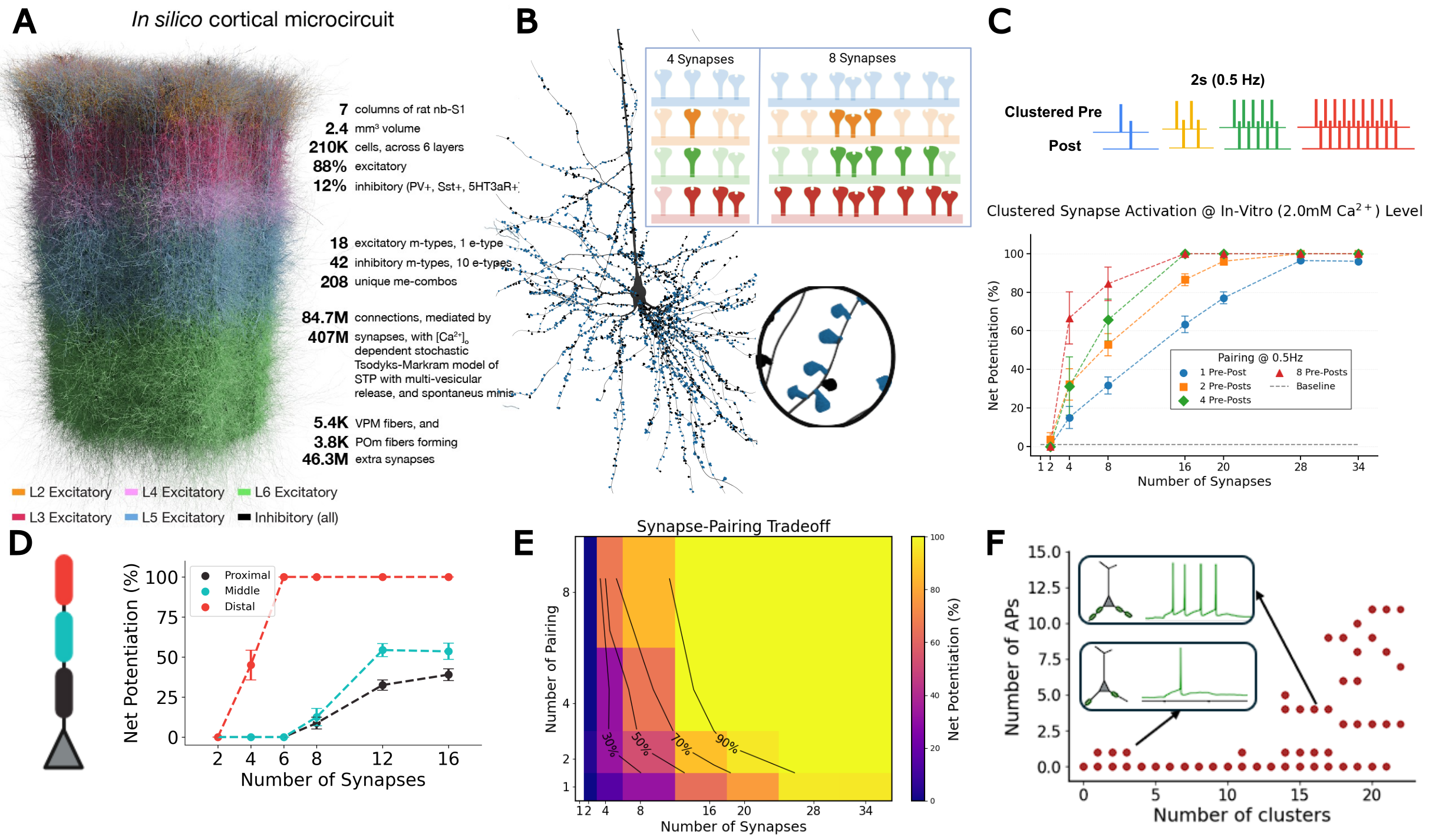
Figure 1. A, In silico cortical microcircuit. B, L5-TTPC with magnified cluster showing plasticity for 4 vs 8 co-active synapses. C, Clustered pre-post pairing (0.5 Hz); net potentiation vs synapse number. D, Spatial learning gradient E, Synapse-pairing tradeoff heatmap. F, Basal cluster coactivation triggers somatic burst.

References
1. Chindemi, G., Abdellah, M., Amsalem, O., Benavides-Piccione, R., Delattre, V., Doron, M., Ecker, A., Jaquier, A. T., King, J., Kumbhar, P., Monney, C., Perin, R., Rössert, C., Tuncel, A. M., Van Geit, W., DeFelipe, J., Graupner, M., Segev, I., Markram, H., & Muller, E. B. (2022). A calcium-based plasticity model for predicting long-term potentiation and depression in the neocortex.
2. Ecker, A., Egas Santander, D., Abdellah, M., Alonso, J. B., Bolaños-Puchet, S., Chindemi, G., Gowri Mariyappan, D. P., Isbister, J. B., Ki

Sunday July 12, 2026 10:40am - 11:10am ADT
Ballroom B1

2:00pm ADT

FO2: Norepinephrine Restores Cortical Dynamics and Enables Machine Learning–Based Severity Mapping in a Multiscale Model of Parkinson’s Disease
Sunday July 12, 2026 2:00pm - 2:30pm ADT
Jeeyune Jung*1,3,   Adam Newton1,3,  Donald Doherty1,3,  Hong-Yuan Chu2,3,  Samuel Neymotin4 , William Lytton1,3
1. Department of Physiology and Pharmacology, SUNY Downstate Health Sciences University, Brooklyn, NY, USA 
2. Department of Pharmacology and Physiology, Georgetown University Medical Center, Washington, DC, USA 
3. Aligning Science Across Parkinson’s (ASAP) Collaborative Research Network, Chevy Chase, MD, USA
Center for Biomedical Imaging & Neuromodulation, Nathan Kline Institute, Orangeburg, NY, USA

*Email: [email protected]


Introduction
Parkinson’s disease (PD) involves not only basal ganglia dopamine loss but also cortical dysfunction, including excessive beta synchronization, abnormal beta–gamma coupling, altered bursting, and impaired corticospinal recruitment. Early locus coeruleus degeneration may reduce cortical norepinephrine (NE), disrupting cell-type-specific gain control in pyramidal tract (PT) and intratelencephalic (IT) neurons. We tested whether experimentally constrained NE modulation restores cortical excitability and network dynamics in an advanced MitoPark motor cortex model and whether NE-sensitive cortical biomarkers support severity mapping and prediction of dopamine-therapy response.


Methods
Whole-cell patch-clamp recordings from Layer 5 PT and IT neurons quantified NE (10 µM)-induced changes in firing–current relationships. Conductance-based single-cell models were fit to baseline and NE responses and embedded in a biophysically detailed laminar M1 network model in NEURON/NetPyNE. Parkinsonian simulations incorporated reduced PT5B intrinsic excitability and reduced thalamocortical drive, with disease-stage-dependent NE scaling from PK/PD modeling. From the resulting simulations, we extracted cortical biomarkers including PT5B firing, IT5B beta-synchronized bursting, IT/PT imbalance, beta power, beta-burst duration, beta–high gamma phase-amplitude coupling, and avalanche slope, which were then used for severity mapping. 

Results
NE exerted opposite intrinsic effects across Layer 5 pyramidal subtypes: PT firing increased, whereas IT repetitive firing decreased. In the Parkinsonian network, NE-dependent conductance changes partially rescued pathological dynamics: PT5B firing increased by ~60–70%, IT5B bursting declined, the IT/PT activity ratio shifted toward control-like values, and pathological beta–high gamma phase-amplitude coupling decreased by ~40%. Across the biomarker set, NE shifted cortical dynamics toward the control regime. In the machine-learning framework, greater deviation from control tracked greater disease severity and predicted progressively shorter and weaker levodopa benefit as NE support declined.
Discussion
These findings identify NE-sensitive intrinsic gain control as a mechanistic bridge between single-cell excitability and pathological cortical state in PD. Loss of noradrenergic modulation may directly contribute to corticospinal under-recruitment, hypersynchronous beta activity, and broader cortical biomarker abnormalities, whereas restoring NE-dependent PT/IT balance may complement dopamine-based therapy. This multiscale framework links cellular mechanisms, network dysfunction, severity mapping, and predicted treatment response, positioning noradrenergic modulation as a promising strategy for advanced PD.

References
1. Dura-Bernal, S., et al. (2023). Multiscale model of primary motor cortex circuits predicts in vivo cell-type-specific, behavioral state-dependent dynamics. Cell Reports.
2. Chu, H. Y., et al. (2024). Dysfunction of motor cortices in Parkinson’s disease. Cerebral Cortex.
3. Doherty, D. W., et al. (2025). Enhanced beta power emerges from simulated parkinsonian primary motor cortex. npj Parkinson’s Disease.

Acknowledgments
This work was supported by the Aligning Science Across Parkinson’s (ASAP) Collaborative Research Network. We thank colleagues for providing experimental data used to constrain the model. Computational resources were provided by SUNY Downstate Health Sciences University.


Sunday July 12, 2026 2:00pm - 2:30pm ADT
Ballroom B1
 
Monday, July 13
 

10:40am ADT

FO3: Gene Gradients Reveal Directed Structural Connectivity Across Species
Monday July 13, 2026 10:40am - 11:10am ADT
Benjamin S. Sipes*1, Ashish Raj1

1Department of Radiology and Biomedical Imaging, University of California, San Francisco, San Francisco, CA, United States

*Email: [email protected]

Introduction
Diffusion MRI (dMRI) tractography estimates the brain's white matter structural connectivity (SC) in vivo, but it cannot resolve the directionality of white matter pathways. Yet, much recent work has shown that genes and gene co-expression maps relate to SC across species [1-4]. Here we test whether gene co-expression gradients can infer connection directionality from undirected structural connectivity using the brain’s structure–function relationship.

Methods
We introduce asymmetry to SC (C) via a similarity transform with a node-level gauge parameterized by genetic gradients: C̃=ACA^-1, where A=diag(e^{Ga}), with G=[g_1,...,g_k] genetic gradient vectors and a=[a_1,...,a_k]^T gradient weights. We learn gradient weights by fitting a higher order network diffusion (HONeD) model of the SC graph Laplacian, ℒ=I-C̃D_{in}^-1, f(ℒ)=-κI-βℒ+ξℒ^2, to the residual of the Lyapunov equation, f(ℒ)^TΣ+Σf(ℒ)+I [5,6], with stationary covariance (Σ) estimated from functional neuroimaging. We compared our model's performance to ground truth directionality in three species: C. elegans, mouse, and macaque [7-10]. We then ran our model on 770 HCP subjects [11,12]. Public datasets supplied gene expression [13-17].

Results
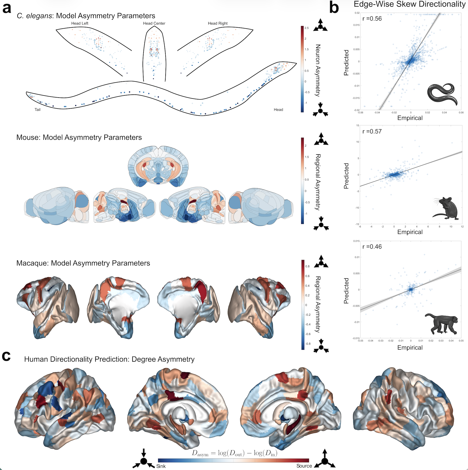
Model-predicted directionality significantly correlated with ground-truth directed edges in all three species. Our model predicted neuron-to-neuron synaptic directionality in C. elegans (r=0.56, p<10^-253) and tracer-based directionality in mouse (r=0.57, p<10^-37) and macaque (r=0.46, p<10^-44) (Fig.1a-b). The optimal numbers of genetic gradients was also different in each species (C. elegans: k=3; Mouse: k=5; Macaque: k=1). We found that humans had optimal test-retest reliability when using k=5 genetic gradients (ICC=0.46). Human predicted degree asymmetry suggests that the hippocampus and posterior cingulate are network sources while temporal poles are network sinks (Fig.1c).

Discussion
Although white matter pathways exhibit directionality, estimating their orientation has largely been restricted to tracer-based experiments and a small number of specialized imaging methods. Our results suggest that gene gradients combined with structure–function modeling provide a biologically grounded framework for inferring directed structural connectivity across species, supporting the idea that molecular gradients may encode directional biases in large-scale brain networks. Estimating human SC directionality is valuable not only for basic neuroscience, but also for evaluating circuit-level models of brain function and for studying diseases such as Alzheimer’s, Parkinson’s, and ALS that may propagate along structural pathways [18].

Figure 1. (a) Model-estimated directionality parameters (e^{Ga}) for the three non-human species: C. elegans (top), Mouse (middle), Macaque (bottom). In the C. elegans plot, each dot represents a single neuron. (b) Scatter plots comparing empirical to predicted skew edges with Pearson correlations listed at the top left (all p<10^{-37}). (c) Predicted human overall degree asymmetry for 414 brain regions.
Speakers
avatar for Benjamin Snow Sipes

Benjamin Snow Sipes

Graduate Student Researcher, University of California, San Francisco
My research develops computational approaches for understanding how brain structure shapes neural function. I use graph signal processing, spectral graph theory, and multimodal neuroimaging—including fMRI, diffusion MRI, and MEG—to study structure–function coupling, network... Read More →
Monday July 13, 2026 10:40am - 11:10am ADT
Ballroom B1

2:00pm ADT

FO4: Selective routing of spatial information in dentate granule cells emerges through disparate combinations of synaptic and intrinsic plasticity
Monday July 13, 2026 2:00pm - 2:30pm ADT

Sanjna Kumari*1 and Rishikesh Narayanan1
1 Cellular Neurophysiology Laboratory, Molecular Biophysics Unit, Indian Institute of Science, Bengaluru 560012, India
*Email: [email protected]

Introduction
Granule cells (GCs) in the dentate gyrus (DG) receive grid-like spatial inputs and contextual inputs from the entorhinal cortex, both broadly tuned to multiple spatial locations. Despite this, GCs elicit sparse spatial firing that is confined to single place fields, thus playing a central role in selective routing of spatial information to the hippocampal circuit. The mechanisms behind the transformation of broadly tuned afferent inputs into sparse and location-specific outputs remains unclear. In this study, we ask if there are physiologically relevant plasticity mechanisms that can mediate selective routing of spatial information towards place-cell emergence and spatial remapping, especially when inhibitory synapses are absent.

Methods
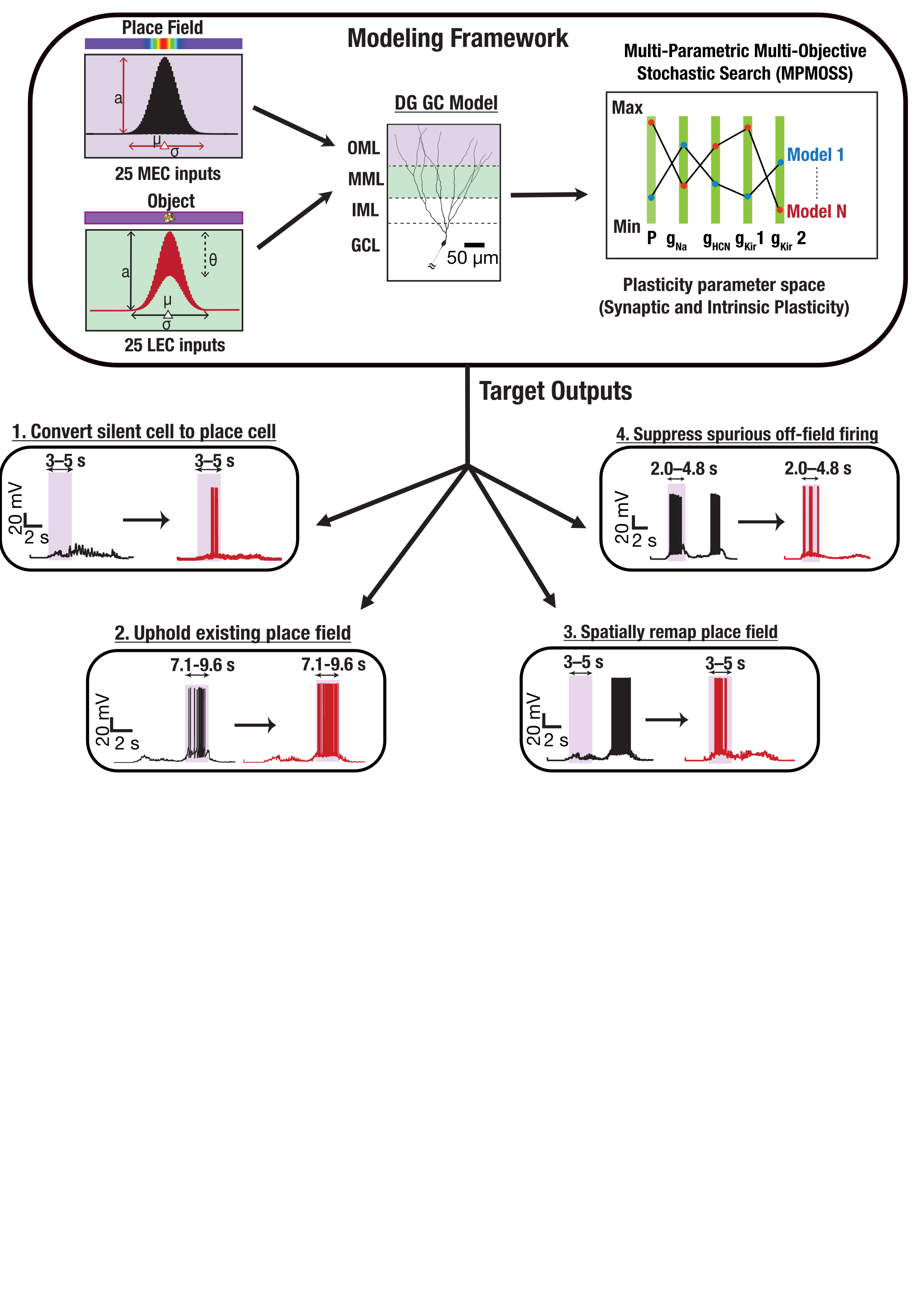
We employed morphologically and biophysically realistic models of DG GCs (Kumari & Narayanan, 2024), receiving grid-like and contextual spatial inputs from the entorhinal cortex. We employed a stochastic search paradigm in the plasticity space involving fold-changes in excitatory synaptic strengths, persistent sodium (NaP), hyperpolarization-activated cyclic nucleotide-gated (HCN), and inward rectifier potassium (Kir) conductances. We validated plasticity combinations that achieved one of four functional targets relevant to DG spatial tuning: conversion of silent neurons to place cells, uphold existing place field firing, spatial remapping to a new location, and suppression of spurious place fields to obtain a single place field (Fig 1).

Results
While excitatory synaptic plasticity alone was insufficient to generate valid spatial tuning, conjunctive synaptic and intrinsic plasticity yielded several valid plasticity combinations for all 4 targets (Valid/Total models for 4 targets: 243/142,000, 325/10,000, 139/5,000, 224/50,000). These valid plasticity combinations manifested pronounced heterogeneity across all fold-changes, unveiling plasticity degeneracy where disparate plasticity combinations yielded similar spatial tuning outcomes. Dimensionality reduction analyses revealed low-dimensional structures in intrinsic measurement and parameter spaces of valid models. In contrast, the plasticity space did not manifest strong constraints on plasticity across different components.

Discussion
While inhibitory synaptic inputs have been studied as mechanisms for sculpting spatial tuning, we show that selective routing of information and suppression of off-field firing can be achieved through intrinsic plasticity. Among intrinsic components, we predict the axonal initial segment Kir conductance as the strongest determinant of spatial selectivity. We demonstrate that disparate combinations of concomitant plasticity in excitatory synaptic and intrinsic conductances can mediate the emergence, refinement, and remapping of place fields. We show that co-dependent plasticity in different neuronal components can enable robust yet flexible spatial representations despite heterogeneities in neuronal composition and plasticity mechanisms.

FIgure 1. Medial and lateral entorhinal cortex inputs impinge on a DG granule cell. Disparate combinations of synaptic and intrinsic plasticity (NaP, HCN, Kir channels) achieved one of four targets: convert silent cell to place cell, uphold existing place field, remap, or suppress spurious firing. Our results show that robust and flexible spatial tuning is achievable through plasticity degeneracy.References
Kumari, S., & Narayanan, R. (2024). Ion-channel degeneracy and heterogeneities in the emergence of signature physiological characteristics of dentate gyrus granule cells. J Neurophysiol, 132(3), 991-1013. https://doi.org/10.1152/jn.00071.2024

Speakers
Monday July 13, 2026 2:00pm - 2:30pm ADT
Ballroom B1
 
Share Modal

Share this link via

Or copy link

Filter sessions
Apply filters to sessions.