Loading…
Type: Posters clear filter
arrow_back View All Dates
Tuesday, July 14
 

5:00pm ADT

P096: Toward Auditory-Like Sparse Representations: Adaptive Central Frequencies Locally Competitive Algorithm for Efficient Neuromorphic Speech Recognition
Tuesday July 14, 2026 5:00pm - 7:00pm ADT
Introduction
The auditory periphery efficiently transmits sparsely encoded information to cochlear nuclei and higher centers while preserving acoustic features. Lateral inhibition among cochlear hair cells and cochlear nucleus neurons increases sparsity, improves frequency selectivity and resolution. Outer hair cells (OHC) dynamically modulate cochlear responses through stiffness changes, hypothesized to refine frequency tuning and receptive fields of inner hair cells (IHC).
Inspired by these mechanisms, we implement a sparse coding approach using a lateral inhibitory neuromorphic network. We propose the Adaptive Central Frequencies Locally Competitive Algorithm (ALCA-CF), which adapts neuronal parameters to optimize acoustic signal representation [1].

Methods
ALCA-CF optimizes offline the IHC gammachirp models by adapting receptive field sensitivity to the acoustic environment. This partially mimics the online adaptability of OHC, where stiffness changes affect IHC responses. Additionally, lateral inhibition in ALCA-CF enhances frequency resolution while maintaining sparse coding, like mechanisms in the cochlear system (see Fig. 1). By separately analyzing the impact of bandwidth, central frequency changes, and sparsity of the IHC models, we quantify their influence on signal quality and recognition. We also show that this processing produces state-of-the-art (SOTA) speech representations enabling SOTA classification with much smaller and simpler spiking neural networks.


Results
ALCA-CF improves reconstruction quality and sparsity over fixed Gammatone filter bank. On Heidelberg Digits, ALCA-CF achieves an SNR of 15.35 dB (+5.52 dB vs. fixed filters) and reduces sparsity by 17.53%. Similar gains are observed on Google Speech Commands (SNR: 23.04 dB). ALCA-CF learns a non-linear frequency resolution, removing information in irrelevant frequency intervals while concentrating coefficients in relevant ones, yielding sparser and more efficient speech representations. On Intel's Loihi 2 [2], a neuromorphic chip, ALCA-CF reaches 94.88% speech classification accuracy at 0.004 W, a 5.25× reduction vs. fixed filters (0.021 W) and 3.75× vs. LAUSCHER silicon cochlea [3] SHD (0.015 W), which achieved a lower accuracy of 83.77%.

Discussion
ALCA-CF provides an adaptive front-end trained independently of any target application, distinguishing it from supervised approaches where the front-end is optimized for a specific task. This independence makes it highly versatile, as the same method has the potential to adapt to acoustic signals of varying nature, including speech, environmental sounds, and music. Furthermore, ALCA-CF offers the ability to modulate the number of active filters based on data complexity and signal nature, enabling a flexible trade-off between representation accuracy and computational efficiency. This flexibility is particularly advantageous for embedded neuromorphic systems, where energy constraints demand sparse and content-adaptive representations.

Figure 1. Overview of the ALCA-CF front-end. Each filter is represented by a neuron with a receptive field. Red arrows are lateral inhibition synapses and blue arrows are the feedback that adapts each neuron's receptive field. Lateral inhibition weights are the correlation between pre- and post-synaptic neuron receptive fields. Neuron activations ai are represented by blue dots in the time-frequency output.

References


[1] Bahadi, S., Plourde, E., & Rouat, J. (2025, April). Adaptive Central Frequencies Locally Competitive Algorithm for Speech. In ICASSP 2025-2025 IEEE International Conference on Acoustics, Speech and Signal Processing (ICASSP) (pp. 1-5). IEEE. https://doi.org/10.1109/ICASSP49660.2025.10887648
[2] Orchard, G., et al. (2021, October). Efficient neuromorphic signal processing with loihi 2. In 2021 IEEE workshop on signal processing systems (SiPS) (pp. 254-259). IEEE. https://doi.org/10.1109/SiPS52927.2021.00053
[3] Cramer, B., et al. (2020). The heidelberg spiking data sets for the systematic evaluation of spiking neural networks. IEEE Transactions on Neural Networks and Learning Systems, 33(7), 2744-2757. https://doi.org/10.1109/TNNLS.2020.3044364

Acknowledgement
Thank the ”Fonds de recherche du Québec - Nature et technologies” and ”Natural Sciences and Engineering Research Council of Canada” for funding this research. We extend our appreciation to NVIDIA for donating the GTX1080 and Titan Xp GPUs. We thank Intel for giving us access to Loihi 2 and Andreas Wild for his insights on the power/energy benchmark.
Tuesday July 14, 2026 5:00pm - 7:00pm ADT
Ballroom B2

5:00pm ADT

P097: How Axon Refractory Dynamics and Ionic Excitability Shape Peripheral Nerve Stimulation Responses
Tuesday July 14, 2026 5:00pm - 7:00pm ADT
Introduction
Optimizing peripheral nerve stimulation is crucial for improving clinical outcomes in neural prosthetics and bioelectronics medicines. The effects of extracellular electrical stimulation of peripheral myelinated axons depend jointly on the stimulus amplitude, waveform, and frequency. This work shows that variation in the waveform configuration (monophasic and biphasic with and without interphase gaps) defines the baseline activation thresholds across monopolar and bipolar electrode configurations using the same cuff structure around the rat sciatic nerve. Further, we explored how pulse-train frequency influences temporal entrainment limits, subharmonic skipping patterns, and intrinsic ionic adaptation kinetics.

Methods
We used the McIntyre-Richardson-Grill model for a myelinated nerve fiber with modified adaptation kinetics [1]. Next, a mesh of a rat sciatic nerve was created with GMESH [2]. We solved spatial potential maps for both monopolar and bipolar electrode cuff setups to isolate electric field effects using the Finite Element Method in FEniCSx [3]. We mapped these potential values onto the axon in NEURON [4] to simulate the nerve behavior. Nodes contained fast transient Na+, persistent Na+ driving afterdepolarization (ADP), and slow adaptation K+ currents. Further, we evaluated baseline thresholds for 12 biphasic waveforms at 5 Hz to ensure intervals exceeded the refractory period. Lastly, frequency adaptation was tested at 5, 100, and 1000 Hz.

Results
At 5 Hz, the activation threshold was observed at 250 µA for the symmetric and 100 µA for the asymmetric pulse because the bipolar configuration constrains the current spread. However, the monopolar configuration distributes the field between source and ground, resulting in variable thresholds ranging from 250 µA to 650 µA. The reduction in activation thresholds was observed with the introduction of an interphase gap. At 100 Hz, bursting emerged due to ionic tug-of-war: ADP drove rapid firing until a slowly developing AHP suppressed activity, creating 60–180 ms silent intervals. At 1000 Hz, axons show spike-skipping (1:3 to 1:5 entrainment) due to the refractory period, with 3–5 ms intra-burst ISIs and 200–400 ms adaptation gaps.


Discussion
Symmetric cathodic-leading pulses show lower baseline thresholds due to rapid depolarization, whereas the anodic-leading pulses show higher baseline thresholds because they hyperpolarize the membrane. However, the interphase gap (IPG) lowers the thresholds further, providing extra time for sodium channels to open more before the charge-reversing phase arrives. These findings show that axonal activation is a highly dynamic process shaped by spatial coupling and stimulation rates. The progression from linear scaling at 5 Hz to bursting at 100 Hz and combined bursting and subharmonic skip firing at 1000 Hz indicates that the behavior of axons under electrical stimulation is highly dependent upon the biophysical properties of ion channels.

References
[1] McIntyre, C. C., Richardson, A. G., & Grill, W. M. (2002). Modeling the excitability of mammalian nerve fibers: Influence of afterpotentials on the recovery cycle. Journal of Neurophysiology, 87(2), 995–1006. https://doi.org/10.1152/jn.00353.2001
[2] Geuzaine, C., & Remacle, J. F. (2009). Gmsh: A three-dimensional finite element mesh generator. International Journal for Numerical Methods in Engineering, 79(11), 1309–1331. https://doi.org/10.1002/nme.2579
[3] Baratta, I. A., et al. (2023). DOLFINx: The next generation FEniCS problem solving environment. Preprint. https://doi.org/10.5281/zenodo.10447666
[4] Hines, M. L., & Carnevale, N. T. (1997). The NEURON simulation environment. Neural Computation, 9(6), 1179–1209. https://doi.org/10.1162/neco.1997.9.6.1179

Acknowledgement
This work was conducted at the Prescott Lab, University of Calgary. Computational resources and simulation infrastructure were provided by the Prescott Lab, with additional laboratory infrastructure and support from the Hotchkiss Brain Institute, University of Calgary.
Tuesday July 14, 2026 5:00pm - 7:00pm ADT
Ballroom B2

5:00pm ADT

P098: CompNeuroVis: Connecting Existing Computational Neuroscience Workflows to Interactive Applications
Tuesday July 14, 2026 5:00pm - 7:00pm ADT
Introduction
Understanding and developing computational models of neurons relies on complementary views, such as voltage traces, activity mapped onto morphology, and channel kinetics. Interactive adjustment of parameters can further expose how model mechanisms shape dynamics. Existing support spans simulator-native interfaces [1], model platforms [2,3], network builders [4], and model-tuning environments [5], but these often require working within a particular simulator, platform, or model representation. CompNeuroVis addresses the gap between general-purpose plotting and specialized tools by helping researchers assemble interactive applications around workflows they already use.


Methods
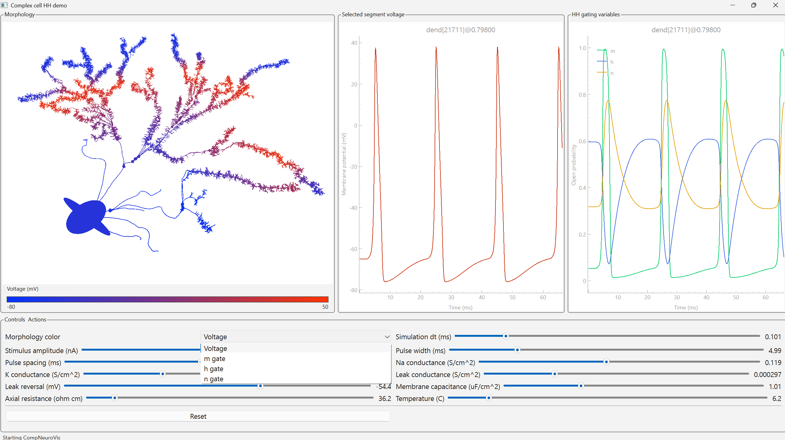
CompNeuroVis is a Python toolkit for building interactive applications around existing models and data. In a compositional style similar to common scientific plotting libraries, the user defines a source, such as a runnable simulation, Python model, or recorded data stream, then adds views and controls. The application is launched with a single call (Fig. 1). Views are extensible: current examples include trace plots, morphology views, and state diagrams, with raster and network views in progress. Controls adjust parameters and other settings live. Each composition reduces to a shared specification of data, views, controls, and synchronizing messages, allowing the same application to run across sources and within notebooks.


Results
We demonstrated the toolkit across several workflow classes. For a compartmental NEURON model with Hodgkin-Huxley dynamics, we built an interactive application with controls that modify parameters during execution and a morphology view that color-codes spatial variables while allowing compartments to be selected for plotting [1]. We reproduced these views for an equivalent Jaxley model, illustrating use across simulators. We also interfaced with user-defined models such as LIF neurons written in Python, embedded a live interface within a Jupyter notebook, and created state diagrams for Markov-based ion channel models and 3D surface plots for higher-dimensional data.


Discussion
CompNeuroVis complements existing computational neuroscience interfaces by emphasizing low-friction integration with the code, models, data, and simulators researchers already have. Rather than introducing a new simulator, platform, or model format, it provides reusable components for assembling individualized interactive applications. Because each application reduces to a shared specification, the same components can extend to networked interfaces and purpose-built tools, from model editors and teaching interfaces to remote simulator dashboards and multi-simulator comparison views. This supports model development, teaching, debugging, and exploratory analysis, all of which help researchers build intuition about neural mechanisms.


Figure 1. A CompNeuroVis application built around a compartmental NEURON model of a reconstructed cell with Hodgkin-Huxley dynamics. The morphology is color-coded by a selected variable, here membrane voltage. For a selected segment, linked plots show membrane voltage and the gating variables m, h, and n. A dropdown sets the mapped variable and sliders adjust stimulus and biophysical parameters.

References

  1. Hines, M. L., & Carnevale, N. T. (1997). The NEURON simulation environment. Neural Computation, 9(6), 1179-1209. https://doi.org/10.1162/neco.1997.9.6.1179
  2. Gleeson, P., et al. (2019). Open Source Brain: A collaborative resource for standardized models. Neuron, 103(3), 395-411.e5.
  3. Cantarelli, M., et al. (2018). Geppetto: A reusable modular open platform for neuroscience data and models. Philosophical Transactions B, 373(1758), 20170380.
  4. Dura-Bernal, S., et al. (2019). NetPyNE, a tool for data-driven multiscale modeling of brain circuits. eLife, 8, e44494.
  5. Makarov, R., Chavlis, S., & Poirazi, P. (2025). DendroTweaks: An interactive approach for unraveling dendritic dynamics. eLife, 13, RP103324.


Acknowledgement
I thank Ethan Irby (Research Assistant, Nathan Kline Institute) for ideas on use cases and capability requirements, and the open-source computational neuroscience community and simulator developers whose tools and discussions informed this work.

Speakers
Tuesday July 14, 2026 5:00pm - 7:00pm ADT
Ballroom B2

5:00pm ADT

P099: Hierarchical Multi-Timescale learning in a mushroom body network model
Tuesday July 14, 2026 5:00pm - 7:00pm ADT
Introduction
In Drosophila melanogaster, associative learning occurs in the mushroom body, where synapses between Kenyon cells (KCs) and mushroom body output neurons (MBONs)[1] are modulated by dopaminergic neurons (DANs) that convey reinforcement signals. KC–MBON–DAN circuits form parallel functional units that influence behaviour [2], and distinct learning properties across compartments allow appetitive and aversive memories for the same stimulus to coexist [3]. However, the interaction between the compartments is not fully understood. Here we implemented a mushroom body network model having parallel MBON units with different time scales and valences to investigate how the interactions between these units help in shaping different behaviours.


Methods
We propose a network model consisting of a KC layer for odor representation, multiple MBONs with short-term (STM) and long-term (LTM) memory, and a set of DANs representing unconditioned stimuli. Synaptic weight between KC and MBON depends on the relative timing between KC and DAN activity. We have implemented cross valence inhibitory modulation and hierarchical interaction between LTM and STM, where strong LTM activity can positively influence STM compartment activity. Behavioural readout is determined by the relative firing rates of the MBON population encoding opposite valences.


Results
The model could reproduce the core features of associative learning including first order conditioning. Parallel MBONs allow associations with opposite valence to coexist for the same stimuli. Due to the presence of cross valence inhibitions, the model can exhibit valence shifting during sequential experiences of opposing reinforcements or when relative influence of other compartment changes over time. Hierarchical LTM-STM interactions further enable second order conditioning, producing a short-term memory.


Discussion
Our results indicate that interactions between parallel memory units can produce flexible behaviour even with a relatively simple plasticity rule. The hierarchical interaction between memory units of different timescales allows stable long-term memories to influence short term memories, which helps in adapting to a dynamic environment. These results highlight how network architecture of the mushroom body can support flexible yet stable behaviours. 
\n


References
  1. Waddell, S. (2013). Reinforcement signalling in Drosophila; dopamine does it all after all. Current Opinion in Neurobiology, 23(3), 324–329. https://doi.org/10.1016/j.conb.2013.01.005
  2. Aso, Y., Hattori, D., Yu, Y., Johnston, R. M., Iyer, N. A., Ngo, T., Dionne, H., Abbott, L., Axel, R., Tanimoto, H., & Rubin, G. M. (2014). The neuronal architecture of the mushroom body provides a logic for associative learning. eLife, 3, e04577. https://doi.org/10.7554/elife.04577 
  3. Aso, Y., & Rubin, G. M. (2016). Dopaminergic neurons write and update memories with cell-type-specific rules. ELife, 5. https://doi.org/10.7554/eLife.16135 

Acknowledgement
This work is supported by the Centre for High Impact Neuroscience and Translational Applications (CHINTA), TCG CREST. I sincerely thank Dr. C. Sivaraju for his valuable discussions and encouragement. 

Tuesday July 14, 2026 5:00pm - 7:00pm ADT
Ballroom B2

5:00pm ADT

P100: JARDESIGNER: Installation-free in-browser modeling of multiscale models with signaling and conductances in multicompartment neurons.
Tuesday July 14, 2026 5:00pm - 7:00pm ADT
Introduction
Neuronal model building remains a challenge despite highly capable simulator platforms [1,2]. This is even more the case for multiscale models spanning molecular to electrophysiological detail, which are increasingly relevant for modeling plasticity, neuromodulation and long time-scale neuronal dynamics. Recent GUIs such as DendroTweaks have simplified electrophysiology modeling [3], but GUIs for multiscale models are scarce. JARDESIGNER is the Javascript App for Reaction-Diffusion and Electrical SIGnaling in NEuRons. It provides three things: a no-installation GUI for building and running models, a JSON file format for defining multiscale models, and a standalone Python library for building and running the models on the MOOSE simulator.

Methods
The Jardesigner GUI is a client-server system. The GUI is implemented in Javascript in the REACT framework on the client browser, and the server backend is coded in Python. It uses the jardesigner Python library to build models running on the C++ codebase of MOOSE to efficiently perform the computations. There is a publicly hosted Jardesigner server at https://www.jardesigner.org/. The Jardesigner GUI does not use cookies or track users, and any files uploaded for model building are deleted as soon as the user quits a modeling session. The complete client-server package can be locally installed using pip install.

Results
Jardesigner enables rapid building of production multiscale neuronal simulations. Successive menu boxes specify morphology, spine distributions, passive and active properties, signaling pathways, and mappings between ephys and signaling entities (Fig. 1). Users load neuronal morphologies, channel mechanisms, and signaling kinetic specifications from standard databases and assemble them in the GUI. Additional menu boxes allow one to plot graphs, set up 3-D animations, and save simulation results. As the simulation is assembled, a 3-D view of the cell grows and provides icons for mechanisms and display elements. It is trivially easy to swap out one morphology for another, even as all the other components of the model are retained.

Discussion
Jardesigner makes it easy to build and run complex, multiscale models entirely in the GUI, using predefined morphology, channel, and signaling building blocks. It is particularly well suited for teaching, and has been used effectively by students with no previous modeling experience. It is also in production use to build and test multiscale models for research simulations. Development directions include video tutorials and better integration with online databases. We encourage user feedback.

Figure 1. Screenshot of Jardesigner GUI. The menu options are in the blue bar above, and the Channel Menu box is open to the left. A snapshot of the current simulation is presented in the 3D display to the right.

References
[1] Hines, M. (2009). NEURON and Python. Frontiers in Neuroinformatics, 3. doi: 10.3389/neuro.11.001.2009
[2] Ray, S., Bhalla, U.S. (2008). PyMOOSE: interoperable scripting in Python for MOOSE. Frontiers in Neuroinformatics, 2, 365 https://doi.org/10.3389/neuro.11.006.2008
[3] Makarov, R., Chavlis, S., Poirazi, P (2025). DendroTweaks, an interactive approach for unraveling dendritic dynamics. eLife, https://doi.org/10.7554/eLife.103324.3

Acknowledgement
The development of Jardesigner was supported by the Kavli Foundation through the EOSS Grant Cycle 6. USB receives research support from NCBS-TIFR through the Department of Atomic Energy, Government of India, under Project Identification No. RTI 4006. SR is supported by CHINTA and IAI under TCG CREST.
Speakers
avatar for Upinder Singh Bhalla

Upinder Singh Bhalla

Professor, NCBS/TIFR
Multiscale modelling of neurons especially in synaptic plasticity: including chemical and electrical signaling, traffic and mechanical changes. Tool development for all of these, including GENESIS, MOOSE, FindSim and more.
Tuesday July 14, 2026 5:00pm - 7:00pm ADT
Ballroom B2

5:00pm ADT

P101: BrainSymphony Reveals Psilocybin-Induced Network Reorganization
Tuesday July 14, 2026 5:00pm - 7:00pm ADT
Introduction
fMRI foundation models have grown rapidly in size [1-2], but scale may be suboptimal for neuroimaging given domain constraints and interpretability needs. BrainSymphony instead embeds neurobiological priors to build a lightweight, parameter-efficient, multimodal model capturing spatiotemporal BOLD dynamics and diffusion-MRI connectivity. We test out-of-distribution generalization on PsiConnect (62 participants; pre/post psilocybin; rest, meditation, music, movie) and relate model-derived dynamics to MEQ-30 mystical-type experience ratings. BrainSymphony reconstructs unseen fMRI time series with high fidelity and exposes fine-grained psychedelic network reorganization beyond classical functional connectivity.

Methods
BrainSymphony uses three fMRI encoders: an ROI-wise Spatial Transformer with mixed positional embeddings (including cortex-gradient priors), a Temporal Transformer with sinusoidal time embeddings, and a 1D convolutional stream for short-range transients. Encoder outputs are fused by a Perceiver module. We applied a frozen model pretrained on HCP/HCP-Aging to PsiConnect (baseline and psilocybin; rest, meditation, music, movie). Reconstruction was quantified by R², Pearson r, and MAE against ROI-shuffled controls. Directed influence matrices were computed from Perceiver attention (incoming/outgoing) and used to decode context and to characterize psilocybin–baseline reorganization, including modulation by experience intensity.

Results
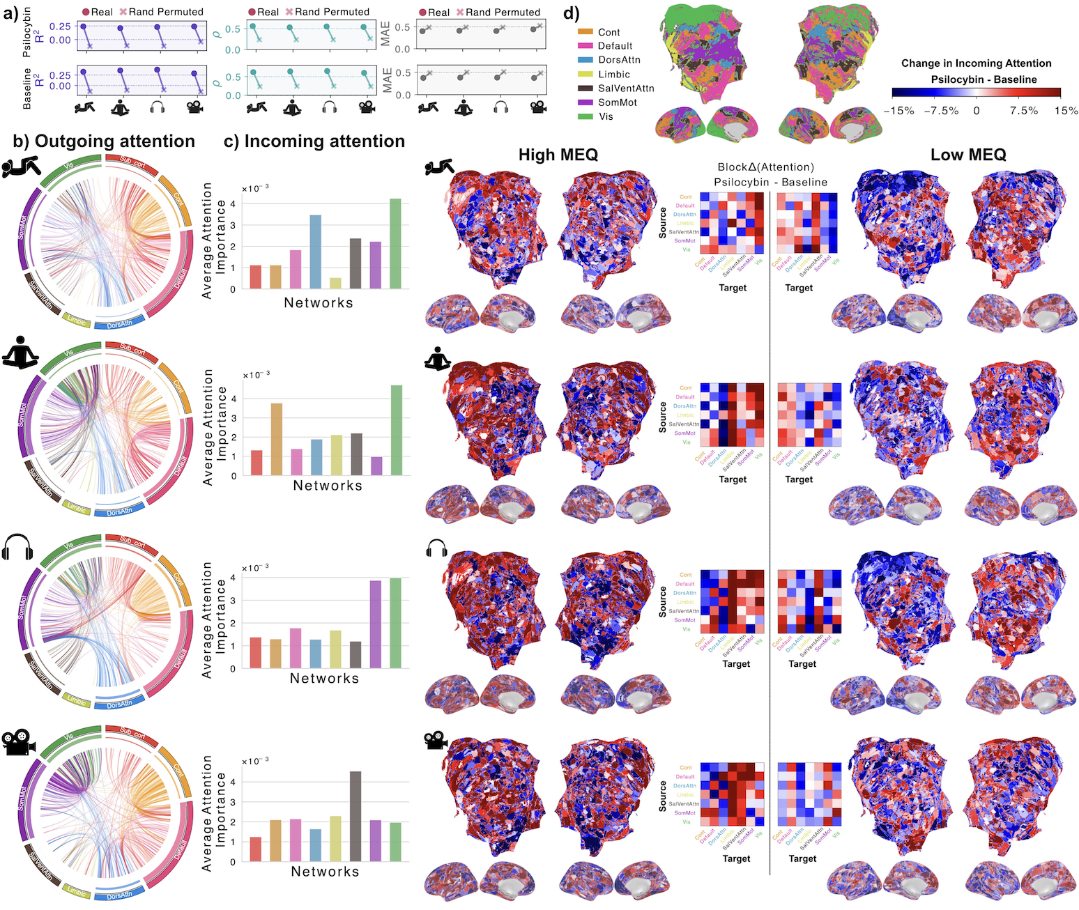
BrainSymphony reconstructed unseen psychedelic fMRI with high fidelity; R² and Pearson r exceeded shuffled controls (Fig. 1). Directed influence matrices decoded rest/meditation/music/movie at baseline (~0.64) and remained above chance under psilocybin (~0.46; chance 0.25), consistent with reduced modular boundaries and increased integration. Psilocybin increased outgoing influence in DMN, Control, and Visual networks, suggesting reduced DMN autonomy. Incoming influence highlighted Visual cortex as a dominant driver even in eyes-closed states, with limbic/salience drivers strongest in affective contexts. These effects scaled with Mystical Experience Questionnaire intensity with conventional functional connectivity failing to identify them.

Discussion
Compact, domain-informed foundation models can be predictive and mechanistically useful. BrainSymphony’s attention-derived directed influence reveals psilocybin-driven redistribution of large-scale communication that tracks behavioral context and subjective intensity of the experience. Visual territories emerge as consistent drivers even with closed eyes, aligning with internally generated imagery; limbic circuits amplify during emotionally rich contexts; DMN/Control/ systems show condition-specific increases in being influenced, reflecting decreased segregation and increased integration. BrainSymphony demonstrates that efficiency and interpretability can coexist with strong generalization.

Figure 1. BrainSymphony reconstruction and attention-based reorganization. (a) Paired dots: real vs permuted ROI series across conditions; higher R²,r, lower MAE. (b) Circos: Admin–Baseline attention Δ (top 500 edges) colored by source network; inner track = total outgoing. (c) Network-mean incoming attention Δ. (d) High vs Low MEQ receptive-attention maps plus inter-network Δ matrices.

References
1. Caro, J. O., Fonseca, A. H. D. O., Averill, C., Rizvi, S. A., Rosati, M., Cross, J. L., ... & van Dijk, D. (2023). BrainLM: A foundation model for brain activity recordings. BioRxiv, 2023-09.
2. Dong, Z., Li, R., Wu, Y., Nguyen, T. T., Chong, J., Ji, F., ... & Zhou, J. H. (2024). Brain-jepa: Brain dynamics foundation model with gradient positioning and spatiotemporal masking. Advances in Neural Information Processing Systems, 37, 86048-86073.
3. Stoliker, D., Novelli, L., Khajehnejad, M., Biabani, M., Barta, T., Greaves, M. D., ... & Razi, A. (2025). Psychedelics align brain activity with context. bioRxiv, 2025-03.

Acknowledgement
A.R. is affiliated with The Wellcome Centre for Human Neuroimaging, supported by core funding from Wellcome [203147/Z/16/Z]. A.R. is a CIFAR Azrieli Global Scholar in the Brain, Mind & Consciousness Program.

Speakers
MK

Moein Khajehnejad

Post-doctoral Research Fellow, Monash University
I am a post-doctoral research fellow in Monash Data Future Institute and Computational & Systems Neuroscience Laboratory at Turner Institute for Brain and Mental Health at Monash University working with Prof. Adeel Razi.
I am passionate about advancing Foundation Models in Neuro... Read More →
Tuesday July 14, 2026 5:00pm - 7:00pm ADT
Ballroom B2

5:00pm ADT

P102: Long-timescale Plasticity Mediated by ER-Dependent Synaptic Stabilization
Tuesday July 14, 2026 5:00pm - 7:00pm ADT
Introduction
Bridging the short-timescale of classic Hebbian learning and long-timescale of behavioral memory remains a challenge in the study of synaptic plasticity. The synaptic tag-and-capture theory proposes that activity-dependent molecular markers enable a state of synaptic stability [1], and it has been shown theoretically that multiple stability states enhance memory capacity [2]. Trafficking of the endoplasmic reticulum (ER) in and out of dendritic spines could viably serve as such a “tag” [3], but its effects on plasticity have yet to be studied in silico. We hypothesize that ER-mediated synaptic stabilization extends memory capacity and supports stable learning in neuronal microcircuits through activity-dependent modulation of plasticity.

Methods
We model ER trafficking in dendritic spines using a piecewise deterministic Markov process with three synaptic states: ER−, ER+, and ER stable. Potentiation increases the probability of ER entry to a given synapse while depression promotes ER exit. Synaptic plasticity follows a calcium-based rule [4] where weight changes decay to baseline within minutes. However, ER+ synapses can transition to ER stable when activity elevates calcium above a threshold, resetting the baseline weight and enabling potentiation to persist over long timescales. The ER model is incorporated into recurrent networks of leaky integrate-and-fire neurons to test its impact on learning and memory through the lens of neuronal assemblies [5]. 

Results
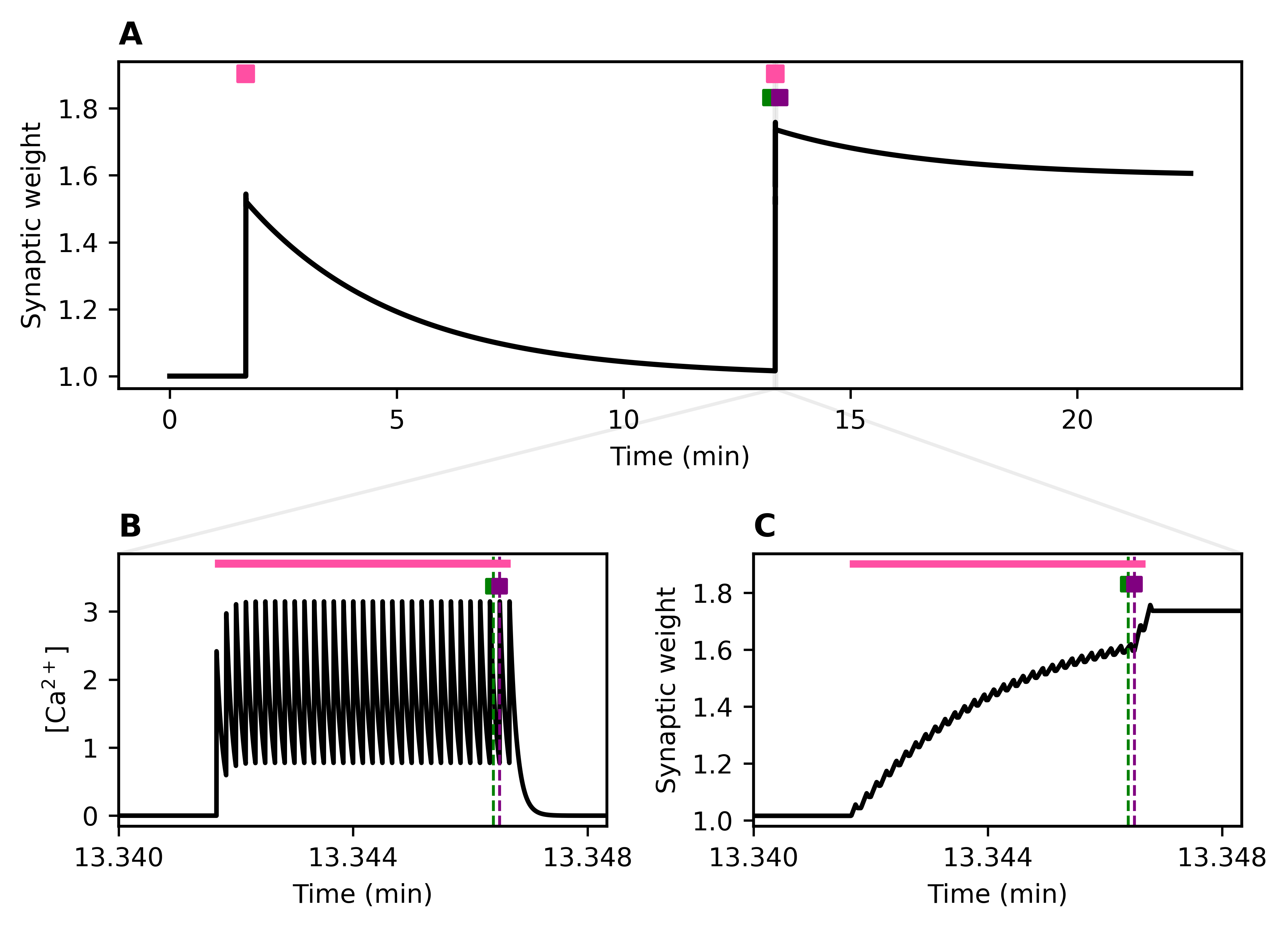
In a single synapse subject to high-frequency stimulation, potentiation from the calcium-dependent plasticity rule will decay on the order of minutes without the presence of ER (Figure 1A). A second high-frequency stimulus first successfully draws the ER into the synapse and subsequently triggers ER stabilization, preserving synaptic potentiation over longer timescales (Figure 1B-C). We expect the differences in these timescales to play a significant role in learning and memory formation in in silico recurrent neuronal microcircuits.

Discussion
Our model of stochastic ER recruitment stabilizes the effects of potentiation in individual synapses in a biologically informed manner [3]. By extending the timescale of potentiation beyond that predicted by calcium-dependent plasticity alone, we expect to similarly affect the timescale of learning and memory in in silico microcircuits. Therefore, our model will generate experimentally testable predictions regarding the effects of ER-mediated stabilization on learning dynamics and long-term memory storage. These conclusions will be augmented by constraining the model parameters for ER visitation with experimentally measured dendritic ER distributions obtained from electron microscopy studies to make brain region specific predictions.

Figure 1. Stochastic activity-driven ER entry and stabilization preserve calcium-dependent plasticity over long timescales. (A) Identical high-frequency pre/post spike trains (pink) produce synaptic weight changes; the first stimulation fails to trigger ER entry, while the second induces ER entry (green) followed by stabilization (purple). (B–C) Insets show calcium (left) and weight (right) dynamicsReferences
1.    Redondo et al. (2011). Making memories last: the synaptic tagging and capture hypothesis. Nat Rev Neurosci 12, 17–30.
2.     Fusi et al. (2005). Cascade models of synaptically stored memories. Neuron, 45(4), 599–611.
3.    Dittmer et al. (2024). L-type Ca2+ channel activation of STIM1-Orai1 signaling remodels the dendritic spine ER to maintain long-term structural plasticity. Proc Natl Acad Sci USA., 121(35), e2407324121.
4.    Moldwin et al. (2025). A generalized mathematical framework for the calcium control hypothesis describes weight-dependent synaptic plasticity. J Comput Neurosci 53, 333–357.
5.     Miehl et al. (2023). Formation and computational implications of assemblies in neural circuits. J physiol, 601(15), 3071–3090.

Acknowledgement
None.
Speakers
avatar for Scott Rich

Scott Rich

Assistant Professor, University of Connecticut
I am an Assistant Professor at the University of Connecticut, having opened my lab in the Department of Physiology and Neurobiology in January 2024.

The Rich Lab uses computational neuroscience in research centered on a fundamental question: How does the brain benefit from biophysical diversity at the level of neurons and microcircuits? The lab utilizes a wealth of tools from computational neuroscience, including the creation... Read More →
Tuesday July 14, 2026 5:00pm - 7:00pm ADT
Ballroom B2

5:00pm ADT

P103: Pathological cortical oscillations disrupted by the cholinergic response to vagus nerve stimulation
Tuesday July 14, 2026 5:00pm - 7:00pm ADT
Introduction
While vagus nerve stimulation (VNS) coupled with motor rehabilitation significantly improves post-stroke recovery [1], its mechanism of action remains poorly understood. Preclinical studies indicate that VNS enhances motor learning when stimulation is precisely paired with task success [2,3], with cholinergic neuromodulation necessary for these effects [4]. These findings collectively suggest that rapid cholinergic signaling may be pivotal in stroke rehabilitation, although this response to VNS has not been well characterized experimentally.
We hypothesize that the immediate cholinergic response to VNS produces rapid changes in cortical oscillatory dynamics through modulation of the muscarinic M-current in cortical neurons.

Methods
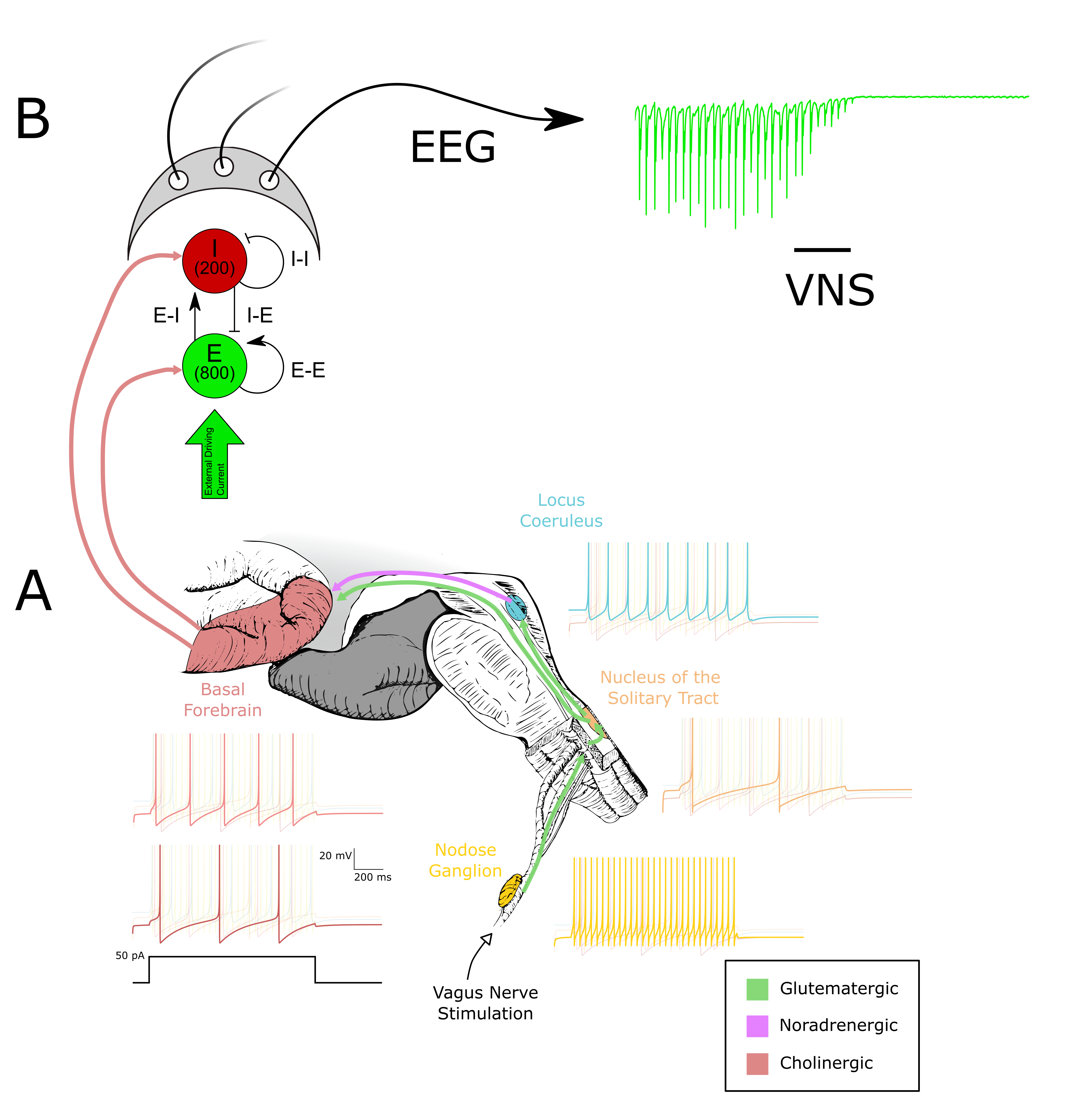
We model the brainstem circuitry connecting the vagus nerve to the cortex with populations of quadratic integrate and fire (Izhikevich) neurons: the nodose ganglion of the vagus nerve, the nucleus of the solitary tract, the locus coeruleus, and cholinergic neurons of the basal forebrain projecting to the cortex (Fig. 1A). Each neuronal population is parameterized using firing rate and adaptation properties fit to published electrophysiological data.
The predicted VNS-triggered cholinergic output derived from basal forebrain neuronal spiking modulates an excitatory-inhibitory (E-I) cortical microcircuit of Hodgkin-Huxley neurons through a conductance-based model of the muscarinic M-current (Fig. 1B).

Results
Preclinical [2] and preliminary clinical EEG recordings show that VNS immediately reduces cortical gamma power, reflecting a decrease in synchronized spiking activity. Our model generates a dynamic cholinergic output which in turn modulates the M-current within an in silico cortical network [5]. The resulting synaptic activity is used to generate a pseudo-EEG signal that can be compared directly to the spectral changes observed in clinical recordings (Fig. 1B). Expanding upon recent in silico work showing that dynamically increasing acetylcholine concentrations desynchronize cortical microcircuits dependent upon the rate of modulation [6], we determine whether the specific cholinergic response to VNS can account for VNS’s effect on EEG.

Discussion
The combination of an experimentally constrained brainstem model and a cortical microcircuit subject to cholinergic neuromodulation allows us to test whether rapid cholinergic signaling can account for the immediate decrease in cortical gamma power observed following VNS. This approach allows for explorations of how stimulation parameters—such as inter-stimulation interval, pulse train duration, and stimulation intensity—influence cholinergic output and cortical dynamics beyond what is currently done clinically. In doing so, the model may help identify stimulation paradigms that maximize beneficial neuromodulatory effects, providing mechanistic insight into how VNS protocols can be optimized to improve stroke rehabilitation.

Figure 1. (A) The circuitry between the vagus nerve and the basal forebrain is modeled using quadratic integrate-and-fire (Izhikevich) neurons fitted to experimental data. Representative voltage responses to a 50 pA injection are shown. (B) The output of the basal forebrain in (A) is used to modulate ACh-sensitive K+ channels in an E–I network. Synaptic activity within this network is used to compute an EEGReferences
1.      Dawson, J., et al. (2021). Vagus nerve stimulation paired with rehabilitation for upper limb motor function... The Lancet, 397(10284), 1545–1553.
2.      Engineer, N. D., et al. (2011). Reversing pathological neural activity... Nature, 470(7332), 101–104. https://doi.org/10.1038/nature09656
3.      Hays, S. A., et al. (2014). The timing and amount of vagus nerve stimulation... Neuroreport, 25(9), 676–682.
4.      Bowles, S., et al. (2022). Vagus nerve stimulation drives selective... Neuron, 110(17), 2867–2885.e7.
5.      Stiefel, K. M., et al. (2009). The effects of cholinergic neuromodulation... Journal of Computational Neuroscience, 26(2), 289–301.
6.      Pandian, S., & Rich, S. (2025). Dynamic cholinergic signaling differentially... (p. 2025.06.20.660675). bioRxiv
Speakers
avatar for Scott Rich

Scott Rich

Assistant Professor, University of Connecticut
I am an Assistant Professor at the University of Connecticut, having opened my lab in the Department of Physiology and Neurobiology in January 2024.

The Rich Lab uses computational neuroscience in research centered on a fundamental question: How does the brain benefit from biophysical diversity at the level of neurons and microcircuits? The lab utilizes a wealth of tools from computational neuroscience, including the creation... Read More →
Tuesday July 14, 2026 5:00pm - 7:00pm ADT
Ballroom B2

5:00pm ADT

P104: Characterization of nontrivial voltage noise in electrosensory pyramidal neurons
Tuesday July 14, 2026 5:00pm - 7:00pm ADT
Introduction
The stochastic flickering of ion channels is known to cause ongoing membrane potential fluctuations in neurons [1]. This channel noise is often considered negligible when compared to synaptic noise, yet it can shape the integrative properties of neurons [2]. Here, we show that valuable information on a neuron's intrinsic dynamics can be extracted by closely inspecting recordings of spontaneous membrane potential fluctuations in the absence of synaptic input.

Methods
We offer a reanalysis of previously published data [3] that consists of in vitro recordings from primary electrosensory pyramidal neurons in weakly electric fish under complete synaptic blockade. Raw voltage traces are segmented based on the applied holding currents, and each segment is decomposed into a fast and slow component. These components are analyzed with a suite of techniques, including wavelet transform, principal component analysis, Hilbert transform, and empirical mode decomposition. 


Results
Our analyses reveal an intrinsic noise structure that is richer than what could be expected based on usual assumptions pertaining to intrinsic voltage noise: we identify rapid, small-amplitude, shot noise-like events, and we quantify how their rate and amplitude are modulated by slower, large-amplitude fluctuations. This cross-relation is evidence that, at the single-neuron level, membrane potential dynamics can exhibit a form of phase-amplitude coupling. We also investigate the appearance of fast, intermittent subthreshold oscillations and investigate whether they are manifestation of stochastic linear dynamics, possibly with time-varying parameters.

Discussion
To our knowledge, this is the first study to explore and quantify a form of cross-frequency phase-amplitude coupling within spontaneous voltage fluctuations in single neurons. Collectively, our results suggest that spontaneous voltage noise at the single-neuron level can be nontrivial and should be closely investigated to fully contextualize the arrival of synaptic input. Given the richness of the intrinsic noise structure that we uncover, we lend concrete support to the notion that “the precise impact of synaptic noise can be evaluated only once channel noise is understood and quantified” [2].


References
1. Faisal, A. A., Selen, L. P. J., & Wolpert, D. M. (2008). Noise in the nervous system. Nature Reviews Neuroscience, 9, 292–303.
2. White, J. A., Rubinstein, J. T., & Kay, A. R. (2000). Channel noise in neurons. Trends in Neurosciences, 23, 131–137.
3. Marcoux, C. M., Clarke, S. E., Nesse, W. H., Longtin, A., & Maler, L. (2016). Balanced ionotropic receptor dynamics support signal estimation via voltage-dependent membrane noise. Journal of Neurophysiology, 115, 530–545.

Acknowledgement
This work was funded by the Natural Sciences and Engineering Research Council of Canada under Grant No. RGPIN-2022-0 531 4.

Tuesday July 14, 2026 5:00pm - 7:00pm ADT
Ballroom B2

5:00pm ADT

P105: Gamma Oscillations in Conductance-Based QIF Networks: A Three-Stage Progression and Its Limits
Tuesday July 14, 2026 5:00pm - 7:00pm ADT
Introduction
Gamma oscillations in cortical circuits are often modeled through excitatory-inhibitory (E-I) interactions underlying Pyramidal-Interneuron Network Gamma (PING) or through inhibitory interactions underlying Interneuron Network Gamma (ING). Quadratic integrate-and-fire (QIF) neuron models are widely used in modeling such dynamics because they support a direct correspondence between spiking-network simulations and tractable mean-field reductions. However, many QIF studies on PING oscillations mix current and conductance drive or use non-physiological reversal potentials, making it unclear whether conductance-based QIF E-I networks can generate biologically realistic gamma under physiological constraints.

Methods
We studied an all-to-all conductance-based QIF E-I spiking network together with a corresponding mean-field firing-rate model. To isolate conductance-induced effects, we varied synaptic conductances and excitatory/inhibitory reversal potentials without additive direct current injection. Parameters were restricted to biologically realistic reversal-potential ranges. Because the resulting network dynamics are analytically difficult to interpret directly, we also analyzed single-neuron QIF responses under different conductance-based inputs and reversal potentials, using their analytical solutions to gain insight into how these parameters shape emergent network behavior.

Results
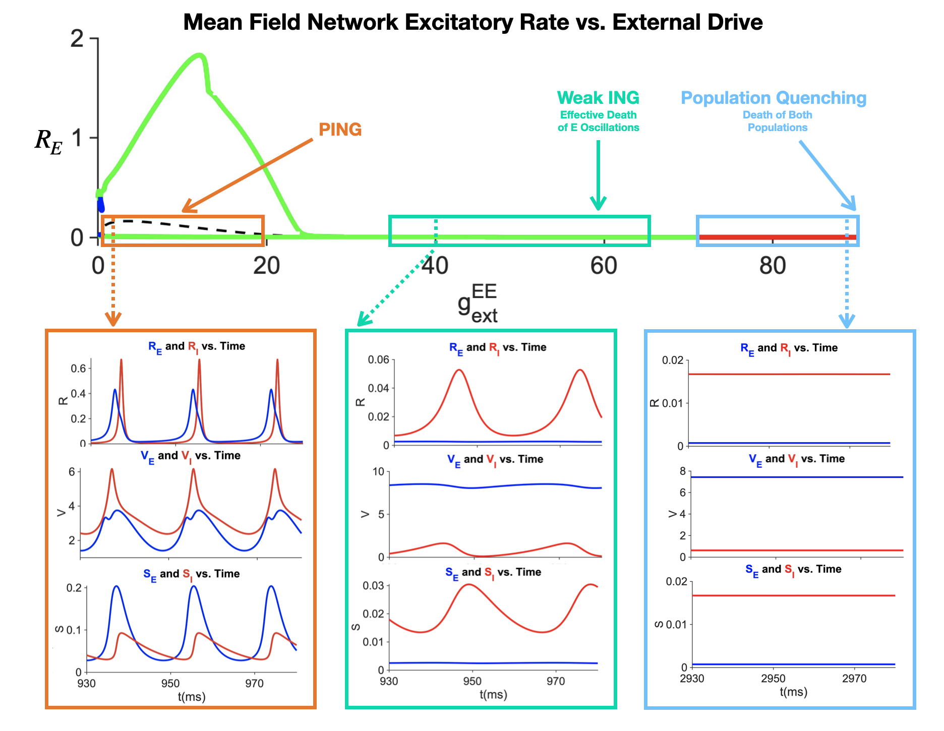
The conductance-based QIF E-I network exhibited gamma-range PING-like oscillations in some parameter regimes. As external drive to the excitatory population increased, the network exhibited a three-stage progression: a PING-like regime, a weak-ING-like intermediate regime marked by suppressed excitatory oscillations and persistent weak inhibitory oscillations, and population quenching at high drive (Fig.1). Under the same physiological constraints, both the spiking network and the mean-field model also displayed systematic non-biological features, including excitatory-cell doublets, weak inhibitory post-hyperpolarization currents, and overshooting inhibitory synaptic currents during depolarization.

Discussion
These results reveal a three-stage dynamical progression in conductance-based QIF E-I networks under physiological synaptic constraints. This progression provides a compact description of how increasing drive reshapes network dynamics. At the same time, the observed anomalous firing patterns and synaptic-current dynamics reveal important limitations under biologically realistic synaptic constraints and suggest that intrinsic properties of the QIF formalism contribute substantially to these deviations, beyond generic E-I network mechanisms alone. Our results clarify when QIF models succeed or fail in capturing realistic gamma dynamics and motivate refinement of reduced spiking models for studying cortical oscillations.

Figure 1. E-I Network dynamics as a function of external excitatory conductance drive in high input regime

References

  1. Coombes, S., & Byrne, Á. (2019). Next generation neural mass models. In F. Corinto & A. Torcini (Eds.), Nonlinear Dynamics in Computational Neuroscience (pp. 1-16). Springer. https://doi.org/10.1007/978-3-319-71048-8_1
  2. Ermentrout, B. (1996). Type I membranes, phase resetting curves, and synchrony. Neural Computation, 8(5), 979-1001. https://doi.org/10.1162/neco.1996.8.5.979
  3. Keeley, S., Byrne, Á., Fenton, A., & Rinzel, J. (2019). Firing rate models for gamma oscillations. Journal of Neurophysiology, 121(6), 2181-2190. https://doi.org/10.1152/jn.00741.2018
  4. Montbrió, E., Pazó, D., & Roxin, A. (2015). Macroscopic description for networks of spiking neurons. Physical Review X, 5(2), 021028. https://doi.org/10.1103/PhysRevX.5.021028

Tuesday July 14, 2026 5:00pm - 7:00pm ADT
Ballroom B2

5:00pm ADT

P106: Controlling the Speed–Accuracy Trade-Off in Brain–Computer Interfaces
Tuesday July 14, 2026 5:00pm - 7:00pm ADT
Introduction
Brain–computer interfaces (BCIs) decode brain signals into device commands [1]. Their performance is limited by a low signal-to-noise ratio, leading to a speed–accuracy trade-off: increasing accuracy through trial averaging requires more trials and reduces communication speed [2]. Existing metrics, such as the Information Transfer Rate (ITR) or BCI-Utility [3], obscure how accuracy depends on the number of trials — potentially introducing biases and limiting explainability. We propose a framework that separates and optimizes speed and accuracy, enabling explicit control of the trade-off and improving user- and experiment-specific adaptation.

Methods
The framework quantifies BCI speed and accuracy through two measures: Gain (relative speed improvement) and Conservation (relative accuracy preservation), defined with respect to a baseline BCI. These are combined into a trade-off equation termed Gain–Cons Balance (GCB), αGain + (1 − α)Cons, where α controls the desired balance. The GCB specifies the target trade-off, while an early-stopping strategy adjusts the number of trials to satisfy the selected α. In this way, the early-stop implements the policy dictated by the GCB. The approach was validated on 63 subjects using two P300 paradigms, three classifiers, and three stopping criteria within a nested leave-one-session-out cross-validation procedure.

Results
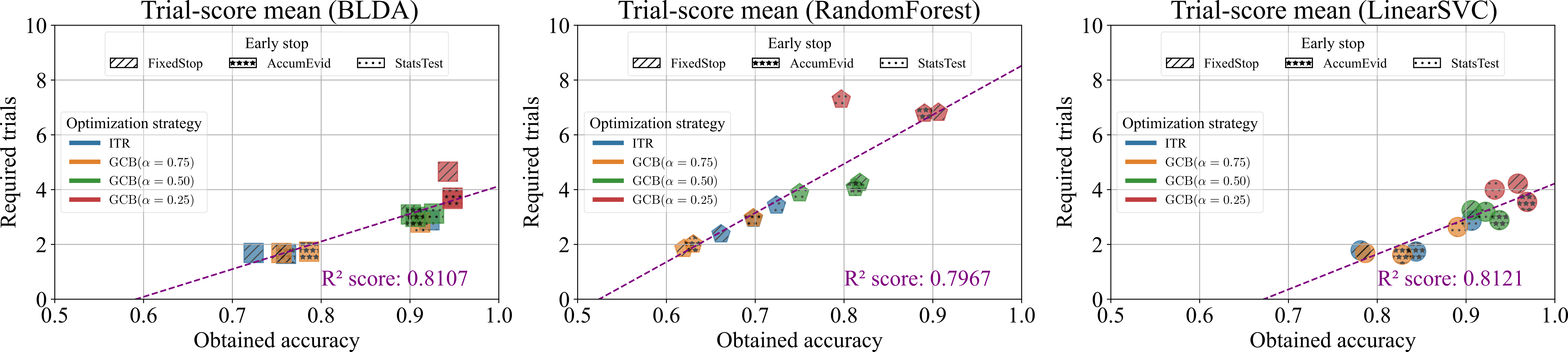
Optimization strategies produced distinct BCI behaviors (Fig. 1). The ITR behaved similarly to GCB(α = 0.75), favoring faster decisions with lower accuracies, whereas GCB(α = 0.25) prioritized accuracy at the cost of more trials. The balanced configuration (α = 0.5) achieved high accuracy with moderate speed. This trend was consistent across paradigms, subjects, sessions, classifiers, and early-stopping methods. These findings demonstrate that tuning α in the Gain–Cons Balance explicitly controls the speed–accuracy trade-off: selecting a desired accuracy determines the required number of trials, and vice versa.

Discussion
The proposed Gain–Cons Balance framework enables explicit control of the speed–accuracy trade-off across Rapid Serial Visual Presentation and Row–Column Paradigm P300 datasets. By tuning the parameter α, the trade-off becomes a controllable design variable rather than an implicit constraint. The framework supports conditional analysis of expected accuracy or required trials, facilitates subject-level performance prediction, and reveals metric biases — particularly ITR’s preference for speed. Overall, the Gain–Cons Balance provides a general and interpretable tool to optimize and compare BCIs across paradigms, users, sessions, classifiers, and early-stopping strategies.

Figure 1. Required trials and obtained accuracies across classifiers, early-stopping strategies, and optimization methods for Hoffmann et. al. Rapid Serial Visual Presentation dataset [1]. Points show mean performance under leave-one-session-out validation. The fitted line illustrates the speed–accuracy trade-off.

References
[1] Hoffmann,U., Vesin,J.-M., Ebrahimi,T., & Diserens,K.(2008).An efficient P300-based brain–computer interface for disabled subjects. Journal of Neuroscience Methods, 167(1):115–125, https://doi.org/10.1016/j.jneumeth.2007.03.005.
[2] Schreuder,M., Höhne,J., Blankertz,B., Haufe,S., Dickhaus,T., & Tangermann,M.(2013).Optimizing event-related potential based brain-computer interfaces: A systematic evaluation of dynamic stopping methods. Journal of Neural Engineering, 10(3):036025, https://doi.org/10.1088/1741-2560/10/3/036025.
[3] Yuan,P., Gao,X., Allison,B., Wang,Y., Bin,G., & Gao,S.(2013).A study of the existing problems of estimating the information transfer rate in online brain-computer interfaces. Journal of Neural Engineering, 10(2):026014, https://doi.org/10.1088/1741-2560/10/2/026014.

Acknowledgement
This work was supported by the Predoctoral Research Grants of the Universidad Autónoma de Madrid (FPI-UAM) and by PID2023-149669NB-I00 (MCIN/AEI and ERDF – “A way of making Europe”).
Tuesday July 14, 2026 5:00pm - 7:00pm ADT
Ballroom B2

5:00pm ADT

P107: Resonance-Driven Phase Locking and Temporal Coding in CA1 Pyramidal Neurons
Tuesday July 14, 2026 5:00pm - 7:00pm ADT
Introduction


Neural oscillations in the hippocampus at theta and gamma bands are believed to support temporal coding in memory and learning [2]. We hypothesize that intrinsic theta resonance in hippocampal CA1 pyramidal neurons facilitate frequency-selective enhancement and phase locking required for precise phase coding. Through computational (biophysically plausible) network models and Simulation Based Inference (SBI) [1], we examine how intrinsic and synaptic dynamics  influence the transition between firing patterns which shape oscillatory synchronization. Our results link biophysical parameters to phase locked firing in the theta and gamma frequency ranges to provide mechanistic insight into underlying temporal coding in hippocampal circuits. 


Methods


We model a simplified hippocampal network of neurons consisting of one excitatory population and two inhibitory populations of neurons. The inhibitory populations consist of slow spiking and fast spiking interneurons. We include key ionic currents (e.g. H-, M-, persistent Na+) and synaptic connections to capture spiking mode transitions and resonant dynamics. To estimate the underlying biophysical parameters we apply SBI, a Bayesian approach used to infer parameter distributions from voltage data using neural density estimators. This method allows us to capture the underlying mechanisms governing the formation of neuronal oscillations, degeneracies, and temporal coding.


Results


Our simulations show that intrinsic theta resonant currents are sufficient for enhancing spike timing and frequency selective phase locking in CA1 neurons. Phase locking analysis reveals a pronounced peak in phase locking value (PLV) near theta resonance, which is diminished with the removal of resonant currents. In addition our results demonstrate that intrinsic theta resonance is able to recruit fast spiking inhibitory neurons in order to modulate phase locking in the gamma ranges. Finally, our results show that by tuning resonant currents the phase at which neurons lock to can be modulated.


Discussion


Synaptic connections are capable of setting temporal windows where excitatory neurons can fire which on its own can produce weak phase locking. However, precise phase coding requires millisecond-scale spike timing that synaptic connections on their own cannot account for due to variability and noise. Intrinsic resonance is therefore required to stabilize spike timing, amplify responses at preferred frequencies, and promote robustness in frequency and phase specific domains. By tuning intrinsic resonant currents we are able to show robust phase locking in both theta and gamma ranges. Therefore, network connection sets firing windows, while resonance determines spike strength and phase by adaptively recruiting network activity. 


References

References 
  1. Boelts, J., et al (2022). Flexible and efficient simulation-based inference for models of decision-making. eLife, 11, e77220. https://doi.org/10.7554/eLife.77220
  2. Lowet, E., et al. (2023). Theta and gamma rhythmic coding through two spike output modes in the hippocampus during spatial navigation. Cell Reports, 42(8), 112906. https://doi.org/10.1016/j.celrep.2023.112906
  3. Rotstein, H. G., et al (2005). Slow and fast inhibition and an H-current interact to create a theta rhythm in a model of CA1 interneuron network. Journal of Neurophysiology, 94(2), 1509–1518. https://doi.org/10.1152/jn.00957.2004


Acknowledgement
Acknowledgements: NSF IOS-2002863 (HGR)

Tuesday July 14, 2026 5:00pm - 7:00pm ADT
Ballroom B2

5:00pm ADT

P108: Computational Phenotyping of Neurotrauma Using High-Throughput Actigraphy-Derived Sleep Signatures
Tuesday July 14, 2026 5:00pm - 7:00pm ADT
Introduction
Sleep disturbance is a common consequence of neurotrauma, including traumatic brain injury (TBI) and spinal cord injury (SCI), yet objective biomarkers capable of distinguishing injury type remain limited. Wearable actigraphy generates high-dimensional physiological time-series data that may contain latent signatures of injury-related sleep disruption. Although sleep disturbances also occur following severe orthopedic injury (SOI), it remains unclear whether computational analysis of actigraphy-derived sleep architecture can detect injury-specific phenotypes. Here, we tested whether supervised machine learning models could classify injury type from wrist actigraphy-derived sleep signatures.


Methods
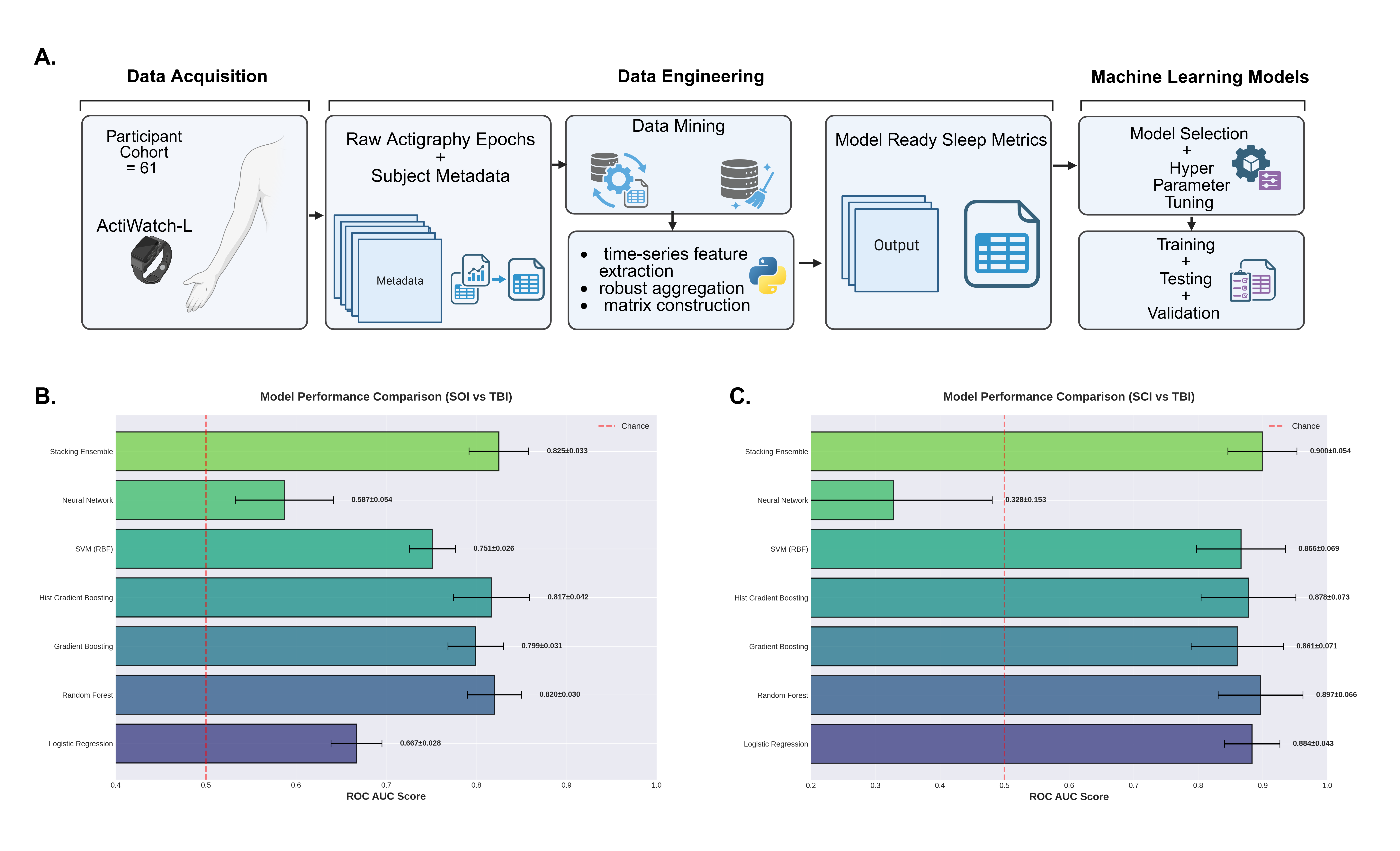
Continuous wrist actigraphy was collected from 61 inpatients (TBI n = 33, SCI n = 12, SOI n = 15). Raw actigraphy time-series were processed using a custom Python-based computational pipeline that performed automated preprocessing, circadian segmentation and high-throughput extraction of sleep–wake metrics. This workflow produced 24 features per observation, including sleep efficiency, fragmentation, and bout structure. Five engineered composite features capturing circadian disruption and sleep consolidation yielded a 31-feature representation of sleep dynamics. Seven supervised machine learning classifiers were evaluated using stratified five-fold cross-validation with performance assessed by AUC and average precision. (Fig. 1)


Results
A stacking ensemble method achieved the strongest performance across both classification tasks. Discrimination was strong for TBI versus SOI (AUC=0.825±0.033; average precision=0.896) and SCI versus TBI (AUC=0.900±0.054; average precision=0.975). Nighttime sleep efficiency emerged as the most informative feature, suggesting that nocturnal sleep consolidation carries strong diagnostic value across injury groups.


Discussion
These findings demonstrate injury-specific sleep signatures and show that actigraphy-derived sleep phenotyping can distinguish central nervous system injury from peripheral trauma using scalable, noninvasive monitoring. This computational framework may enable objective clinical stratification in neurotrauma and provides a foundation for future machine learning approaches to track recovery trajectories and identify sleep-based biomarkers of injury burden.

Figure 1. Actigraphy-based sleep feature pipeline and machine learning model performance for TBI classification.

References
1. Courville, E., Kazim, S. F., Vellek, J., Tarawneh, O., Stack, J., Roster, K., Roy, J., Schmidt, M., & Bowers, C. (2023). Machine learning algorithms for predicting outcomes of traumatic brain injury: A systematic review and meta-analysis. Surgical Neurology International, 14, 262. https://doi.org/10.25259/SNI_312_2023
2. Tunthanathip, T., & Oearsakul, T. (2021). Application of machine learning to predict the outcome of pediatric traumatic brain injury. Chinese Journal of Traumatology, 24(6), 350–355. https://doi.org/10.1016/j.cjtee.2021.06.003
 

Acknowledgement
Acknowledgements: A.L. was supported by the National Institutes of Health Training Program in Sleep and Circadian Biology (T32HL149646). Additional thanks to the Sleep, Inflammation, and Neuropathology Lab members for the support.
Tuesday July 14, 2026 5:00pm - 7:00pm ADT
Ballroom B2

5:00pm ADT

P109: Imperfectly synchronous dynamics of gamma rhythms and its response to network inputs
Tuesday July 14, 2026 5:00pm - 7:00pm ADT
Introduction
Temporal patterning of neural synchronization was observed to be related to symptoms of multiple neurological and neuropsychiatric disorders. In particular, alterations in the patterns of intermittent synchrony in multiple spectral bands (including gamma band) were found in autism spectrum disorder (ASD), Alzheimer’s disease (AD), and frontotemporal dementia (FTD) as demonstrated in recent electrophysiological studies with resting state EEG recordings [1,2,3]. Computational models may provide mechanistic insights into how synaptic parameters and network properties shape properties of intermittent synchronization dynamics and might underlie the neural dynamics observed experimentally in pathological conditions.


Methods
We used a conductance-based models of pyramidal neurons and interneuron to simulate systems of synaptically connected randomly organized networks of excitatory and inhibitory neurons, that exhibits gamma-band activity, and studied how changes in the local vs long-range connectivity impact the temporal patterning of intermittent gamma synchrony, following the recent model developments presented in [4,5]. The intermittently synchronized dynamics was characterized using measures (such as measures of the distributions of the desynchronization intervals duration) similar to those used in the experimental investigations of human EEG recordings as mentioned above.


Results
Numerical simulations showed that synaptic strength affected not only the average synchronization strength but also the distribution of desynchronization intervals durations (including situations where the former is not substantially different, but the latter is markedly altered). This happened in a way that stronger local connectivity tended to prolong desynchronization intervals, whereas stronger long-range connectivity tended to shorten them across a fairly broad parameter range. The changes in the temporal patterning of synchronized dynamics in these networks may lead to the changes in how these networks respond to common input signals affecting mutual synchronizability of connected networks.


Discussion
Our results suggest that synaptic parameters not only exert control over the strength of gamma synchrony but also its fine temporal structure over relatively short time scales. The differences in synchronizability properties between the networks may be responsible for the differences in the information processing in these networks and their effective communication efficiency. It is plausible to hypothesize that the experimentally observed differences in the patterning of the synchronous dynamics (as mentioned in the Introduction) may be related to the synaptically-induced changes in the temporal patterning of the synchronized dynamics and changes in synchronizability of the networks as those found in numerical simulations.


References
1.      Malaia, E. A., Ahn, S., & Rubchinsky, L. L. (2020). Autism Research, 13, 24-31. https://doi.org/10.1002/aur.2219
2.      Ahn, S., Malaia, E. A., & Rubchinsky, L. L. (2025). Clinical Neurophysiology, 177, 2110931. https://doi.org/10.1016/j.clinph.2025.2110931
3.      Ahn, S., Rubchinsky, L. L., & Malaia, E. A. (2025). Biological Psychology, 199, 109077. https://doi.org/10.1016/j.biopsycho.2025.109077
4.      Nguyen, Q. A., & Rubchinsky, L. L. (2021). Chaos, 31, 043134. https://doi.org/10.1063/5.0042451
5.      Nguyen, Q. A., & Rubchinsky, L. L. (2024). Cognitive Neurodynamics, 18, 3821–3837. https://doi.org/10.1007/s11571-024-10150-9

Acknowledgement
 
Tuesday July 14, 2026 5:00pm - 7:00pm ADT
Ballroom B2

5:00pm ADT

P110: The thalamocortical spiking model demonstrating the kinetics of sleep spindle suppression upon noradrenergic neuromodulation
Tuesday July 14, 2026 5:00pm - 7:00pm ADT
Introduction
The sleep spindles are a characteristic oscillatory EEG pattern occurring during NREM and are their presence is controlled by the noradrenaline released from the Locus Coeruleus (LC). Current consensus attributes the noradrenaline effects to slow depolarization caused by a reduction in the potassium leak current conductance mediated by the α1 adrenergic receptors, although role of β1 receptors affecting the I_h current was also reported. In our study we investigate molecular mechanisms underlying noradrenergic modulation in thalamus and address the discrepancy between the observed time course of the sleep spindle suppression and the reported kinetics the K+ leak channels.


Methods
The ECoG and LC recordings from adult male Sprague-Dawley rats were reanalyzed from previous study [1]. The sleep spindles were extracted from band-pass (11–16 Hz) filtered ECoG power according to the previously published methods [2]. We used existing spiking sleep network model that comprises four distinct populations: 500 pyramidal neurons, 100 inhibitory interneurons, 100 thalamocortical relay neurons, and 100 thalamic reticular neurons, with the details of the implementation are in the original paper [3]. Our most recent extension of this model featured time-dependent noradrenergic modulation mediated by α1 and β1 adrenergic receptors.


Results
Following the onset of the LC burst, after the short time lag the complete spindle suppression is achieved. Our simulations demonstrate the role of individual adrenergic receptors activation in the enzymatic pathways leading to this rapid suppression of the sleep spindles. Furthermore, we show that the investigated pathways are independent of each other, accumulating to an additive effect. Finally, we establish which downstream secondary messengers cascades operate on the timescales required to account for the rapid onset of spindle suppression.


Discussion
While there are many models involving the effects of the noradrenergic modulation on the thalamocortical system, no  concise framework explains how the noradrenaline - induced molecular mechanisms translate to network-level thalamic dynamics. The rapid time course of the observed spindle reduction indicates the prominent role of the initial products of adrenergic receptor activation such as G-protein subunits and diglyceride. Our simulation results provide mechanistic explanation of how sleep - related events are affected during NREM sleep.


References
1. Yang, M., & Eschenko, O. (2025). Differential locus coeruleus–hippocampus interactions during offline states. eLife, 14, Article e109159. https://doi.org/10.7554/eLife.109159.1
2. Durán, E., Pandinelli, M., Logothetis, N. K., & Eschenko, O. (2023). Altered norepinephrine transmission after spatial learning impairs sleep-mediated memory consolidation in rats. Scientific Reports, 13, Article 4231. https://doi.org/10.1038/s41598-023-31308-1
3. Krishnan, G. P., Chauvette, S., Shamie, I., Soltani, S., Timofeev, I., Cash, S. S., Halgren, E., & Bazhenov, M. (2016). Cellular and neurochemical basis of sleep stages in the thalamocortical network. eLife, 5, Article e18607. https://doi.org/10.7554/eLife.18607

Acknowledgement
This work was supported by ERDF-Project No. CZ.02.01.01/00/22_008/0004643
Tuesday July 14, 2026 5:00pm - 7:00pm ADT
Ballroom B2

5:00pm ADT

P111: VIP-Mediated Attentional Modulation of Persistent Activity in a Cortical Microcircuit Model
Tuesday July 14, 2026 5:00pm - 7:00pm ADT
Introduction
Persistent neuronal activity is a proposed neural mechanism supporting the maintenance of information in working memory (WM) [1]. Attention is known to influence WM performance and the stability of internal representations [2]. However, how attentional signals modulate the circuit mechanisms that generate persistent activity remains insufficiently explored in computational models. In this study, we investigate whether modulatory input to vasoactive intestinal peptide (VIP) interneurons can regulate the emergence of persistent activity in a biologically constrained cortical microcircuit model.


Methods
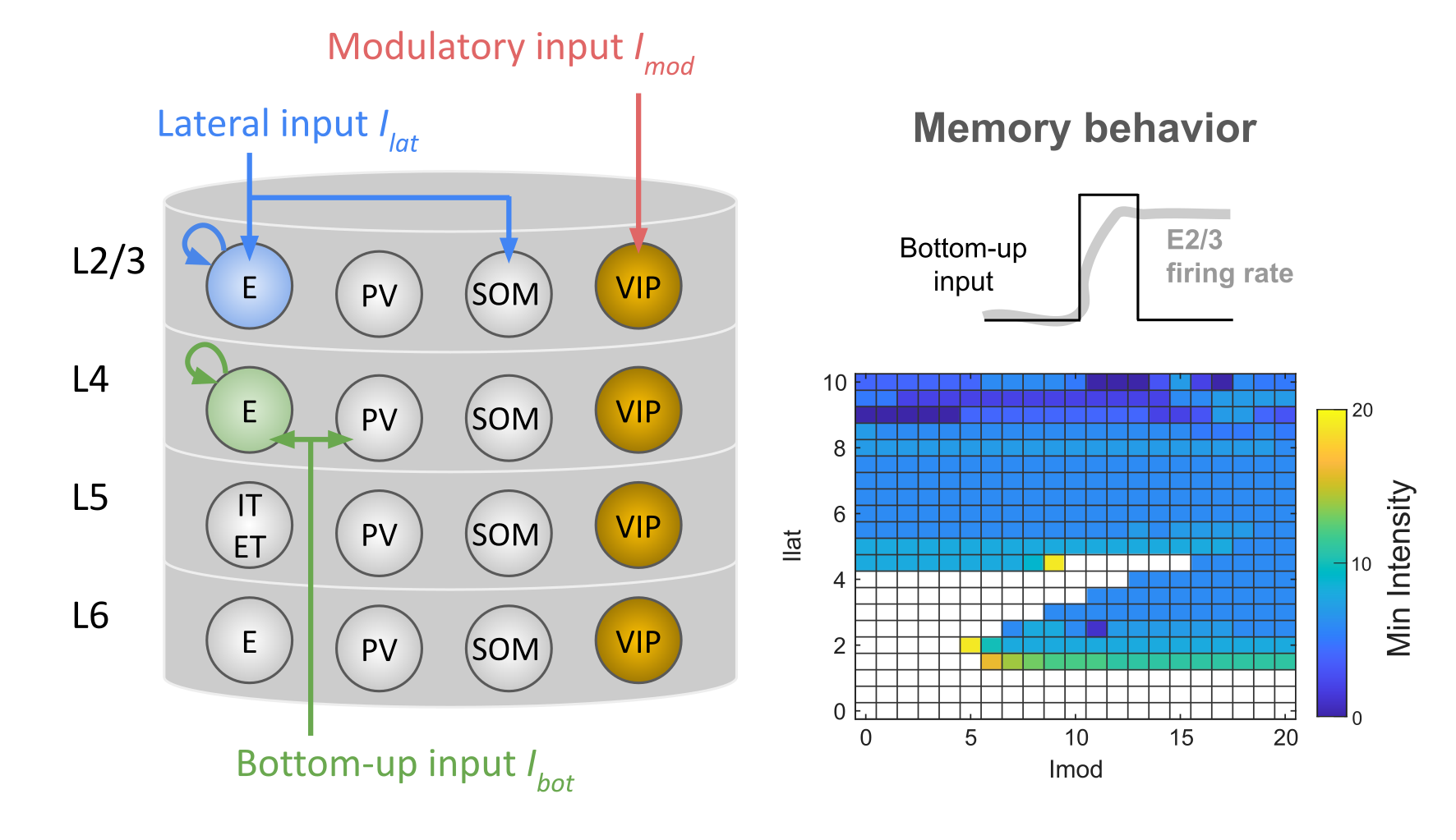
We employed a cortical microcircuit model consisting of excitatory (E) and inhibitory interneuron populations (PV, SOM, and VIP) distributed across cortical layers L2/3, L4, L5, and L6 [3]. The model incorporates biologically informed parameters, including connection probabilities, synaptic strengths, neuronal densities, and firing rate functions for each cell type. Three classes of long-range inputs were implemented: (i) lateral input to E2/3 and SOM2/3 populations, (ii) modulatory input targeting VIP2/3 neurons, and (iii) bottom-up input to E4 and PV4 populations. We systematically varied key parameters to examine their influence on the emergence of persistent activity (Fig. 1).


Results
The model exhibits bistability with respect to bottom-up input. Bistability emerges across a range of parameter configurations, including variations in VIP interneuron cell count, recurrent connectivity within the E2/3 population, recurrent connectivity within the E4 population, and the strength of modulatory input. The size of the bistable region is sensitive to these parameters, particularly the modulatory input to VIP2/3 neurons. Notably, stronger modulatory input reduces the minimum bottom-up input required to sustain persistent activity.


Discussion
VIP interneurons form a canonical disinhibitory circuit motif and are frequently associated with attentional modulation. Our simulations suggest that attentional signals targeting VIP interneurons can facilitate the emergence and maintenance of persistent activity in cortical microcircuits. These findings provide a potential circuit-level mechanism by which attention may enhance working memory stability.

Figure 1. The cortical microcircuit model and its memory behavior. Key model parameters include VIP interneuron cell count (yellow), recurrent connectivity within the E2/3 population (blue), and recurrent connectivity within the E4 population (green). The minimum bottom-up input intensity for a memory behavior varies across different levels of lateral and modulatory inputs.

References
1. Kamiński, J., & Rutishauser, U. (2020). Between persistently active and activity-silent frameworks: novel vistas on the cellular basis of working memory. Annals of the New York Academy of Sciences, 1464(1), 64-75. https://doi.org/10.1111/nyas.14213
2. Gazzaley, A., & Nobre, A. C. (2012). Top-down modulation: bridging selective attention and working memory. Trends in cognitive sciences, 16(2), 129-135. https://doi.org/10.1016/j.tics.2011.11.014
3. Chien, V. S., Jiříček, S., Knösche, T. R., Hlinka, J., & Schmidt, H. (2025). Long-Range Input to Cortical Microcircuits Shapes EEG-BOLD Correlation. bioRxiv, 2025-06. https://doi.org/10.1101/2025.06.06.658058

Acknowledgement
The work was supported by a Lumina-Quaeruntur fellowship (LQ100302301) by the Czech Academy of Sciences (awarded to HS) and ERDF-Project Brain Dynamics, No. CZ.02.01.01/00/22_008/0004643.

Speakers
HS

Helmut Schmidt

Scientific researcher, Institute of Computer Science, Czech Academy of Sciences
JH

Jaroslav Hlinka

Senior researcher, Institute of Computer Science of the Czech Academy of Sciences
Currently                                I am leading the and also serve as the Head of the Department of Complex Systems and as the Chair of the Council of the of the Czech Academy of Sciences.
Brief bio After obtaining master degrees in Psychology from Charles University (2005) and in Mathematics from Czech Technical University (2006), I went on the quest of applying mathematics in helping to understand the complex activity of human brain through neuroimaging data analysis... Read More →
Tuesday July 14, 2026 5:00pm - 7:00pm ADT
Ballroom B2

5:00pm ADT

P112: Biologically Realistic Models of Synaptic Release at Human Cortical Synapses
Tuesday July 14, 2026 5:00pm - 7:00pm ADT
Introduction
Synapses are integral to information processing and plasticity in the brain. Their functional properties, such as their strength and plasticity profiles, are determined by their underlying synaptic structure and organization. A recent study by Rollenhagen and colleagues (unpublished data) reconstructed the structure and vesicle organization of cortical layers 1-6 presynaptic boutons in the human temporal lobe neocortex (hTLN) using transmission electron microscopy and quantitative 3D volume reconstructions. The data reveal variations in vesicle numbers, sizes, and positions within each bouton, as well as differences in bouton organization, such as active zone areas, across cortical layers. 


Methods
Using these ultrastructural measurements, we develop biophysically detailed, spatially explicit, stochastic models of hTLN presynaptic boutons across cortical layers, implemented in STEPS (Stochastic Engine for Pathway Simulation, https://steps.sourceforge.net/), and simulate neurotransmitter release to investigate how synaptic transmission is shaped by bouton organization.


Results
We observe that vesicle size diversity within a presynaptic bouton enhances variability in excitatory postsynaptic current amplitudes, and that smaller vesicles exhibit higher fusion propensity due to faster diffusion compared to larger vesicles. Additionally, our modeling framework allows us to reconcile structural measurements and electrophysiological recordings from similar synapses, suggesting layer-specific differences in voltage-dependent calcium channel expression in hTLNboutons.  


Discussion
These findings suggest that nanoscale structural heterogeneity within presynaptic boutons can shape synaptic transmission dynamics and variability. Moreover, our modeling approach provides a quantitative framework linking ultrastructural measurements to functional synaptic outputs.


References
Wils, Stefan, and Erik De Schutter. "STEPS: modeling and simulating complex reaction-diffusion systems with Python." Frontiers in neuroinformatics 3 (2009): 374. 

\nHepburn, Iain, et al. "Vesicle and reaction-diffusion hybrid modeling with STEPS." Communications Biology 7.1 (2024): 573. 
\nGallimore, Andrew R., et al. "Dynamic regulation of vesicle pools in a detailed spatial model of the complete synaptic vesicle cycle." Science Advances 11.22 (2025): eadq6477. 

Acknowledgement
Astrid Rollenhagen and Joachim Lübke, Institute of Neurosciences and Medicine (INM-10), Forschungszentrum Jülich, for sharing unpublished ultrastructural data and helpful discussions; and the Scientific Computing and Data Analysis Section at OIST for access to the Deigo supercomputing cluster. This work was supported by OIST Graduate University.

Tuesday July 14, 2026 5:00pm - 7:00pm ADT
Ballroom B2

5:00pm ADT

P113: Model of Mossy Fiber Bouton (MFB) in hippocampus- the detonator synapse with vesicle release in realistic EM morphology
Tuesday July 14, 2026 5:00pm - 7:00pm ADT
Introduction


The mossy Fiber Bouton (MFB) in hippocampus acts as a detonator synapse with sophisticated morphology, large number of vesicles and multiple active zones (AZs), which provide the physical basis for the detonator properties [1]. However, how presynaptic MFB terminals decode the frequency and number of action potentials to transmit information remains poorly understood.  Due to its complicated morphology, by far there has been no detailed simulation on the presynaptic neurotransmission in a realistic MFB morphology.

Methods


Here, we utilize experimental 3D electron microscopic (EM) data from our collaborators of Prof. Dr. Joachim Lübke unit in Juelich Germany to investigate the basic mechanism of how vesicle docking and release machinery within complicated morphology contributes to MFB’s detonator property. Based on the detailed kinetics of vesicle docking process and rich distribution of mitochondria for uptake calcium in MFB, by implementing the calcium sensor synaptotagmin 1(syt1) and synaptotagmin 7 (syt7) into the exocytosis machinery, we simulate the presynaptic vesicle release of MFB by STochastic Engine for Pathway Stimulation (STEPS)(https://steps.sourceforge.net/manual/) [2,3].

Results


Our results show that surprisingly, all the eight morphologically distinct MFBs consistently exhibit the detonator properties, despite the diversity and variety of vesicle number, bouton size, AZ distributions from each bouton. MFB boutons exhibit frequency independent neural transmission with fast vesicle docking mechanism (spike counting strategies). The release event per pulse highly depends on calcium channel density yet did not change the intrinsic Short Time Facilitation (STF) feature.  Active transport of vesicles can facilitate the fast vesicle docking in some boutons enhancing the detonator properties; for other boutons, active transport did not show difference from pure diffusion probably due to the high density of vesicles.

Discussion


Importantly, our results demonstrate that in certain bouton uptake of calcium by mitochondria plays an important role in regulating precise signal transduction, while for other bouton the effect is not so obvious, though mitochondria still play a role in regulating calcium homeostasis. Finally, we show that spike-locked synchronous release by syt1 dominate over occasional asynchronous releases by syt7, which is consistent to experimental results. In conclusion, a STEPS model of neural transmission in giant MFB with realistic morphology and molecular details provides insights of why MFBs show detonator properties.

References


[1] Nicoll, R. A., & Schmitz, D. (2005). Synaptic plasticity at hippocampal mossy fibre synapses. Nature Reviews Neuroscience, 6(11), 863–876. https://doi.org/10.1038/nrn1786
[2] Gallimore, A. R., Hepburn, I., Georgiev, S. V., Rizzoli, S. O., & De Schutter, E. (2025). Dynamic regulation of vesicle pools in a detailed spatial model of the complete synaptic vesicle cycle. Science Advances, 11(22), eadq6477. https://doi.org/10.1126/sciadv.adq6477
[3] Hepburn, I., Lallouette, J., Chen, W., Gallimore, A. R., Nagasawa-Soeda, S. Y., & De Schutter, E. (2024). Vesicle and reaction-diffusion hybrid modeling with STEPS. Communications Biology, 7(1), 573. https://doi.org/10.1038/s42003-024-06276-5

Acknowledgement


We thank our collaborator Prof. Dr. Joachim H. R. Lübke from Juelich Germany to kindly provide EM data of MFB: Rollenhagen, A., Sätzler, K., Rodríguez, E. P., Jonas, P., Frotscher, M., & Lübke, J. H. R. (2007). Structural Determinants of Transmission at Large Hippocampal Mossy Fiber Synapses. The Journal of Neuroscience, 27(39), 10434–10444. https://doi.org/10.1523/JNEUROSCI.1946-07.2007.
Tuesday July 14, 2026 5:00pm - 7:00pm ADT
Ballroom B2

5:00pm ADT

P114: The effect of weak electric deep brain stimulation fields on the synchronization of multi-compartment neuron models
Tuesday July 14, 2026 5:00pm - 7:00pm ADT
Introduction
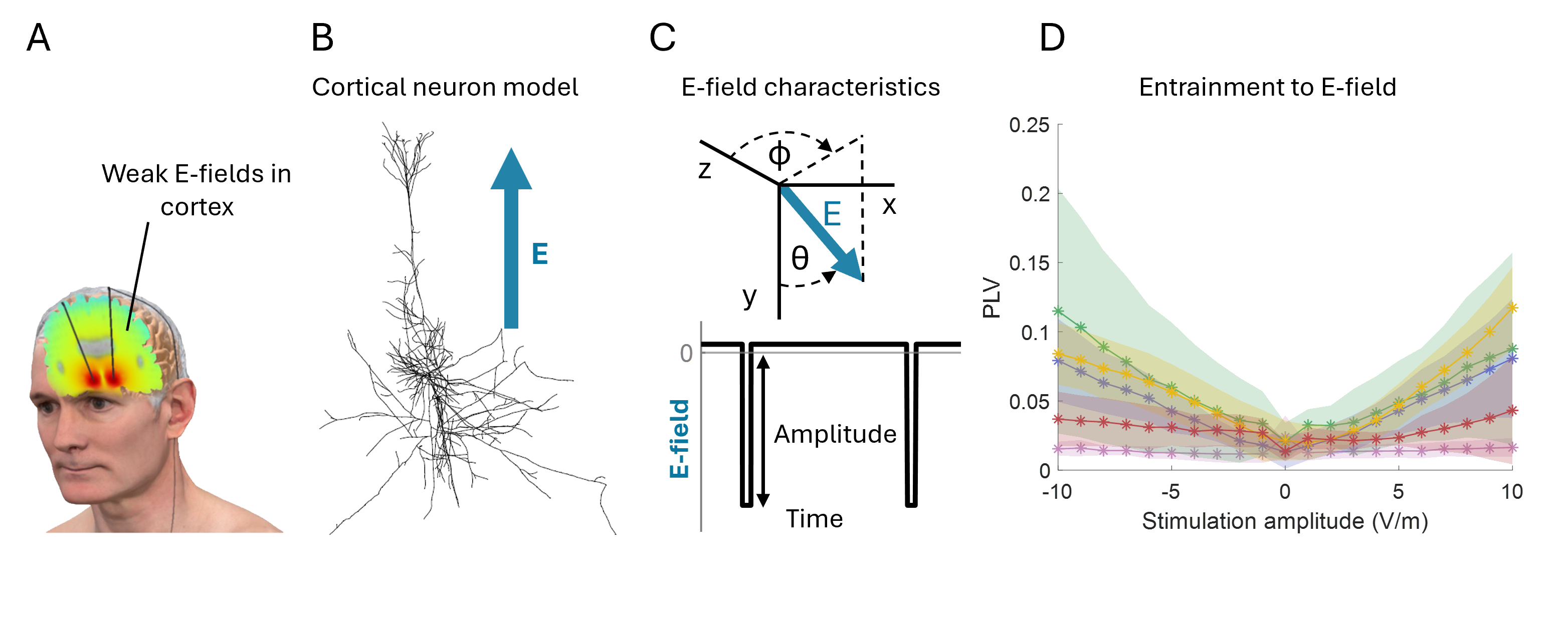
Deep brain stimulation (DBS) is widely used to treat motor symptoms of Parkinson’s disease, but the mechanism by which it alters neural dynamics is still not fully understood. DBS typically consists of short, high-frequency electrical pulses delivered through an implanted electrode, and its effects are attributed to strong electric fields near the electrode. However, studies of non-invasive brain stimulation show that weak sinusoidal electric fields below 1V/m can modulate spike timing and neural synchronization [1]. This raises the possibility that weak electric fields during DBS (Fig. 1A) can affect cortical neuron dynamics. We test this hypothesis by investigating entrainment of multi-compartment neuron models under weak DBS fields.

Methods
We employed morphologically realistic multi-compartment models of cortical neurons (Fig 1B), comprising five cell types from different cortical layers [2]. The models were subjected to an externally applied electric field (Fig. 1C) designed to reproduce high-frequency DBS pulses. The orientation of the electric field was varied relative to neural morphology using the polar angle θ. Entrainment was quantified using the phase-locking value across a range of electric field amplitudes and orientations.

Results
Weak DBS fields can entrain the spike timing of single cortical neurons (Fig. 1D). Spikes tend to cluster at specific phases of the applied DBS waveform, producing a non-uniform spike-time distribution and an increase in phase-locking value. The preferred entrainment phase varies across neuron types and stimulation amplitudes. Differences in neural morphology and electric field orientation contribute to heterogeneous entrainment patterns across cells. However, when the electric field is aligned with each neuron’s preferred orientation, consistent and stronger entrainment emerges.

Discussion
Our results suggest that electric fields with amplitudes below 10 V/m can synchronize the spike timing of individual cortical neurons with the applied stimulation. This finding implies that weak fields, despite being far smaller than those near the electrode, may still influence cortical activity during DBS. Such effects could interact with the effects of stronger fields present at the stimulation site and potentially contribute to both therapeutic outcomes and stimulation-related side effects. Future work will investigate synchronization in network models composed of two-compartment neurons to determine whether network interactions enhance these effects and whether they can promote synchronization at the population level.

Figure 1. Cortical neuron entrainment by weak electric DBS fields. (A) Illustration of electric fields generated in the brain during DBS. (B) Multi-compartment models of cortical neurons. (C) Electric field orientation relative to the neural morphology. Waveform of the applied field, representing a DBS-like stimulus. (D) Mean phase-locking value increases when amplitude increases.References
[1] Krause, M. R., Vieira, P. G., Thivierge, J. P., & Pack, C. C. (2022). Brain stimulation competes with ongoing oscillations for control of spike timing in the primate brain. PLoS Biology, 20(5), e3001650. https://doi.org/10.1371/journal.pbio.3001650
[2] Tran, H., Shirinpour, S., & Opitz, A. (2022). Effects of transcranial alternating current stimulation on spiking activity in computational models of single neocortical neurons. NeuroImage, 250, 118953. https://doi.org/10.1016/j.neuroimage.2022.118953

Acknowledgement
This work is part of a project  that has received funding from the European Research Council (ERC StG DECODE, grant number 101116047, to B.C.S). We would like to thank Ciska Heida for valuable discussions and guidance.   
Tuesday July 14, 2026 5:00pm - 7:00pm ADT
Ballroom B2

5:00pm ADT

P115: Digital Twins Enable Early Alzheimer’s Disease Diagnosis
Tuesday July 14, 2026 5:00pm - 7:00pm ADT
Introduction
Early detection and prognostic prediction of Alzheimer’s disease (AD) is essential for timely intervention and improved patient outcomes. However, current diagnostic methods, including cerebrospinal fluid (CSF) analysis and neuroimaging techniques, are often invasive, costly, and unsuitable for early screenings. Non-invasive neural recordings like electro- or magnetoencephalography (EEG or MEG) provide a non-invasive alternative, yet they often struggle to identify cortical alterations associated with AD at preclinical stages. To address these limitations, we propose a novel approach based on digital twin models that extract personalized digital biomarkers reflecting individual neurodegeneration levels from non-invasive neural recordings.


Methods
We developed the DADD (Digital Alzheimer’s Disease Diagnosis) digital twin model to derive digital biomarkers from non-invasive neural recordings [1, 2]. DADD reconstructs personalized levels of neurodegeneration from individual neural recordings via biophysical modeling of AD-related functional alterations. DADD parameters reconstruct patient-specific levels of synaptic degeneration, network-level brain disconnection and neuroplastic rewiring. DADD biomarkers were used to predict evolution of cognitive decline in 459 participants with prodromal AD, also testing their cross-center and cross-modal generalizability on additional two cohorts totaling 46 HC and 76 MCI participants undergoing different types of neural recordings.


Results


The DADD model significantly outperformed standard EEG analysis in predicting follow-up results in concurrent machine-learning classifications (AUC=0.71 vs AUC=0.62, p<0.00001). Combining DADD digital biomarkers with CSF biomarkers significantly increased the prediction of future conversions (AUC=0.81 vs AUC=0.74, p=0.009). A Cox Proportional Hazard model found that digital biomarkers had the highest predictive power for future conversions in SCD participants (HR=1.94, p=0.003), also outperforming CSF biomarkers (HR=1.40, p=0.013). In the cross-center classification, digital biomarkers obtained consistent cross-center classification (77–78% accuracy), while standard biomarkers performed poorly in the generalization attempt (56–65%) [3].

Discussion
These findings suggest that DADD might be a powerful tool for early AD diagnosis and prognosis based on non-invasive recordings. Our approach supports accurate and generalizable classification of dementia staging, combined with accurate estimation of future cognitive decline risk. The ability of DADD to reconstruct individual neurodegeneration levels provides deeper insights into disease progression, bridging the gap between network structure and cognitive outcomes. This method represents a scalable, generalizable and cost-effective solution for early AD detection, potentially facilitating widespread clinical implementation and improving patient management strategies.


References


[1]  Amato, L. G., et al. (2024). Personalized modeling of Alzheimer’s disease progression estimates neurodegeneration severity from EEG recordings. Alzheimer’s & Dementia: Diagnosis, Assessment & Disease Monitoring, 16(1), e12526. https://doi.org/10.1002/dad2.12526
[2]  Amato, L. G., et al. (2025). Digital twins and non-invasive recordings enable early diagnosis of Alzheimer’s disease. Alzheimer’s Research & Therapy, 17(1), 125. https://doi.org/10.1186/s13195-025-01765-z
[3]  Amato, L. G., et al. (2026). Digital twins support cross-modal and cross-centric classification of mild cognitive impairment. Communications Medicine, 6(1), 30. https://doi.org/10.1038/s43856-025-01281-z

Acknowledgement
This work was supported by the Italian Ministry of Research, in the context of the project NRRP “Fit4MedRob-Fit for Medical Robotics” Grant (# PNC0000007)

Tuesday July 14, 2026 5:00pm - 7:00pm ADT
Ballroom B2

5:00pm ADT

P116: Vasoactive intestinal polypeptide-expressing neurons (VIPs) as a mechanism for flexible cognitive control
Tuesday July 14, 2026 5:00pm - 7:00pm ADT
Introduction
Working memory and decision making are mediated by common cortical areas [1], but they make conflicting demands of circuit dynamics. Competition between neural populations is essential for decision making, but exacerbates working memory storage limitations. This discrepancy implies a mechanism for mediating between dynamic regimes. VIPs are strong candidates for this role, as they receive long-range cortical and modulatory inputs, and selectively target classes of inhibitory interneurons with varying connectivity profiles [2,3]. As such, VIPs are well positioned to integrate a range of task factors and to control the spatial structure of inhibition in a context-dependent manner, providing a rich mechanism for cognitive flexibility.

Methods
We developed a cortical circuit model comprising pyramidal neurons, large basket cells (broad, indiscriminate inhibition) and small basket cells (local surround inhibition) [4], connected by AMPA, NMDA, and GABA receptor synapses with conductance strengths informed by electrophysiological data. VIP control was simulated by independently modulating inhibitory conductance onto each interneuron class. We simulated a luminance-contrast visual discrimination task in which the network identified a target among distractors and a visuospatial working memory task in which the network maintained stimulus information across a delay. Both tasks received identical stimuli to enable direct comparison between the dynamics imposed by inhibitory structure.

Results
Under a single set of parameter values, our model accounts for neural and behavioural signatures of decision making and working memory. On our decision task, these data include target-selective and distractor-selective neural activity, and psychometric and chronometric curves. On our working memory task, they include statistics of persistent mnemonic activity and storage limitations. Local inhibition has a strong stabilizing effect in the model, attenuating the competitive dynamics engendered by large basket cells. As such, VIPs establish a decision regime by preferentially targeting small basket cells (local disinhibition) and establish a working memory regime by targeting large basket cells (broad disinhibition).

Discussion
Our findings support the hypothesis that VIPs mediate flexible cognitive control by selectively targeting inhibitory cell classes with different connectivity profiles, effectively determining the spatial structure of inhibition in cortical circuits. This mechanism enables the rapid switching between task-appropriate dynamic regimes for decision making (competition) and working memory (stability with minimal competition) as well as fine-tuning the dynamics within each regime. The model makes testable predictions for neural and behavioural data from visual discrimination and working memory tasks.

References
  1. Murray, J. D., Jaramillo, J., & Wang, X. J. (2017). Working memory and decision-making in a frontoparietal circuit model. Journal of Neuroscience, 37(50), 12167-12186.
  2. Pi, H. J., Hangya, B., Kvitsiani, D., Sanders, J. I., Huang, Z. J., & Kepecs, A. (2013). Cortical interneurons that specialize in disinhibitory control. Nature, 503(7477), 521-524.
  3. Wall, N. R., De La Parra, M., Sorokin, J. M., Taniguchi, H., Huang, Z. J., & Callaway, E. M. (2016). Brain-wide maps of synaptic input to cortical interneurons. Journal of Neuroscience, 36(14), 4000-4009.
  4. Wang, Y., Gupta, A., Toledo-Rodriguez, M., Wu, C. Z., & Markram, H. (2002). Anatomical, physiological, molecular and circuit properties of nest basket cells in the developing somatosensory cortex. Cerebral cortex, 12(4), 395-410.
Tuesday July 14, 2026 5:00pm - 7:00pm ADT
Ballroom B2

5:00pm ADT

P117: Neurite heterogeneity controls signal propagation in a model of the ctenophore syncytial nerve net
Tuesday July 14, 2026 5:00pm - 7:00pm ADT
Introduction

Recent work [1] on the ctenophore M. leidyi has shown that the standard architecture of excitable neurons connected by neurites terminated by synapses is not universal. The ctenophore subepithelial nerve net (SNN) is a continuous cytoplasmic network lacking chemical or electrical synapses. Electron micrographs revealed a nerve net with a beaded or “pearls-on-a-string” morphology in which spherical swellings alternate with thin cylindrical constrictions [1]. This morphology appears to be heterogeneous, with varying degrees of pearls (or beads) and connectors between them. The overall function and electrical measurements of the M. leidyi SNN are not yet known, inviting investigation into basic models of possible signal propagation modes.


Methods

We model the structure of the SNN as consisting of a variable number of polygons embedded on a circular disk, respecting the organism’s physical dimensions. The aboral organ and the comb rows are modeled as excitable tissue with the help of a standard diffusively coupled neuron model. The neurites connecting neurons are modelled as only partially excitable, with two parameters controlling their excitability, reflecting the special ‘blebbed’ morphology of the SNN. We then study activity spread on the computational domain by numerically solving the underlying partial differentials equations. We also derive an effective cable equation taking into account the blebbed morphology.


Results

A central function of the SNN is the conduction of a swimming or reversal signal from the aboral organ to all eight comb rows [2]. We examine the conditions under which this can occur using our model, and derive constraints on neurite heterogeneity. We discuss neuropeptides and other biophysical stimuli as drivers for the beaded morphology. Using simulations of excitable and diffusive elements on a network, we parameterize the model with a modified cable equation and compare the conduction speed and directionality in such networks to the observed ciliary beating. We use our modified cable equation to gauge the accuracy of parameters used in our simulations and suggest that the SNN uses neurite morphology to adjust propagation delays.


Discussion

Taken together we theoretically investigate SNN neurite structure and heterogeneity for its consequences for signal propagation on multiple scales. Notably, the same organism at a later developmental stage possesses a second syncytial nerve net - the mesogleal nerve net - whose neurites display a distinct cylindrical morphology [4]. The coexistence of two nerve nets with morphology in the same animal suggests a link between SSN morphology and function. Indeed, in mammalian central nervous system axons, remodelling of an analogous beaded morphology fine-tunes action potential conduction velocity and overall neuronal function was recently discussed [3]. Yet the connection to the synapse-free ctenophore SNN has not been exploreduntil now. 


References

  1. Burkhardt, P., et al (2023). Syncytial nerve net in a ctenophore adds insights on the evolution of nervous systems. Science, 380(6642), 293–297. https://doi.org/10.1126/science.ade5645
  2. Tamm, S. L. (2014). Cilia and the life of ctenophores. Invertebrate Biology, 133(1), 1–46. https://doi.org/10.1111/ivb.12042
  3. Griswold, J. M., et al (2025). Membrane mechanics dictate axonal pearls‑on‑a‑string morphology and function. Nature Neuroscience, 28(1), 49–61. https://doi.org/10.1038/s41593-024-01813-1
  4. Jokura, K., Jasek, S., Niederhaus, L., Burkhardt, P., & Jékely, G. (2026). Neural connectome of the ctenophore statocyst. eLife, 14, e108420. https://doi.org/10.7554/eLife.108420


Acknowledgement

JS acknowledges funding by the European Union (ERC, SYNNEURO, 101163768). Views and opinions expressed are however those of the author(s) only and do not necessarily reflect those of the European Union or the European Research Council. Neither the European Union nor the granting authority can be held responsible for them.

Speakers
JS

Jan Steinkuehler

Assistant Professor, Kiel University
avatar for Wilhelm Braun

Wilhelm Braun

Junior Research Group Leader, Kiel University (CAU Kiel), Faculty of Engineering, Department of Electrical and Information Engineering
Early nervous systems, functional neuronal networks, stochastic neural dynamics, animal behavior, reinforcement learning, network reconstruction
Tuesday July 14, 2026 5:00pm - 7:00pm ADT
Ballroom B2

5:00pm ADT

P118: Development and Validation of a Multi-scale Model of Cerebellar Transcranial Magnetic Stimulation
Tuesday July 14, 2026 5:00pm - 7:00pm ADT
Introduction
Transcranial magnetic stimulation (TMS) is a promising technique to alleviate symptoms related to cerebellar pathologies. However, the outcomes of TMS are variable and it is not understood how exactly neuronal activity is affected by it. Many models, at different scales, have been developed to try to address the open questions on the functioning of TMS. The state of the art are multi-scale models, which integrate MRI generated head models of the electric field induced by TMS and biologically realistic neuron models. A limitation of multi-scale models lies in their validation [1]. In this work we present a framework to model and validate TMS targeting the cerebellum.


Methods

We calculated the mean electric field induced by TMS in the cerebellar vermis using the SimNIBS simulator. We developed a biologically realistic model of the cerebellar cortex [2] and coupled the TMS induced electric field to its neurons using NEURON’s extracellular mechanism.
We generated the input-output curve of the cerebellar network, in baseline and in stimulated conditions, by increasing the input mossy fibre frequency from 0 to 150 Hz in 3 Hz steps. 
We normalised the input-output curves in the two conditions and utilised them in a vertical oculomotor system model [3] to predict eye movements. We compared predicted eye movements in the baseline and stimulated conditions with experimental data to validate the TMS model.


Results

The maximum mean electric field induced by TMS in the cerebellum vermis is 30.4 V/m. The stimulation does not alter the mean firing rate of the neurons, but it alters the spike timing and the instantaneous firing rate. 
We reproduced the eye movements of a healthy subject with the network in baseline conditions. Eye movements in the stimulated condition show a drift during gaze holding. Saccades are not affected by the stimulation.
Experimentally it was found that the application of TMS generated an increase in the amplitude of ipsilateral reflexive saccades and in the acceleration of ipsilateral smooth pursuit movements.


Discussion

The vertical oculomotor system model produces observable differences in predicted eye movements following stimulation. This suggests that the model can serve as a validation framework for transcranial magnetic stimulation models targeting cerebellar circuits involved in saccades and gaze holding.
Predicted eye movements do not accurately reproduce the experimental findings. This may suggest that the employed model is not detailed enough to reproduce experimental results. It may also suggest eye movement changes seen experimentally  are not primarily driven by neuronal activity within the targeted cerebellar region, but may also arise from activation of more superficial neural structures or neighbouring regions in the cerebral cortex.


References

  1. Shahid, S. S., Bikson, M., Salman, H., Wen, P., & Ahfock, T. (2014). The value and cost of complexity in predictive modelling: Role of tissue anisotropic conductivity and fibre tracts in neuromodulation. Journal of Neural Engineering, 11(3), 036002. https://doi.org/10.1088/1741-2560/11/3/036002 
  2. Bernasconi, E., Yousif, N., & Steuber, V. (2024). 32nd Annual Computational Neuroscience Meeting: CNS*2023 - P194 Development of a biologically realistic cerebellar model to study the effects of non-invasive stimulation. Journal of Computational Neuroscience, 52(1), 3–166. https://doi.org/10.1007/s10827-024-00871-5 
  3. Glasauer, S., & Rössert, C. (2008). Modelling drug modulation of nystagmus. Progress in Brain Research, 171, 527–534. https://doi.org/10.1016/S0079-6123(08)00675-4


Acknowledgement
-
Tuesday July 14, 2026 5:00pm - 7:00pm ADT
Ballroom B2

5:00pm ADT

P119: Low-Effort Attention as Free-Energy Optimization via Cingulo–Autonomic Control
Tuesday July 14, 2026 5:00pm - 7:00pm ADT
Introduction


Active inference models of attention and self-control typically emphasize effortful precision weighting implemented through executive control. However, empirical findings indicate that attentional stability and self-regulation can improve under minimal cognitive effort. This poses a challenge for effort-centric interpretations of control cost in predictive systems [1]. We address this gap by proposing a regulatory framework in which attention optimization emerges through reductions in expected free energy mediated by brain–body coupling, rather than increased top-down control. The model reframes low-effort regulation as efficient precision allocation across interoceptive and exteroceptive hierarchies.

Methods


We formalize low-effort attention as an active inference process in which expected free energy is minimized via autonomic and cingulate-mediated precision control. The model centers on an anterior cingulate–posterior cingulate–striatal (APS) circuit interacting with parasympathetic regulation to modulate policy selection and control cost. Empirical constraints include observed shifts in midline theta–alpha dynamics, heart-rate variability, and network reconfiguration following brief low-effort training paradigms [2]. Control is modeled as adaptive precision modulation rather than sustained executive signaling.

Results


The framework predicts that reducing expected free energy can be achieved by stabilizing interoceptive predictions and lowering control-related metabolic demand. Simulated dynamics reproduce empirical signatures of low-effort regulation, including increased midline coherence, parasympathetic dominance, and transitions from frontoparietal engagement to cingulo-striatal coordination. The model accounts for rapid plasticity in cingulate pathways and the emergence of automaticity without increased policy complexity. These results suggest that attentional efficiency arises from optimized precision weighting rather than enhanced effort.

Discussion


This work situates low-effort attention and self-control within an active inference framework, offering a computational account of how regulation can improve while control cost decreases. By treating attention as precision optimization coupled to autonomic regulation, the model reconciles empirical findings with free-energy principles and generates testable predictions for electrophysiological and interoceptive markers [1, 2]. The framework generalizes across training paradigms and informs theories of embodied cognition, adaptive control, and energy-efficient artificial agents.

References


1. Tang, Y.Y., Tang, R, Posner, M. I., & Gross, J. J. (2022). Effortless training of attention and self-control: mechanisms and applications. Trends Cogn Sci. 26(7), 567-577. https://doi.org/10.1016/j.tics.2022.04.006
2. Friston, K. (2010). The free-energy principle: a unified brain theory?. Nat Rev Neurosci 11, 127–138. https://doi.org/10.1038/nrn2787

Acknowledgement


This work is supported by the ONR N000142412270 and NIH R33 AT010138.
Tuesday July 14, 2026 5:00pm - 7:00pm ADT
Ballroom B2

5:00pm ADT

P120: Adaptive Multisensory Support for Low-Effort Attention in Embodied Agents
Tuesday July 14, 2026 5:00pm - 7:00pm ADT
Introduction


Attention regulation depends on how neural systems balance internal control with sensory input. When this balance becomes rigid, regulation requires greater effort and becomes unstable. Empirical work shows that a brief low-effort training paradigm, the integrative body–mind training (IBMT) improves attention by inducing a low-effort regulatory state [1], yet individuals vary in their ability to access and stabilize this state. Existing accounts do not explain how such low-effort regulation is learned and generalized at the level of control dynamics. We propose a theoretical framework in which embodied AI supports this learning by stabilizing a measurable target regulatory state via adaptive multisensory input.

Methods


We model attention regulation using predictive coding and the free energy principle [2], treating control as precision-weighted inference. Effortful states arise from excessive precision on self-related control priors, increasing energetic cost. Low-effort states correspond to reduced control precision and efficient integration of interoceptive and exteroceptive signals. We introduce an AI-based multisensory support architecture operating as a closed-loop system layered onto IBMT. The AI estimates regulatory state from physiological and attentional markers and adaptively modulates basic sensory parameters (timing, intensity, variability) to bias precision allocation without explicit instruction or reinforcement.

Results


The framework predicts that adaptive multisensory support facilitates access to low-effort regulatory states in individuals with high attentional noise or rigid control. Predicted outcomes include reduced energetic cost, reflected in stabilized autonomic markers and decreased indices of cognitive effort. With repeated practice, the model predicts internalization of the target state, leading to reduced dependence on external modulation. The framework also predicts failure regimes, including destabilization under excessive modulation and limited benefit when control precision is already low, highlighting the need for individualized adaptation.

Discussion


By treating low-effort attention as a configuration of regulatory dynamics rather than a subjective state, this framework bridges contemplative neuroscience and embodied active inference. Individual differences in attention training are reframed as differences in precision dynamics and learning trajectories. The model generates testable predictions that adaptive sensory environments accelerate stabilization and effortless re-entry into low-effort regulation, and suggests general design principles for embodied agents that support regulation by shaping sensory context rather than increasing control intensity.

References


1. Tang, Y.Y., Tang, R, Posner, M. I., & Gross, J. J. (2022). Effortless training of attention and self-control: mechanisms and applications. Trends Cogn Sci. 26(7), 567-577. https://doi.org/10.1016/j.tics.2022.04.006
2. Friston, K. (2010). The free-energy principle: a unified brain theory?. Nat Rev Neurosci 11, 127–138. https://doi.org/10.1038/nrn2787

Acknowledgement


This work is supported by the ONR N000142412270 and NIH R33 AT010138.
Tuesday July 14, 2026 5:00pm - 7:00pm ADT
Ballroom B2

5:00pm ADT

P121: Optogenetic WiChR based seizure control in a potassium driven epilepsy model
Tuesday July 14, 2026 5:00pm - 7:00pm ADT
Introduction
Optogenetic modulation of pyramidal cells using potassium selective opsins has been shown to inhibit neuronal activity, making it a promising strategy for suppressing seizures [1]. A prior study in a point‑neuron hippocampal model showed seizure termination after brief opsin activation, but different seizure generation mechanisms may alter intervention efficacy [2]. In this study, we examine optogenetic modulation in a multicompartment model based on the potassium hypothesis, in which elevated extracellular potassium drives seizure initiation and maintenance.

Methods
We extended the potassium based seizure model of Gentiletti et al. (2022) by adding a synaptically connected cell (0) without extracellular coupling and incorporating the WiChR opsin model proposed by Weyn et al. (2025) into all principal cell (1-4) compartments, as schematically shown in (Fig.\u202f1A) [1,3]. We illuminated different subsets of principal cells for varying illumination durations (tI) and quantified (1) mFRall, the mean firing rate of cells\u202f1-4; (2) FR0, the firing rate of cell\u202f0 as a measure of SLE related synaptic propagation; (3) seizure-like event (SLE) duration, defined as periods where mFRall\u202f>\u202f2.5\u202fHz for\u202f>\u202f7\u202fs; and (4) SLE end time.

Results
The results are shown in Fig.\u202f1B. mFRall was consistently reduced during illumination, with stronger suppression when larger fractions of the onset zone were targeted. FR0 was highly sensitive to the number of illuminated cells and returned to pre‑SLE (<1\u202fHz) levels only when 75% of the zone was targeted. Longer tI further decreased firing, although similar reductions also occurred without stimulation due to intrinsic SLE dynamics. Despite transient suppression, the increased SLE end time shows that activity resumed after illumination. Only prolonged, centered (cells 2/3) partial or full onset‑zone (cells\u202f1-4) targeting fully abolished the SLE, reducing both duration and end time.

Discussion
In potassium driven SLEs, inhibiting a large proportion of the onset zone is necessary to prevent synaptic propagation during illumination. Furthermore, transient complete pyramidal inhibition is insufficient for SLE termination, as activity reliably resumes once illumination ceases. Effective suppression depends on the target zone and requires prolonged illumination that outlasts the potassium driven dynamics sustaining SLEs. These findings contrast with the earlier point‑neuron study, where short optogenetic intervention fully stopped SLEs, suggesting that the efficacy of optogenetic strategies critically depends on the underlying biophysical mechanism and the need for mechanism specific intervention design.

Figure 1. A. Schematic representation of the model and example output. B. Impact of illumination duration (tI) and targeted cell subsets (stimCells) on firing rates during illumination (mFR_all, mFR0) and on SLE duration and end time.

References
[1] Weyn, L., Tarnaud, T., Schoeters, R., De Becker, X., Joseph, W., Raedt, R., & Tanghe, E. (2025). Computational analysis of optogenetic inhibition of CA1 neurons using a data‑efficient potassium‑ and chloride‑conducting opsin model. Journal of Neural Engineering, 22(4), 046051. https://doi.org/10.1088/1741-2552/adf94a
[2] Weyn, L., Tarnaud, T., Joseph, W., Raedt, R., & Tanghe, E. (2026). Optogenetic inhibition of a hippocampal network model. Journal of Computational Neuroscience, 54(Suppl 1). https://doi.org/10.1007/s10827-025-00915-4
[3] Gentiletti, D., de Curtis, M., Gnatkovsky, V., & Suffczynski, P. (2022). Focal seizures organized by feedback between neural activity and ion concentration changes. eLife, 11, e68541. https://doi.org/10.7554/eLife.68541 

Acknowledgement
This work is supported by BOF project SOFTRESET.
Speakers
avatar for Thomas Tarnaud

Thomas Tarnaud

Associate professor, INTEC WAVES, University of Ghent - IMEC
Tuesday July 14, 2026 5:00pm - 7:00pm ADT
Ballroom B2

5:00pm ADT

P122: Computational modeling of ultrasonic neuromodulation in realistic cortical cells
Tuesday July 14, 2026 5:00pm - 7:00pm ADT
Introduction
Transcranial ultrasound stimulation (TUS) is an emerging neuromodulation technique offering millimeter‑scale precision and non‑invasive stimulation. While experimental studies show that ultrasound can modulate neural activity across multiple brain regions, the underlying biophysical mechanisms remain incompletely understood, limiting optimal protocol design. Among the proposed mechanisms, including flexoelectricity, mechanosensitivity, thermodynamic effects, and membrane displacement, intramembrane cavitation (IC) has gained particular attention. IC describes the oscillating gas cavities between the phospholipid membrane leaflets, generating capacitive currents and membrane charge oscillations that can alter neuronal excitability.

Methods
Early computational work using the neuronal intramembrane cavitation excitation (NICE) model demonstrated the potential of this mechanism but suffered from large computational cost [1]. Subsequent developments, such as the multi‑scale optimized SONIC model and the spatially extended SECONIC model, introduced precomputation, hybrid integration, and Fourier analysis to accelerate simulations and incorporate charge redistribution dynamics [2,3]. However, these models were limited to one-dimensional representations of neurons, restricting their ability to capture realistic spatial activation patterns. In this work, we extend IC‑based ultrasonic neuromodulation modeling to morphologically realistic neurons across different cortical layers.

Results
By integrating detailed neuronal, we investigate how ultrasound parameters such as frequency, pressure amplitude, pulse repetition frequency (PRF), duty cycle (DC), and sonophore radius, shape neuronal responses. Fig. 1 shows how the firing rate of a L2/3 pyramidal cell depends on the pressure amplitude and the duty cycle. This approach enables localization of activation sites, assessment of the role of higher‑order charge overtones, and evaluation of whether IC can account for experimentally observed cell‑type specificity and protocol sensitivity. For instance, across multiple cortical cell types, we found that activation consistently originated at terminal nodes.

Discussion
By integrating advanced biophysical models with detailed, realistic neuronal morphologies, this study advances a mechanistic understanding of how TUS influences neural activity and offers a foundation for accurate, in‑silico optimization of stimulation strategies.

Figure 1. The intramembrane cavitation model in a pyramidal cell (top left), a schematic of the implementation of ultrasound field – neuron coupling (top right) and the firing rate of a L2/3 pyramidal cell as a function of amplitude and duty cycle.

References
[1] Plaksin, M., Kimmel, E., & Shoham, S. (2016). Cell-Type-Selective Effects of Intramembrane Cavitation as a Unifying Theoretical Framework for Ultrasonic Neuromodulation. eNeuro, 3(3), ENEURO.0136-15.2016. https://doi.org/10.1523/ENEURO.0136-15.2016
[2] Lemaire, T., Neufeld, E., Kuster, N., & Micera, S. (2019). Understanding ultrasound neuromodulation using a computationally efficient and interpretable model of intramembrane cavitation. Journal of neural engineering, 16(4), 046007. https://doi.org/10.1088/1741-2552/ab1685
[3] Tarnaud, T., Joseph, W., Schoeters, R., Martens, L., & Tanghe, E. (2020). SECONIC: Towards multi-compartmental models for ultrasonic brain stimulation by intramembrane cavitation. Journal of neural engineering, 17(5), 056010. https://doi.org/10.1088/1741-2552/abb73d

Acknowledgement
/
Speakers
avatar for Thomas Tarnaud

Thomas Tarnaud

Associate professor, INTEC WAVES, University of Ghent - IMEC
Tuesday July 14, 2026 5:00pm - 7:00pm ADT
Ballroom B2

5:00pm ADT

P123: Population-based Functional Source Separation enables identification of cortical sources in new individuals
Tuesday July 14, 2026 5:00pm - 7:00pm ADT
Introduction
Functional Source Separation (FSS) extends Blind Source Separation (BSS) by incorporating prior knowledge about the functional characteristics of neural responses during source extraction. FSS has previously been used to estimate primary motor (FS_M1) and somatosensory (FS_S1) cortical representations from stimulus-evoked EEG responses [1], and to identify FS_M1 in passive subjects [2]. Here we propose a population-based FSS approach that estimates spatial filters from pooled cross-subject data. We test whether these population-derived filters can identify motor and sensory cortical sources in new individuals using resting-state EEG


Methods
EEG recordings were obtained from 22 healthy subjects during median nerve stimulation (Sensory condition), weak isometric handgrip (Motor condition), and resting state with eyes open and closed. FSS was applied in two ways: (i) FSSindividual, computed separately for each subject, and (ii) FSSpopulation, estimated from pooled cross-subject data. Both approaches were used to identify FS_S1 and FS_M1 sources in both hemispheres. The similarity between sources extracted with the two approaches was assessed using mutual information (MI), time correlation coefficient (TCC), and Kullback–Leibler (KL) divergence. Cortico-muscular coherence (CMC) was computed in the Motor condition to evaluate the functional relevance of the motor sources.


Results
In the Motor condition, CMC between the population-derived motor source and the prime mover muscle during isometric handgrip did not differ from the coherence obtained using the individual motor source in either hemisphere (p = 0.3). Similarly, the population-derived sensory source showed responses to median nerve stimulation comparable to those obtained with the individual sensory source (p = 0.1). Across all four sources, the broadband activity of each population-derived source showed higher MI and TCC, and lower KL divergence, with its corresponding individual source than with other sources (p < 0.03).

Discussion
These results show that motor and sensory cortical sources can be identified from resting-state EEG using population-derived FSS filters that generalize to new subjects. This approach may be particularly useful in clinical settings where task-related activity is difficult to obtain, such as in patients with stroke or limb amputation [3,4]. Because FSS is deterministic, it may also improve the reproducibility of EEG-based studies of regional neuronal activity. Future work should extend this framework to larger populations, pathological conditions, and additional cortical regions such as auditory and visual cortices.


References
1. Barbati, G., Sigismondi, R., Zappasodi, F., Porcaro, C. et al. (2006). Functional source separation from magnetoencephalographic signals. Hum. Brain Mapp. doi:10.1002/hbm.20232
2. Porcaro, C., Cottone, C., Cancelli, A., Salustri, C., Tecchio, F. (2018). Functional semi-blind source separation identifies primary motor area without active motor execution. Int. J. Neural Syst. doi:10.1142/S0129065717500472
3. Delcamp, C., Srinivasan, R., Cramer, S. C. (2024). EEG provides insights into motor control and neuroplasticity during stroke recovery. Stroke. doi:10.1161/STROKEAHA.124.048458
4. Liu, S., Fu, W., Wei, C., Ma, F. et al. (2022). Interference of unilateral lower limb amputation on motor imagery rhythm and remodeling of sensorimotor areas. Front. Hum. Neurosci.doi:10.3389/fnhum.2022.1011463


Acknowledgement
The authors thank Annalisa Pascarella, Gian Luca Loli, Rosario Ribecco, and Filippo Zappasodi for their scientific contributions over the years, and the participants who took part in the recordings.
Tuesday July 14, 2026 5:00pm - 7:00pm ADT
Ballroom B2

5:00pm ADT

P124: Power Spectrum Harmonics Provide a Signature of Balanced Excitation-Inhibition Across Cortical Scales
Tuesday July 14, 2026 5:00pm - 7:00pm ADT
Introduction
Transcranial alternating current stimulation (tACS) is widely used to modulate brain activity, yet its mechanisms remain incompletely understood and its effects vary across individuals and studies [1]. When nonlinear neural circuits are driven by sinusoidal input, responses often contain harmonic frequencies in addition to the stimulation frequency [2]. Although commonly observed in neural recordings, the dynamical origin of these harmonics remains unclear. We hypothesized that harmonic structure may reflect the balance of excitation and inhibition in the stimulated network.

Methods
We used a Wilson–Cowan mean-field model to examine how recurrent connectivity, transfer-function gain, and input amplitude shape harmonic responses under periodic drive. Model predictions were evaluated using broadband LFP recordings from macaque V4 during prefrontal tACS (N = 17 sessions; randomized blocks at 5 and 10 Hz; [3]). Power spectra were computed from 60-s segments (frequency resolution ≈ 0.017 Hz), with all harmonics well below the Nyquist limit. Results were consistent using both fast Fourier transform (FFT) and Welch spectra. For comparison, we also implemented a spiking leaky integrate-and-fire (LIF) network with balanced connectivity.


Results
In the Wilson–Cowan model, harmonic structure depended on the population’s operating regime, determined by net E–I connectivity and input amplitude. Activity near the transfer function’s inflection region produced predominantly odd harmonics, whereas excursions toward ceiling or floor regions generated even harmonics (Fig. 1). Across 193 experimental epochs, 88% showed entrainment to the input frequency. Among these, 84% exhibited odd harmonics, 16% were restricted to the fundamental frequency, and none displayed even harmonics. The LIF network reproduced both odd and even harmonic patterns under comparable connectivity regimes.

Discussion
Our results show that harmonic structure during rhythmic stimulation reflects the circuit operating point, determined by E–I balance and input strength. Changes in gain modify the width of odd-dominant regions but preserve the overall pattern of responses. The predominance of odd harmonics in the experimental data is consistent with model regimes associated with balanced excitation–inhibition dynamics typical of healthy cortical networks. Together, these findings suggest that harmonic analysis could provide a non-invasive probe of cortical state, and that physiological or behavioral changes affecting network gain may alter harmonic profiles.

Figure 1. (A) Power spectrum of experimental recordings during SHAM and 5 Hz sinusoidal stimulation. Stimulation induces odd harmonics (15–35 Hz). (B) Log10-OEHR heatmaps from the Wilson–Cowan model showing harmonic dominance across recurrent connectivity (red = odd, dark blue = even). Connectivity sets the net input to the sigmoid transfer function and thus the operating regime (circles).References
1. Krause MR, Vieira PG, Pack CC. (2023). Transcranial electrical stimulation: How can a simple conductor orchestrate complex brain activity?. PLOS Biology 21(1): e3001973. https://doi.org/10.1371/journal.pbio.3001973
2. Jin C, Wang X, Chen B, Wan H, Lu Z, Sun Y, Sun Y, Bu J. (2026). Media Transmission Characteristics of Harmonic Signatures in tACS: From Simple Linear Phantoms to Complex Biological Systems, Brain Stimulation, https://doi.org/10.1016/j.brs.2026.103052.
3. Krause MR, Vieira PG, Thivierge JP, Pack CC. (2022). Brain stimulation competes with ongoing oscillations for control of spike timing in the primate brain. PLOS Biology 20(5): e3001650. https://doi.org/10.1371/journal.pbio.3001650

Acknowledgement
Thank you to C. C. Pack, P. Vieira and M. R. Krause for the experimental data. 
Tuesday July 14, 2026 5:00pm - 7:00pm ADT
Ballroom B2

5:00pm ADT

P125: Learning Barrel Cortex Representations from Whisker Dynamics and Multimodal Convergence
Tuesday July 14, 2026 5:00pm - 7:00pm ADT
Introduction
Rodents can discriminate object shape, size, and texture through active whisker contacts. Accordingly, rodent primary somatosensory cortex (S1) contains topographic representations of the whiskers, and neurons in S1 represent multiple stimulus features associated with whisker based touch. Although these representations have been characterized experimentally, the computational mechanisms that generate them remain unclear. In addition, there is substantial convergence between whisker-based touch and other sensory modalities in multiple cortical areas. We test whether training whisker and visual input representations to align in a self-supervised way can reproduce representations in barrel cortex.


Methods
Using a morphologically accurate model of the mouse whisker array [2], we simulated whisker contact sequences in terms of the radial distance and angle of contact. These signals were encoded using per-whisker temporal units; these units also integrated the signals over the whisk cycle. Learned whisker embeddings were then arranged into a 5×7 topographic grid approximating barrel cortex. The encoder was trained using a contrastive objective that aligned whisker embeddings with visual embeddings produced by MouseNet [1]. Condition-averaged embeddings were used to construct representational dissimilarity matrices (RDMs), which were compared to neural population RDMs using Pearson correlation and established noise correction procedures.


Results
The anatomically structured whisker network produced embeddings aligned with neural population geometry. Representational similarity analysis (RSA) yielded a raw RSA of 0.54 between model and neural dissimilarity matrices across stimulus conditions, with a noise-corrected estimate of 1.14 relative to the neural reliability ceiling. Alignment was reduced when temporal integration was removed or when whisker embeddings were not arranged in a topographic grid, indicating that both recurrent dynamics and barrel-like spatial organization contribute to emergent representational structure.


Discussion
These results suggest that biologically relevant representations may arise from temporally integrated whisker signals constrained by anatomical hierarchy, even when the model is trained using a multimodal contrastive objective rather than explicit supervision on the whisker discrimination task, as in [3]. The dependence of alignment on both recurrent dynamics and topographic organization supports the hypothesis that barrel organization and recurrent dynamics are key determinants of tactile shape coding. This framework provides a principled foundation for extending biologically constrained models toward multimodal cortical integration.


References
1. Shi, J., Tripp, B., Shea-Brown, E., Mihalas, S., & A. Buice, M. (2022). MouseNet: A biologically constrained convolutional neural network model for the Mouse Visual Cortex. PLOS Computational Biology, 18(9). https://doi.org/10.1371/journal.pcbi.1010427
2. Bresee, C. S., Belli, H. M., Luo, Y., & Hartmann, M. J. Z. (2023). Comparative morphology of the whiskers and faces of mice (Mus musculus) and rats (Rattus norvegicus). Journal of Experimental Biology, 226(19), jeb245597. https://doi.org/10.1242/jeb.245597
3. Chung, T., Shen, Y., Kong, N. C. L., & Nayebi, A. (2025). Task-optimized convolutional recurrent networks align with tactile processing in the rodent brain. arXiv. https://doi.org/10.48550/arXiv.2505.18361


Acknowledgement
CN was supported by NSERC RGPIN-2025-04919 and Alliance International Catalyst. KK and MJH were supported by NIH award R01 NS-116277. 

Tuesday July 14, 2026 5:00pm - 7:00pm ADT
Ballroom B2

5:00pm ADT

P126: Remapping Convolutional Layers to Fit Cortical Connectivity
Tuesday July 14, 2026 5:00pm - 7:00pm ADT
Introduction
Convolutional networks are commonly used in brain models [e.g. 3, 5]. Convolutions assume a uniform connection structure from source to target. However, a recent analysis of mesoscale connectivity in the mouse cortex [4] showed that many connections over and under-represent parts of the source and/or target area, and may project distant parts of a source into nearby parts of a target. Because multiple connections converge in each cortical area, these complex projection structures constrain which signals converge in the brain. Here we analyze the structural error inherent in using standard convolutional networks to model the mouse cortex, and develop an extension that has a more accurate structure.

Methods
We first calculated 2D cortical-surface coordinates for each voxel of each mouse neocortical area, as in [4], and normalized them to have standard deviations (SD) of 1. For each projection, we used voxel-voxel connection density estimates from [2] to calculate the weighted-mean source-area coordinates that provided input to each target-area voxel. We evaluated potential remapping functions (f) that mapped target coordinates (t) to corresponding source coordinates (s), such that s = f(t). We first fitted a hierarchy of constrained geometric models, including rotation/flip, linear, and affine remapping. We also introduced a more flexible nonlinear remapping model based on a radial basis function interpolator with thin-plate spline kernel [1].

Results
We calculated the mean Euclidean distance dμ between actual source coordinates and those predicted by each function. Rotation/flip remapping, which is consistent with standard convolution (accounting for different map orientations in cortical coordinates), gave the poorest fit (dμ = 0.62 over all connections, vs. coordinate SD=1). Linear and affine remapping, which correspond to convolution with non-integer stride and offset, reduced the mean error to 0.17 and 0.10, respectively. However, large errors remained in some voxels for each of these models. The general RBF model further reduced mean error to < 0.001, with no large errors. This model corresponds to convolution layers followed by general data-driven remapping layers.

Discussion
This analysis shows that standard convolutional networks are inconsistent with the mesoscale connectivity of mouse cortex. However, realistic connectivity can be recovered by inserting remapping layers into convolutional networks. This proposed extension improves approximation of a wide range of complex cortical structures, including diverse subfields in higher visual areas, low-level multisensory connections, and complex convergence patterns in prefrontal areas.

References
[1] GE Fasshauer. “Meshfree Approximation Methods with Matlab, World Sci”. In: Publishing Co, Singapore (2007).
[2] Joseph E Knox et al. “High-resolution data-driven model of the mouse connectome”. In: Network Neuroscience 3.1 (2018), pp. 217–236.
[3] Jonathan A Michaels et al. “A goal-driven modular neural network predicts parietofrontal neural dynamics during grasping”. In: Proceedings of the national academy of sciences 117.50 (2020), pp. 32124–32135.
[4] Kinjal Patel et al. “Spatial organization of multisensory convergence in mouse isocortex”. In: bioRxiv (2024), pp. 2024–12.
[5] Jianghong Shi et al. “MouseNet: A biologically constrained convolutional neural network model for the mouse visual cortex”. In: PLOS Computational Biology 18.9 (2022), e1010427.

Acknowledgement
This work was supported by NSERC Discovery Grant RGPIN-2025-04919. 
Tuesday July 14, 2026 5:00pm - 7:00pm ADT
Ballroom B2

5:00pm ADT

P127: Distinguishing between fetal alcohol spectrum disorders and attention-deficit/hyperactivity disorder using diffusion modelling and spiking neurons
Tuesday July 14, 2026 5:00pm - 7:00pm ADT
Introduction
Drift Diffusion (DD) models and spiking neural networks have been effective in predicting and simulating disordered behaviour. They offer deep insight into behavioural processes and spiking neuronal models understand the relationship between such processes and the underlying biological system. A joint approach is valuable as DD models reflect the entirety of performance data and spiking networks mimic neuronal communication. This offers a more detailed analysis that is biologically-plausible and gives a deeper insight into neural dynamics and cognitive processes. Thus, our work uses both models to identify differences among fetal alcohol spectrum disorders (FASD), attention-deficit/hyperactivity disorder (ADHD), and comorbid presentations.

Methods
DD models assume, following encoding, that decisions are made via a noisy process where evidence for a response is accumulated until it crosses a response boundary, after which a corresponding motor response is initiated. DD analysis is used to identify how attention processes differ and to inform spiking Search over Time and Space (sSoTS) model parameters. sSoTS is a spiking neuronal model including several synaptic components (AMPA, NMDA, GABA) and frequency adaptation mechanisms. Pool coupling parameter is changed to simulate differences in encoding between FASD and ADHD. Furthermore, we extend prior research in visual search [1,2] by simulating visual selective attention processes to distinguish FASD, ADHD, and comorbid presentations.

Results
DD analysis showed ADHD favoured accuracy, whereas FASD (without ADHD) had a similar boundary separation to controls on easy search. ADHD and controls had a similar drift rate, whereas FASD (with and without ADHD) had a slower drift rate on easy visual search. On difficult search, all participants had a similar boundary separation favouring accuracy. All diagnostic groups had similar drift rates on difficult search, which was slower than controls. Our results suggest that FASD affect bottom-up attention processes but an ADHD comorbidity may buffer some effects. To further understand how attention processes differ, data from DD analysis was used to inform sSoTS parameters and changes were successfully simulated. 

Discussion
Results from our visual search paradigm demonstrated the importance of combining computational models to distinguish different patterns of attention deficit among individuals with FASD, ADHD, and comorbid presentations. Incorporating coupling parameter changes into the sSoTS model successfully simulated the observed behaviours. Our outcomes provide an initial demonstration of how integrating computational methodologies can further enhance our understanding of how attention processes differ across different disorders. Our work underscores that FASD, ADHD, and when both disorders present comorbidly may be able to be distinguished based on the efficiency of bottom-up attention processes.

References
[1]        Mavritsaki, E., Heinke, D., Allen, H., Deco, G., & Humphreys, G. W. (2011).        Bridging the Gap Between Physiology and Behavior: Evidence from the sSoTS         Model of Human Visual Attention. Psychological Review, 118(1), 3–41. https://doi.org/10.1037/a0021868
[2]        Mavritsaki, E., & Humphreys, G. (2016). Temporal Binding and Segmentation in Visual Search: A Computational Neuroscience Analysis. Journal of Cognitive            Neuroscience, 28(10), 1553–1567. https://doi.org/10.1162/jocn_a_00984

Acknowledgement
I would like to thank my supervisory team and all collaborators for their support in preparing this abstract.
Tuesday July 14, 2026 5:00pm - 7:00pm ADT
Ballroom B2

5:00pm ADT

P128: Ion Channels Tune Population-Level Intrinsic Biophysical Heterogeneity
Tuesday July 14, 2026 5:00pm - 7:00pm ADT
Introduction

Human neurons show remarkable variation, even among the same type. Contrary to the belief that it is noise, within-cell-type heterogeneity enhances information coding and, as our recent work has shown, endows dynamical resilience to insults [1]. Furthermore, our work has shown that heterogeneity is malleable and is reduced in seizures and [1] under correlated inputs [2,3].

We focus on intrinsic biophysical heterogeneity--relating to passive and firing properties of a neuron--which is influenced by ion channels. We have discovered that blocking one type of ion channel, the HCN channel, can restore heterogeneity under certain conditions [2]. This study aims to clarify the mechanism and conditions under which the effect emerges.

Methods
Following common approaches, large ensembles of human L5 pyramidal neuron models were built from a pre-tuned model, sampling ion channel conductances across empirically defined ranges and validating the resulting populations against experimental datasets. Intrinsic heterogeneity was quantified through variance of normalized spiking metrics and information-theoretic measures analogous to those used in our experimental work, so that the results may be directly compared.

Results
Our results at this time show that upregulation of specific ion channels, particularly HCN, strongly influences population heterogeneity ofsome intrinsic properties such as resting membrane potential (RMP) and input resistance (Fig. 1). While trends appear modest across the full log-scale conductance range, focusing on the physiological regime (10-4–10⁻³) reveals a sharp decline in variance. In contrast, transient sodium and M-type potassium channels show weak correlations with the variance of most intrinsic properties.

Discussion
We hypothesize that HCN channels’ unique influence on population-level heterogeneity arises from their involvement in a voltage-dependent negative feedback, such that increased expression of HCN channels reduces the range of potential intrinsic properties. However, the counterargument is that, despite similar feedback, M-type potassium channels do not show this effect. Owing to inter-channel interactions and degeneracy, further systematic investigation is needed to understand these non-linear relationships fully. Given the established role of ion channels in diseases and our finding that reduced heterogeneity drives pathology, this work will inform strategies for modulating intrinsic heterogeneity as a novel therapeutic approach.

Figure 1. Scatter plot of the (A) resting membrane potential (RMP) and (B) input resistance of a population of ~8000 neurons sorted in increasing order of apical H-current (Ih) maximal conductance, with the coefficient of variation (CV) of RMP (C) and input resistance (D) respectively plotted on the right.

References
1. Rich, S., Moradi Chameh, H., Lefebvre, J., & Valiante, T. A. (2022). Loss of neuronal heterogeneity in epileptogenic human tissue impairs network resilience to sudden changes in synchrony. Cell Reports, 39(8), 110863.
2. Chameh, H. M., Falby, M., Yang, Y., Movahed, M., Arbabi, K., Sarathy, C., Tripathy, S. J., Zhang, L., Lefebvre, J., & Valiante, T. A. (2025). Hyperpolarization-activated cation channel mediated intrinsic plasticity changes underlie the malleability of within cell-type electrophysiological heterogeneity. In Neuroscience. bioRxiv.
3. Trotter, D., Valiante, T., & Lefebvre, J. (2026). Intrinsic plasticity underlies the malleability of neural network heterogeneity. PRX Life, 4(1), 013023.


Acknowledgement
This work is supported by the Ontario Graduate Scholarship and the Canada Graduate Research Scholarship through Canadian Institutes of Health Research (CIHR).

Tuesday July 14, 2026 5:00pm - 7:00pm ADT
Ballroom B2

5:00pm ADT

P129: Sequential variability of Steady State Visually Evoked Potentials and its relation with BCI performance
Tuesday July 14, 2026 5:00pm - 7:00pm ADT
Introduction
Recent work highlights the importance of identifying and assessing sequential neural activity. Here we introduce a novel approach to characterize sequential patterns in EEG data, focusing on steady-state visually evoked potentials (SSVEPs) that appear as oscillations in the occipital lobe when attending to a flickering light. This phenomenon is widely used in brain-computer interfaces [1], and is often characterized solely in terms of oscillations and their frequency components, a framework that may overlook critical aspects of neural dynamics [2]. Using spectrograms and wavelet transforms, we examine the temporal evolution of frequency and power in an SSVEP BCI speller dataset and assess its relationship with BCI performance.

Methods


The EEG recordings used in our analysis come from the BETA dataset [3], which includes recordings from 70 subjects in a BCI speller experiment with 40 different stimulus frequencies and 4 trials per frequency. We used spectrograms and wavelet transforms to examine the evolution of the signal’s frequency content and power over time. In this analysis, we aimed to assess how long a response attributable to the stimulus frequency is maintained throughout the trial, as well as how long it takes for the response to appear (latency). These results were obtained for all subjects, averaged across trials, and compared with their respective performance using a traditional canonical correlation analysis (CCA) detection method.

Results
After characterizing the EEG signals in terms of three metrics (active time percentage (ATP) in the wavelet transform, active time percentage (ATP) in the spectrogram, and response latency), we computed the average results for each subject across four trials.  We then examined these results and compared them with each subject’s CCA performance. We found a highly significant positive correlation between the CCA performance and the two metrics evaluating the percentage of active time: CWT ATP (r = 0.896), and Spectrogram ATP (r = 0.943). On the other hand, we found a highly significant negative correlation with the response latency metric (r = –0.907).

Discussion
Our characterization of the evolving response reveals that SSVEPs are not universally steady as their name suggests. High-performing subjects react to the stimulus earlier and tend to maintain a response throughout the trial.  In contrast, subjects with poor performance exhibit unstable responses with sequential activations across different frequency bands contributing to detection errors. We also found that response latency is linked to performance: a subject who responds correctly but reacts slowly will perform poorly in the BCI task. Our analysis suggests that poor performance may not always result from an inherent inability to respond to stimuli, but rather from visual attention issues in the context of simultaneous visual stimuli.

References
[1] Liu,  S., Zhang, D.,  Liu,  Z.,  Liu,  M.,  Ming,  Z.,  Liu,  T.,  Suo, D., Funahashi, S., and Yan, T. (2022).   Review of brain–computer in- terface based on steady-state visual evoked  potential.   Brain  Science Advances,   8(4): 258–275, https://doi.org/10.26599/BSA.2022.9050022.
[2] Labecki, M., Nowicka, M. M., Wrobel,  A., and Suffczynski, P. (2024). Frequency-dependent  dynamics of  steady-state visual  evoked pottials under sustained flicker stimulation.  Scientific Reports, 14(1):9281, https://doi.org/10.1038/s41598-024-59770-5.
[3] Liu, B., Huang, X., Wang, Y., Chen, X., and Gao, X. (2020). BETA: A Large Benchmark Database Toward SSVEP-BCI Application.  Frontiers in Neuroscience, 14, https://doi.org/10.3389/fnins.2020.00627.

Acknowledgement
Research funded by grants PID2024-155923NB-I00 and CPP2023-010818 (MCIN/AEI and ERDF- "A way of making Europe").

Speakers
TR

Tania Romero-Segura

PhD student, Universidad Autonóma de Madrid
Tuesday July 14, 2026 5:00pm - 7:00pm ADT
Ballroom B2

5:00pm ADT

P130: Burst-to-burst information resetting in Central Pattern Generators
Tuesday July 14, 2026 5:00pm - 7:00pm ADT
Introduction
Bursting neural activity is widely expressed in many neural systems and has been proposed to underlie reliable information multiplexing, propagation, and execution. This type of neural activity typically involves interactions at slow and fast timescales. In the context of central pattern generators (CPGs), bursting is part of the sequential motor control mechanisms responsible for respiration, locomotion, chewing, and other key motor tasks. In various CPGs, sequential dynamical invariants in the form of cycle-by-cycle relationships between specific intervals have been identified. These invariants are thought to balance sequence robustness with flexibility by creating timing constraints to the events that compose the sequence [1,2,3].

Methods
We combined models and experiments to examine how modulations introducing trends in the CPG rhythm shape the cycle-by-cycle and cross-cycle structure of these invariants. We quantified the variability of the intervals forming the sequence between the LP and PD neurons, as well as their pairwise invariant relationships in the pyloric CPG and also in a conductance-based model [4]. Relationships between intervals belonging to the same and to different cycles were assessed in experiments and in the model where ionic and synaptic currents were linked to interval durations. Invariants were identified with pairplots and quantified using the coefficient of determination R².

Results
We show that, both in computational models and electrophysiological experiments, sequential dynamical invariants are generally present only between intervals that compose the sequence within the same cycle. Only when a slow external modulation introduces a trend can intervals from different cycles become related. This is observed when modulation arises naturally or is experimentally induced in the pyloric CPG, and when a slow modulating current is injected into the model CPG. Model ionic and synaptic currents reset in every cycle and do not influence intervals in the next one, but modulating the CPG causes intervals to be related across cycles.

Discussion
Assessing sequential dynamical invariants is key to understanding the autonomous coordination of rhythmic motor sequences produced by CPGs. Not all intervals in the sequence are related, which contributes to the timing flexibility within the constraints of the invariants. The persistence of relationships between intervals across cycles can serve as a means to characterize CPG modulation. Combining models and electrophysiological experiments, we established the conditions under which sequential intervals can be related both cycle-by-cycle and across cycles when perturbations introduce trends in the CPG rhythm. These results are also relevant for bio-inspired robotics, where such principles could support autonomous motor control.

References
[1] Elices et al (2019). doi:https://doi.org/10.1038/s41598-019-44953-2.
[2] Berbel et al (2025). doi:https://doi.org/10.1016/j.neucom.2025.130218.
[3] Garrido-Peña et al (2021). doi:https://doi.org/10.1016/j.neucom.2020.08.093.
[4] Deistler et al (2022). doi:https://doi.org/10.1073/pnas.2207632119.

Acknowledgement
Research funded by grants PID2024-155923NB-I00, PID2023-149669NB-I00, CPP2023-010818 (MCIN/AEI and ERDF- "A way of making Europe").
Tuesday July 14, 2026 5:00pm - 7:00pm ADT
Ballroom B2

5:00pm ADT

P131: Decreased alpha/theta temporal ExSEnt of left prefrontal cortex in dementia: a robust biomarker
Tuesday July 14, 2026 5:00pm - 7:00pm ADT
Introduction
Dementia involves progressive cognitive decline, with Alzheimer’s disease (AD) and frontotemporal dementia (FTD) as major subtypes. Electroencephalography (EEG) provides a noninvasive and accessible measure of brain dynamics and able to capture pathological slowing, often reflected by increased theta-to-alpha power ratio (TAR) [1]. Reduced signal complexity has also been reported in dementia. We tested whether Extrema-Segmented Entropy (ExSEnt), which separates temporal and amplitude irregularity, improves discrimination and interpretability beyond conventional electroencephalography biomarkers [2].

Methods
We analyzed resting-state EEG from 88 subjects, including 36 with AD, 23 with FTD, and 29 controls [3]. After preprocessing and independent component analysis, we classified brain sources into four clusters: right and left prefrontal cortices (R/LPFC) and right and left visual associations (R/LVA). Then we computed a set of classical spectral and complexity features. We also measured the ExSEnt metrics, which quantifies the entropy of extrema-based segment durations, amplitudes, and their pair. To find table features for dementia detection, we performed stability selection with elastic-net logistic regression and nested leave-one-subject-out validation [4].


Results
ExSEnt improved discrimination mainly in the LPFC, where balanced accuracy increased from 71.5% to 82.6%. In that region, ExSEnt features showed high selection stability and replaced conventional measures as dominant predictors in 3 out of 4 brain areas. The most informative variables were temporal and joint temporal-amplitude entropy measures in alpha and theta bands, with negative coefficients indicating reduced irregularity and reduced dynamical variability in dementia. Other regions showed weaker or inconsistent gains, suggesting a localized effect rather than a global one.

Discussion
ExSEnt provides a compact and interpretable measure for EEG-based dementia classification. The results suggest that dementia is associated not only with spectral slowing but also with reduced diversity of extrema timing and temporal-amplitude variability in LPFC dynamics. Because ExSEnt is explainable by design, when combined with stability selection and sparse linear classification, it yields robust biomarkers with direct physiological interpretation. These findings support ExSEnt as a promising candidate for explainable dementia screening and motivate future validation against cognitive severity measures.


References
[1] M. Penttil¨a, J. V. Partanen, H. Soininen, P. Riekkinen, Quantitative analysis of occipital EEG in different stages of Alzheimer’s disease, Electroencephalography and Clinical Neurophysiology 60 (1985) 1–6. doi:10.1016/0013-4694(85)90942-3
[2] S. Kamali, F. Baroni, P. Varona, Exsent: Extrema-segmented entropy analysis of time series, arXiv (2025). doi:10.48550/arXiv.2509.07751.
[3] A. Miltiadous, et al., A dataset of scalp EEG recordings of Alzheimer’s disease, frontotemporal dementia and healthy subjects from routine EEG, Data 8 (2023) 95. doi:10.3390/data8060095.
[4] N. Meinshausen, P. B¨uhlmann, Stability selection, Journal of the Royal Statistical Society Series B: Statistical Methodology 72 (2010) 417–473. doi:10.1111/j.1467-9868.2010.00740.x.

Acknowledgement
This work was supported by grants PID2024-155923NB-I00 and CPP2023-010818.


Tuesday July 14, 2026 5:00pm - 7:00pm ADT
Ballroom B2

5:00pm ADT

P132: A Knowledge Integration Workflow to Define Functional Interactomes in Neural Systems
Tuesday July 14, 2026 5:00pm - 7:00pm ADT
Introduction
This study addresses the need for standard workflows and best practices in developing evidence-based computational tools and models in neuroscience [1]. Linking multiscale molecular and cellular interactions (e.g., between neurons and glia) are yet to be mainstream in Systems Neuroscience. As a step in this direction, we present an organized workflow to consolidate causal evidence of intermolecular, multicellular and multifunctional crosstalk, and to develop novel functional interactomes in neural systems (FINS). As proof-of-concept, we apply our workflow to test the hypothesis that neuroinflammatory and excitability functions are in a closed loop of multicellular molecular interactions between neurons and microglia.


Methods
Our workflow involves 1) screening primary research articles (PRAs), 2) extracting structured meta-summaries (SMS), 3) generating the FINS network graph model. We screened >120 heterogenous published studies from 2002-2025, of which 65 reporting validated causal functional associations were included. Diverse features of biomolecules such as functional identity, cell type, experimental methodology, species, and brain regions were curated. We identified pleiotropic actions of activator molecules on targets at the molecular (expression and function), cellular (excitability, cell survival), and neural circuit levels (synaptic effects). We then assembled pairwise interactors to create a network graph using the open-source Cytoscape software.

Results
The resulting FINS network revealed a non-random, hub-like topology (see Figure. 1). We introduce a Functional Interaction Score (FIS) that encodes edge thickness to capture the magnitude and direction of literature evidence between two nodes/interactors. Thicker edges correspond to greater reproducibility of empirical effects. Furthermore, edge annotations encode activator-mediated increase (solid lines) v/s decrease (dashed lines) in target function. Node size encodes total PRAs that report the node. Overall, the proinflammatory cytokines, particularly TNF-α, emerge as activator hubs with ion channel proteins mediating neural excitability (e.g., Nav1.8), as convergence points of these inflammatory mediators between neurons and microglia.

Discussion
The systematic pipeline of activities can be widely adopted to develop multiscale network models of molecular interactions that integrate diverse evidence into unified network graphs. Unlike predicted networks, our FINS model represents a validated network of molecular interactors across the brain scale. The case of neuroimmune crosstalk demonstrated in this study identifies significant neuroimmune modulation of neural excitability by microglial cytokines, which alter the expression and function of intrinsic and synaptic ion channels in neurons. This framework provides a new direction for Systems Neuroscience to combine neural circuit-level biomolecular interactors to investigate nervous system function and dysfunction.

Figure 1. A FINS model of neuroimmune crosstalk. Nodes represent biomolecules, node shape indicates the cell type in which one or more studies reported its localization. The node size corresponds to the number of PRAs in which the node was studied. The node color correspond to the functional category assigned in the nervous system. Edge represents pairwise interaction. Their color shows interaction types, aReferences
McDougal RA, Bulanova AS, Lytton WW (2016). Reproducibility in Computational Neuroscience Models and Simulations. IEEE Trans Biomed Eng. 2016 Oct;63(10):2021-35. doi: 10.1109/TBME.2016.2539602.


Acknowledgement
This work was partly supported by the Leslie K\u202fWynston\u202fSummer Research Assistantship, awarded to MZY by the California State University Long Beach (CSULB) College of Natural Sciences and Mathematics (CNSM), and by the CSULB CNSM new faculty startup funds to SV. We thank many student contributors for their assistance in data consolidation.

Tuesday July 14, 2026 5:00pm - 7:00pm ADT
Ballroom B2

5:00pm ADT

P133: A Learning-Rule-Independent Method for Evaluating Memory Capacity in Biophysical Neuron Models
Tuesday July 14, 2026 5:00pm - 7:00pm ADT
Introduction
The spatial distribution of parallel fiber (PF) synaptic inputs to a Purkinje cell (PC) induces complex intracellular dynamics along the dendrites, enabling nonlinear pattern discrimination [1]. However, the relationship between PC morphology and memory capacity remains unclear. Although simulations of biophysical neuron models incorporating morphology are effective for such investigations, current models lack long-term depression (LTD) implementation. Therefore, we propose a learning-rule-independent method to evaluate memory capacity. In this study, we apply the "Survival Game [2]" from machine learning to biophysical models to evaluate neuronal memory capacity without explicit learning rules.

Methods
To evaluate the PC model\'s memory capacity, we presented input patterns by activating 30 of 100 dendritic PF synapses, using the following procedure: (1) 50,000 PC models were prepared with 100 random PF synaptic weights. (2) 30 synapses were randomly activated for current injection, and a teacher label was randomly assigned. (3) Models whose outputs (firing or not) matched the label "survived"; others were eliminated. (4) All models received the same 30 inputs to determine survival. (5) Steps (2)-(4) were repeated until all models were eliminated. Theoretically, with a sufficiently large number of models, the number of rounds corresponds to the memory capacity.


Results
We constructed the PC model using morphology data [3] and ion channel data [4], and performed simulations on the supercomputer "Fugaku." First, the simulation results showed that the PC model memorized an average of 6.7 patterns (SD 1.9) when selecting 30 active synapses out of 100. Next, as the number of PF synapses increased from 100 to 400, the memory capacity increased in proportion to the input dimension. This trend follows the theoretical predictions in [2], validating our evaluation method. Furthermore, evaluating memory capacity using a random subset of 500 models yielded results nearly identical to the full-scale simulation of 50,000 models.


Discussion
Our proposed method offers two primary advantages. First, it is independent of specific neuronal learning rules. While biophysical simulations are effective, the realistic implementation of diverse learning rules (e.g., Hebbian, STDP, LTP/LTD, or dopaminergic modulation) for different neuron types is difficult. Our method bypasses that complexity, allowing for a direct evaluation of the fundamental relationship between neuronal morphology and memory capacity. Second, the memory capacity for complex tasks, such as the 30-out-of-100 synapse activation, can be estimated with hundreds of simulations. Using parallel simulators and GPUs, these simulations can be completed within several hours without requiring a supercomputer.


References
[1] Tamura, K., et al. (2023). Discrimination and learning of temporal input sequences in a cerebellar Purkinje cell model. Front. Cell. Neurosci. 17:1075005.
[2] Okada, M. (2005). [Statistical mechanics of ensemble learning]. Ansanburu gakushu no toukei rikigaku (in Japanese). Watanabe, S. (Eds). [Theory and implementation of learning systems] Gakushu shisutemu no riron to jitsugen (pp. 132-159). Morikita Publishing Co., Ltd
[3] Nedelescu, H., et al. (2018). Regional differences in Purkinje cell morphology in the cerebellar vermis of male mice. Neurosci. Res. 96:1476–1489.
[4] Masoli, S., et al. (2024). Human Purkinje cells outperform mouse Purkinje cells in dendritic complexity and computational capacity. Commun. Biol. 7:5. 

Acknowledgement
This research was supported by AMED under Grant Number JP25wm0625418h0001.

Tuesday July 14, 2026 5:00pm - 7:00pm ADT
Ballroom B2

5:00pm ADT

P134: Coupling TMS induced electric fields to neural state variables
Tuesday July 14, 2026 5:00pm - 7:00pm ADT
Introduction
Transcranial magnetic stimulation (TMS) of the human primary motor cortex (M1) is well studied, aided by abundant possible experimental readouts, such as EEG and motor evoked potentials (MEP). One key readout are DI-waves, reflecting the TMS-evoked output of M1 and characteristically depend on TMS parameters (e.g., coil orientation and pulse strength). Previous modeling studies approximate TMS inputs in a more abstract way as currents or synaptic inputs, thereby missing the biophysical coupling between TMS induced electric fields and the neural states. This study uses cable simulations of realistic neuron morphologies to couple electric fields (and thereby stimulation parameters) to the firing patterns of cortical output neurons [1].


Methods
The coupling model leverages a number of realistic neuronal morphologies for simulating the elicitation of action potentials in upstream cells projecting onto the cortical output neurons, the spreading of the activation across the axonal arbors of these cells, the synaptic coupling onto the output neurons, and the dendritic dynamics in these cells. Averaging over cell morphologies and orientations leads to a statistical kernel representation linking the field to the average input kernel entering the somata of the output neurons. An example M1 cortical circuit is studied, in which TMS stimulates layer 2/3 excitatory and inhibitory neurons that project synapses onto layer 5 corticospinal neurons.


Results
Results indicate that TMS induces unique directionally sensitive distributions of synaptic outputs in time and space for each cell type. Directional and dosage sensitivity carries forward to the dendritic current flowing into layer 5 cells, which can then be translated into cortical output firing patterns.


Discussion
The proposed coupling model provides a novel architecture to translate electric fields from TMS into activation functions that alter neural states and may serve as inputs to cortical circuit modeling. The study of other brain regions is achievable through an alternate choice of cell morphologies, cell locations, and circuit design.


References


1.     Miller, A., Knösche, T.R., Weise, K. (2026). A coupling model of transcranial magnetic stimulation induced electric fields to neural state variables. Brain Stimulation, in press (preprint at https://www.biorxiv.org/content/10.1101/2025.08.11.669601v1)

Acknowledgement
The authors thank Torge Worbs for consultation and support in developing the model interface to 610 NEURON. This study has received support from BMBF grant 01GQ2201.

Tuesday July 14, 2026 5:00pm - 7:00pm ADT
Ballroom B2

5:00pm ADT

P135: A Dendritic Brunel Network for Studying NMDA-Driven Working Memory
Tuesday July 14, 2026 5:00pm - 7:00pm ADT
Introduction
Maintaining a working memory of sensory inputs is a fundamental neural computation, widely thought to rely on synaptic plasticity or dedicated attractor dynamics [1,2]. A complementary substrate is provided by dendritic subunits equipped with NMDA receptors, whose slowly decaying, voltage-gated conductances allow information to persist within individual neurons beyond the membrane timescale. To study how NMDA-driven dendritic dynamics contribute to working memory in a recurrent network, we introduce a dendritic extension of the classical Brunel network [3], yielding a minimal system with realistic NMDA kinetics that preserves the well-characterized asynchronous irregular dynamics of the original model.


Methods
Excitatory neurons are modeled with one somatic and five dendritic compartments incorporating AMPA/NMDA receptor kinetics. In this neuron model, NMDA receptors provide voltage-gated, slowly decaying conductances. Excitatory neurons are organized into clusters with tunable inter- and intra-cluster connection probabilities; inhibitory neurons provide global inhibition targeting random dendritic compartments. MNIST digit images are presented by converting pixel intensities to Poisson spike train firing rates. Network activity serves as a reservoir, and a linear readout trained on spike rates classifies digit identity. Decoding accuracy is evaluated across post-stimulus time windows to quantify the temporal persistence of stimulus information.


Results
Simulations are ongoing; early observations suggest that excitatory clustering modulates the emergence of NMDA-driven activity in the dendritic compartments and extends the temporal persistence of stimulus-specific population dynamics beyond the timescales seen in the standard Brunel network. Preliminary decoding results indicate that stimulus information is retained over longer post-stimulus intervals, with above-chance classification accuracy persisting at delays where the standard network falls to chance.


Discussion

References
[1] Mongillo, G., Barak, O., & Tsodyks, M. (2008). Synaptic theory of working memory. Science. https://www.science.org/doi/10.1126/science.1150769
[2] Amit, D. J., & Brunel, N. (1997). Model of Global Spontaneous Activity and Local Structured Activity During Delay Periods in the Cerebral Cortex. Cerebral Cortex. https://doi.org/10.1093/cercor/7.3.237
[3] Brunel, N. (2000). Dynamics of Sparsely Connected Networks of Excitatory and Inhibitory Spiking Neurons. Journal of Computational Neuroscience. https://doi.org/10.1023/A:1008925309027

Acknowledgement
NeuroSys as part of the initiative “Clusters4Future” is funded by the Federal Ministry of Education and Research BMBF (03ZU2106CB).

Tuesday July 14, 2026 5:00pm - 7:00pm ADT
Ballroom B2

5:00pm ADT

P136: Implementation of Dendritic Hierarchial Scheduling for the Neulite kernel
Tuesday July 14, 2026 5:00pm - 7:00pm ADT
Introduction
Neulite is a lightweight simulator designed for biophysically detailed neuron and network models. It consists of a frontend module, Bionetlite, and a numerical kernel. While the original kernel was optimized for Japan's flagship Supercomputer Fugaku, a CPU-based system, most contemporary supercomputers use CPU/GPU hybrid architectures. To adapt to these environments, we have developed a new kernel specifically for GPUs.

Methods
We implemented Dendritic Hierarchical Scheduling (DHS), a GPU-based scheduling algorithm designed to solve linear equations on tree structures, such as dendritic trees [2]. To evaluate its performance, we utilized a balanced random network model, varying both the number of neurons and the threads per neuron. The excitatory-to-inhibitory ratio was maintained at 4:1. Benchmarks were conducted on a desktop system equipped with an Intel Core i5-14600KF CPU and an NVIDIA GeForce RTX 3070 Ti GPU.


Results
Initially, we evaluated the effect of thread count per neuron by fixing the total number of neurons at 8,192. Under this condition, the optimal performance was achieved with 16 threads per neuron. Subsequently, using this optimal thread count, we varied the number of neurons to assess scalability. The results demonstrated that the GPU-based version consistently outperformed the CPU version as the network size increased. Specifically, at a scale of 8,192 neurons, the GPU implementation achieved a 16-fold speedup compared to the CPU version.


Discussion
These results suggest that DHS is an efficient algorithm for simulating biophysically detailed neuron models, offering significant performance gains on GPU architectures. As a next step, we plan to evaluate the benchmark performance on more complex and biologically realistic network structures, such as cortical column models.

References

Acknowledgement
Part of this study was supported by AMED Brain/MINDS 2.0 (JP25wm0625406). We would like to thank Drs. Kaaya Akira and Rin Kuriyama for technical discussions.
Tuesday July 14, 2026 5:00pm - 7:00pm ADT
Ballroom B2

5:00pm ADT

P137: A Spiking Neural Network Model of Hierarchical Reinforcement Learning in a Maze Navigation Task
Tuesday July 14, 2026 5:00pm - 7:00pm ADT
Introduction
Reinforcement learning (RL) is a learning mechanism that allows animals to acquire behaviors through trial and error, and it is thought to be implemented in the brain's neural circuits. Hierarchical reinforcement learning (HRL), in which multiple RL systems are organized hierarchically, has been hypothesized to improve learning efficiency by decomposing long action sequences into shorter subgoal-directed processes [1]. However, whether such hierarchical organization improves learning efficiency in spiking neural network models has not been fully examined. In this study, we constructed an HRL model based on spiking neurons [2], and examined whether hierarchical organization improves learning efficiency in a two-dimensional maze task.


Methods
The hierarchical model consisted of a meta-controller as the higher-level process and a controller as the lower-level process. The meta-controller selected a subgoal from multiple candidates, whereas the controller generated actions to reach it. These two processes were associated with the basal ganglia and cerebellum, respectively, based on their functional roles in action selection and motor control. External and internal rewards were given for reaching the final goal and subgoal, respectively. To evaluate the effect of hierarchy, we compared this model with a non-hierarchical condition in which only the controller learned to reach the final goal.

Results
The hierarchical model solved the continuous two-dimensional maze task more efficiently than the non-hierarchical model. In the hierarchical condition, the agent gradually acquired subgoal-directed behavior and reduced the number of steps required to reach the final goal. In contrast, in the non-hierarchical condition, the controller alone had to learn actions toward the final goal from sparse external rewards, resulting in inefficient exploration and slower improvement. These results indicate that introducing hierarchy enabled the agent to learn an effective route to the goal more efficiently.


Discussion
The improved performance of the hierarchical model suggests that decomposing a long navigation task into shorter subgoal-directed processes can facilitate learning in a spiking RL framework. By selecting intermediate subgoals, the higher-level process reduced the difficulty of learning long action sequences from sparse rewards, while the lower-level process learned actions for reaching each subgoal. This supports the hypothesis that hierarchical organization may contribute to efficient behavior acquisition in the brain. The model also provides a computational framework for examining how brain-inspired action-selection and control processes can support hierarchical learning.


References
[1] Kulkarni, T. D., Narasimhan, K. R., Saeedi, A., & Tenenbaum, J. B. (2016). Hierarchical Deep Reinforcement Learning: Integrating Temporal Abstraction and Intrinsic Motivation (arXiv:1604.06057). arXiv. https://doi.org/10.48550/arXiv.1604.06057

[2] Frémaux, N., Sprekeler, H., & Gerstner, W. (2013). Reinforcement Learning Using a Continuous Time Actor-Critic Framework with Spiking Neurons. PLoS Computational Biology, 9(4), e1003024. https://doi.org/10.1371/journal.pcbi.1003024

Acknowledgement
This study was supported by MEXT/JSPS KAKENHI Grant Number 22H05161.

Tuesday July 14, 2026 5:00pm - 7:00pm ADT
Ballroom B2

5:00pm ADT

P138: The Functional Contribution of Synaptic Plasticity in the Deep Cerebellar Nuclei to Real-Time Adaptive Robot Control
Tuesday July 14, 2026 5:00pm - 7:00pm ADT
Introduction
Although considerable progress has been made, robotic motor control still lacks the adaptability of biological motor systems in dynamic environments [1]. Replicating the flexibility of biological systems remains a challenge in neurorobotics. The cerebellum is crucial for motor control [2], suggesting it can inform the design of adaptive robotic controllers. Since Marr’s seminal theory [3], most cerebellar models focus on parallel fibre (PF)–Purkinje cell (PC) plasticity. However, other forms of cerebellar plasticity exist [4] and remain underexplored in real-time robotic implementations. We investigated the functional contribution of synaptic plasticity in the deep cerebellar nuclei (DCN) in real-time cerebellum-based robotic control.


Methods
We extended a validated cerebellar spiking network model for robotic control [5]. The model comprises 61,200 leaky integrate-and-fire neurons: 60,000 granule cells, 600 PCs and 600 DCN neurons. Mossy fibres (MFs) encode desired and actual kinematic signals, while climbing fibres convey error signals. The controller operates in real time with a compliant Baxter robot via intermediate modules converting analogue signals into spikes and vice versa. The robot acquires the target trajectory through activity-dependent plasticity at PF–PC synapses. As experimental studies suggest interactions between PF–PC learning and plasticity in the deep cerebellar nuclei, we implemented synaptic plasticity at MF–DCN and PC–DCN synapses.


Results
We compared the mean absolute error (MAE) of different target trajectories across several network configurations: a homosynaptic learning rule at MF–DCN synapses, a heterosynaptic learning rule at MF–DCN synapses, and a homosynaptic learning rule at PC–DCN synapses. These were evaluated against a baseline network in which only PF–PC plasticity was active. The three experimental conditions were assessed during the acquisition of a circle-like trajectory. The results show that incorporating plasticity in the DCN improves performance when the initial MF–DCN or PC–DCN synaptic weights are not optimally tuned. Networks with DCN plasticity achieved lower MAE without requiring pre-optimisation of synaptic weights.


Discussion
These results suggest that plasticity in the deep cerebellar nuclei contributes to motor adaptation alongside cortical learning mechanisms. When DCN plasticity is absent, achieving low error requires careful pre-optimisation of synaptic weights to match the robot’s joint dynamics and actuator properties. In contrast, enabling DCN learning allows the cerebellar controller to adapt synaptic weights online, reducing the need for manual parameter tuning. This supports the hypothesis that cerebellar nuclear plasticity plays a functional role in adaptive motor control.


References
[1] Husbands, P., Shim, Y., Garvie, M., Dewar, A., Domcsek, N., Graham, P., ... & Philippides, A. (2021). Recent advances in evolutionary and bio-inspired adaptive robotics: Exploiting embodied dynamics. Applied Intelligence, 51(9), 6467-6496.
[2] Glickstein, M., & Doron, K. (2008). Cerebellum: connections and functions. The Cerebellum, 7(4), 589-594.
[3] Marr, D. (1969). A theory of cerebellar cortex. The Journal of physiology, 202(2), 437-470.
[4] Gao, Z., Van Beugen, B. J., & De Zeeuw, C. I. (2012). Distributed synergistic plasticity and cerebellar learning. Nature Reviews Neuroscience, 13(9), 619-635.
[5] Abadía, I., Naveros, F., Garrido, J. A., Ros, E., & Luque, N. R. (2019). On robot compliance: A cerebellar control approach. IEEE Transactions on Cybernetics, 51(5), 2476-2489.

Acknowledgement
NY was funded by a grant from the Academy of Medical Sciences.
Tuesday July 14, 2026 5:00pm - 7:00pm ADT
Ballroom B2

5:00pm ADT

P139: Spatial neuroaesthetics in perception of topological and visual characteristics of natural landscapes and in route decision-making
Tuesday July 14, 2026 5:00pm - 7:00pm ADT
Introduction
A comprehensive behavioural decision can be made based on a preliminary aesthetic assessment that harmonizes a variety of factors. Subjective prediction can cover at least 20-30% of brain regions, from sensory and motor areas to the cerebral cortex [1]. To study such processes, neuroaesthetic tools are being developed that combine computational rating models with neurophysiological methods [2].

In our work, we analysed how an aesthetic assessment of a location affects the choice of route for a traveller through natural landscapes. We have shown that computational neuroaesthetics, supplemented by spatial trajectory analysis, can be used to identify significant landscape factors and to predict attractiveness of tourist destinations.

Methods
Our goal was to identify indicators that determine the aesthetic appeal of natural attractions. We calculated topological and visual characteristics for three different tourist sites near Bishkek, Kyrgyzstan, with different Google Maps ratings and different frequencies of mentions on Internet (FM) (Figure 1):

A) topological features of routes and average densities of tourist flows (attendance) based on GPS tracks of activity tracking service (https://www.strava.com);
B) visibility pools and visual perception trails, softness of relief lines and spot colour balance based on remote sensing data.
The analysis was performed using QGIS spatial tools (https://qgis.org).

Results
We performed calculations for three tourist locations and compared the results with 6 non-tourist locations near Bishkek. Then we identified topological and visual indicators that differed by at least 25% between tourist sites and other locations.

Based on our analysis, we determined the "comfortable" properties of the locations, such as: visual openness of space, softness of lines, neutral natural colours.
Similar quality metrics of visual walkability perception in urban pedestrians have been found by Li Y et al. using panoramic street view images, virtual reality, and deep learning [3].
The applied methods and identified indicators can be used in machine learning tasks of artificial neural networks to detect high-rated tourist areas.

Discussion
When receiving information from different sensory systems, the brain processes it, forming a complex response to multi-layered data sets.

Aesthetic ratings of natural scenes can correlate with empirical data on visual comfort, while aesthetic preferences may be driven by optimization of decision-making processes in favour of lower-energy states of brain activity [4].
This is consistent with tourists\' reviews of natural attractions near Bishkek as "soothing" and "relaxing" locations.
In future studies, we plan to continue studying the influence of aesthetic characteristics of complex spatial stimuli on route selection, including comparing data on brain activity and data on movement trajectories.
Figure 1. A1, A2: GPS tracks and photo of the location "Ala-Archa" (FM – 1.6 million, attendance 215 people/hour, rating 4.7), B1, B2: GPS tracks and photo of the location "Sky Bridge" (FM – 39.8 thousand, attendance 36 people/hour, rating 4.5). C1, C2: GPS tracks and a photo of the location "Raspberry Gorge" (FM 2.9 thousand, attendance 4 people/hour, rating 4.0).References
1. Findling, C., et al. (2025). Brain-wide representations of prior information in mouse decision-making. Nature, 645, 192–200. https://doi.org/10.1038/s41586-025-09226-1

2. Li, R., & Zhang, J. (2020). Review of computational neuroaesthetics: bridging the gap between neuroaesthetics and computer science. Brain Informatics, 7, 16. https://doi.org/10.1186/s40708-020-00118-w
3. Li, Y., et al. (2022). Measuring visual walkability perception using panoramic street view images, virtual reality, and deep learning. Sustain Cities Soc, 86, 104140. https://doi.org/10.1016/j.scs.2022.104140
4. Tang, Y., et al. (2025). Less is more: Aesthetic liking is inversely related to metabolic expense by the visual system. PNAS Nexus, 4, pgaf347. https://doi.org/10.1093/pnasnexus/pgaf347


Acknowledgement
We gratefully acknowledge the applications and tools provided by QGIS. This platform is constantly evolving, expanding the possibilities of spatial analysis for all fields of knowledge.

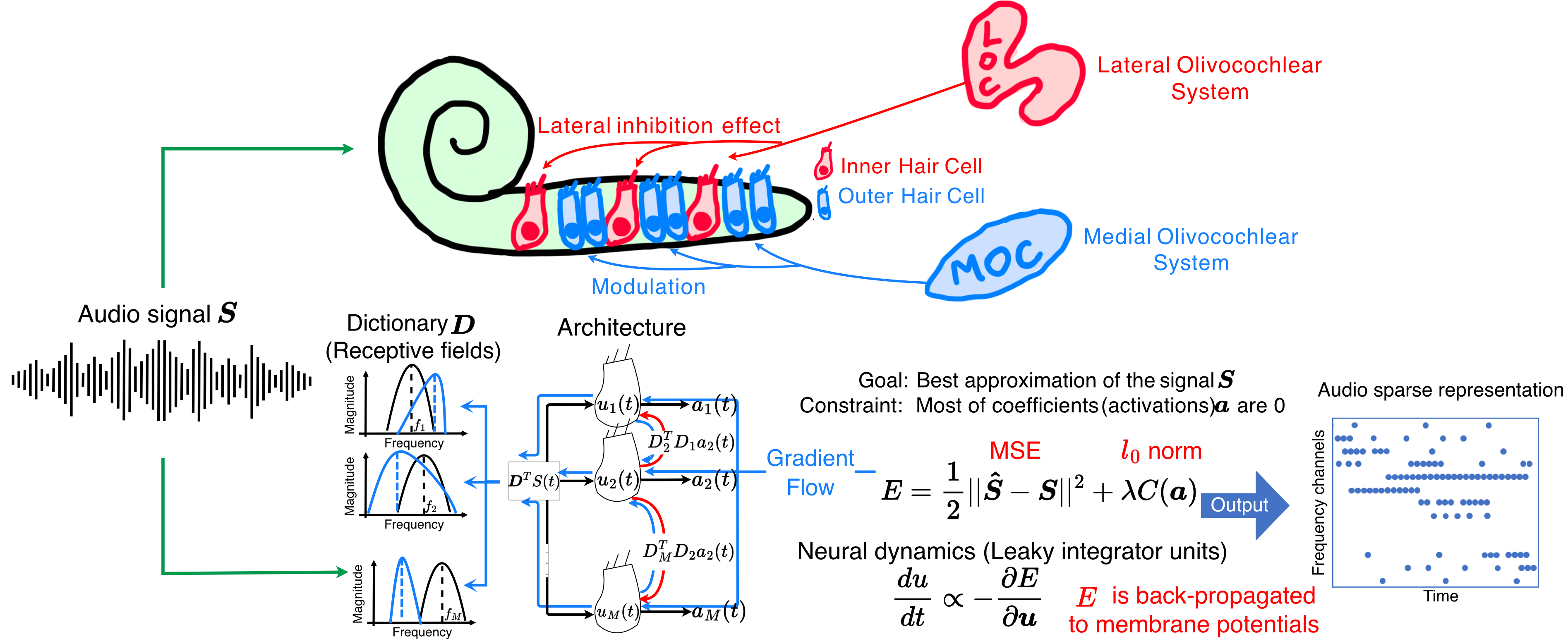
Tuesday July 14, 2026 5:00pm - 7:00pm ADT
Ballroom B2

5:00pm ADT

P140: Unsupervised Machine Learning Analysis of Extracellular Waveforms
Tuesday July 14, 2026 5:00pm - 7:00pm ADT
Introduction
Advances in transcriptomic, morphological, and electrophysiological techniques have led to more comprehensive characterization of cortical cell types. However, in the absence of “ground truth” labels, it is difficult to classify single units into neuron types based on extracellular action potentials (EAPs) recorded from high-density electrode arrays. Previously published classifiers rely on extracted features, machine learning (ML) algorithms, and dimensionality reduction approaches that produce a range of EAP type classes (Haynes, 2024; Jia, 2019; Lee, 2021). We extend these approaches by developing an unsupervised ML framework that relies on spatiotemporal patterns of multi-channel waveforms.


Methods
We developed an unsupervised ML framework to analyze spiking events from extracellular recordings. For each event, a spatiotemporal kernel waveform is constructed with a 1.5 ms temporal window that is centered on a spike time, based on waveforms from channels 200 um above and below the channel with the largest peak amplitude. We analyzed thousands of spiking events and applied KMeans clustering on two different representations of the data: 1) extracted spatiotemporal features and 2) vectorized multi-channel waveforms. We applied our framework to extracellular recordings in the auditory cortex of an awake common marmoset (Callithrix jacchus). We compare these results to previously published studies that cluster EAPs from rodent (Jia, 2019).


Results
Both the feature-based and the vectorized data representations produced well-defined separation of clusters of spiking events recorded from marmoset auditory cortex. For the feature based approach, EAP duration and vertical spread across channels were the most distinguishing features across clusters. Clustering based on vectorized multi-channel waveforms resulted in greater variability in feature distributions, cortical depth profiles, and spatiotemporal patterns across cluster groups.


Discussion
Together, these findings suggest the presence of diverse neural subtypes in the marmoset auditory cortex that exhibit distinct extracellular signatures. Our data-driven framework demonstrates that unsupervised machine learning approaches reveal physiologically meaningful variation in EAP waveforms, offering a scalable approach to comparative analysis of cortical organization and function. Moreover, this framework is applied to extracellular recordings from other species to identify distinct waveform features and identify which aspects of  spatiotemporal patterns are conserved across species.


References

Haynes, V. R., Zhou, Y., & Crook, S. M. (2024). Discovering optimal features for neuron-type identification from extracellular recordings. Frontiers in Neuroinformatics, 18, 1303993. https://doi.org/10.3389/fninf.2024.1303993
Jia, X., Siegle, J. H., Bennett, C., Gale, S. D., Denman, D. J., Koch, C., & Olsen, S. R. (2019). High-density extracellular probes reveal dendritic backpropagation and facilitate neuron classification. Journal of Neurophysiology, 121(5), 1831–1847. https://doi.org/10.1152/jn.00680.2018
Lee, E. K., Balasubramanian, H., Tsolias, A., Anakwe, S. U., Medalla, M., Shenoy, K. V., & Chandrasekaran, C. (2021). Non-linear dimensionality reduction on extracellular waveforms reveals cell type diversity in premotor cortex. eLife, 10, e67490. https://doi.org/10.7554/eLife.67490


Acknowledgement
This research is support by the NIDCD (R01DC019278).
Tuesday July 14, 2026 5:00pm - 7:00pm ADT
Ballroom B2

5:00pm ADT

P141: Different training paradigms yield distinct learned structures in recurrent neural network models
Tuesday July 14, 2026 5:00pm - 7:00pm ADT
Introduction
RNNs trained with backpropagation through time (BPTT) solve tasks through reliance on global error signals [1], while evolutionary algorithms and activity-dependent plasticity rules are gradient-free alternatives to solve the same tasks. How the choice of training paradigm biases the solutions that models arrive at remains unclear. Here we compare four paradigms—BPTT, evolution strategies (ES) [2], a genetic algorithm (GA), and GA with Oja’s Hebbian plasticity (GA+Oja)—on n-back recall, in which the network reports which of five symbols appeared n steps earlier. Beyond task accuracy, we analyze the connectivity changes that occur under each paradigm, finding that training method shapes structure far more than it determines performance.


Methods
We trained RNNs (32, 128, 256 units) on a 5-symbol n-back task. BPTT employed gradient descent with the Adam optimizer (2000 iterations). ES estimated gradients from 128 perturbed copies of the network (perturbation σ = 0.02, 500 generations). GA used tournament selection with neuron-level crossover and self-adaptive mutation (128 individuals, 500 generations). GA+Oja co-evolved two Oja's rule parameters (learning rate and weight bound) which update recurrent weights via correlated activity, enabling within-lifetime adaptation atop evolved connectivity. We tested n-back 1–4 steps, measuring accuracy (chance accuracy = 20%), per-layer weight-change fractions, effective rank of the recurrent weight matrix, and total weight-change magnitude.

Results
All methods converged to ~100% at 2-back, although ES showed high variance at 1-back (~85%; Fig. 1A). At 4-back, ES maintained ~100%, whereas GA declined to ~80% and GA+Oja to ~89%; BPTT remained at ~100%. Connectivity analysis revealed a strikingly different structure despite comparable accuracy. BPTT produced low effective rank recurrent matrices (~8 at 1-back) that increased with difficulty (~26 at 4-back). In contrast, evolutionary methods clustered at a higher rank (~16) with less variation across levels (Fig. 1B). BPTT increasingly allocated weight changes to the output layer with difficulty (~29% to ~43%). In comparison, all evolutionary methods remained flat at ~21% (Fig. 1C).


Discussion

Gradient-based and evolutionary training produce comparably accurate networks with fundamentally different connectivity. BPTT finds low-rank recurrent solutions and progressively shifts learning toward output weights as difficulty increases, consistent with efficient credit assignment [3]. Evolutionary methods converge on distributed, high-rank solutions with uniform layer allocation — a qualitatively different regime. These results caution that studies inferring biological principles from trained RNNs may reflect the training algorithm rather than computational necessity. BPTT’s concentration of learning in output weights could mislead analyses of how recurrent dynamics support memory if the readout is not examined separately.

Figure 1. (A) Accuracy vs. n-back level: methods converge at 2-back; ES maintains ~100% at 4-back, whereas GA declines to ~80%. (B) BPTT effective rank increases from ~8 to ~26 with difficulty; evolutionary methods cluster at ~16. (C) BPTT shifts learning to the output layer (+14%); evolutionary methods stay flat. 32 neurons, mean ± std, 3 seeds.


References

1. Neftci, E. O., Mostafa, H., & Zenke, F. (2019). Surrogate gradient learning in spiking neural networks. IEEE Signal Processing Magazine, 36(6), 51–63. https://doi.org/10.1109/MSP.2019.2931595 
2. Salimans, T., Ho, J., Chen, X., Sidor, S., & Sutskever, I. (2017). Evolution strategies as a scalable alternative to reinforcement learning. arXiv:1703.03864. https://arxiv.org/abs/1703.03864 
3. Izhikevich, E. M. (2007). Solving the distal reward problem through linkage of STDP and dopamine signaling. Cerebral Cortex, 17(10), 2443–2452. https://doi.org/10.1093/cercor/bhl152 


Acknowledgement
We thank Dora Jiayue Li and the Pomona College Summer Undergraduate Research Program (SURP) for supporting early exploration on this project, as well as the Pomona College Department of Neuroscience for their senior thesis support.

Speakers
YZ

Yuqing Zhu

Assistant Professor of Neuroscience, Pomona College
Tuesday July 14, 2026 5:00pm - 7:00pm ADT
Ballroom B2

5:00pm ADT

P142: Building neural manifolds from membrane biophysics and circuit topology
Tuesday July 14, 2026 5:00pm - 7:00pm ADT
Introduction
Neural population activity is often described as evolving on low-dimensional manifolds that structure neural computation and behaviour. These manifolds are typically identified empirically using dimensionality reduction techniques applied to large-scale neural recordings (Gallego et al., 2017). While such approaches successfully reveal structured population dynamics, they are largely agnostic to the underlying biophysical mechanisms that generate neural activity. Consequently, the relationship between neural manifolds and the building blocks of neural tissue – morphology, ion channel, connectivity, synapse physiology, etc. – remains undefined.

Methods
Here, we propose a framework that constructs neural population dynamics from biophysical elements. Combing cable theory with Hodgkin-Huxley membrane dynamics, axial current along neuronal processes can be balanced by total membrane current across each membrane segment. Linearizing over axial and membrane kinetic elements governed by experimentally measurable conductances and gating variables, the total membrane current is expressed by contributions from voltage-gated ion channels, ligand-gated synaptic channels, electrical synapses, and electrogenic transport mechanisms, including ion pumps and exchangers. Thus longer-timescale membrane dynamics may be explicitly taken into account.

Results
We validate this framework in the compact, stereotypic, and connectomically-defined nervous system of C. elegans (White et al., 1986). Our analysis focuses on the locomotor rich club neurons, which coordinate transitions between forward and reverse states (Fieseler et al., 2025). Biophysical parameters are estimated from current-clamp electrophysiological recordings that constrain passive membrane properties and voltage-gated conductances. Population activity in this circuit is further constrained using whole-brain Ca²⁺ imaging measurements of the same neurons during behaviour (Fieseler et al., 2025). Together, these data provide experimentally grounded estimates of the membrane and coupling parameters governing the dynamical system.


Discussion
By deriving network dynamics directly from experimentally measurable cellular and circuit properties, this framework provides a mechanistic bridge between membrane biophysics and population-level neural dynamics. In this view, neural manifolds are not only empirical low-dimensional decompositions of neural activity, but also naturally emerging physical organizations of neural tissue: a graphical interaction between membrane dynamics and neuronal morphology. This approach therefore links cellular physiology, circuit topology, and population dynamics within a unified dynamical system to provide a principled foundation for interpreting neural manifolds as geometric consequences of biophysical mechanisms.


References
  1. Gallego, J. A., Perich, M. G., Miller, L. E., & Solla, S. A. (2017). Neural Manifolds for the Control of Movement. Neuron, 94(5), 978-984. https://doi.org/10.1016/j.neuron.2017.05.025
  2. White, J. G., Southgate, E., Thomson, J. E., & Brenner, S. (1986). The structure of the nervous system of the nematode Caenorhabditis elegans. Philosophical Transactions of the Royal Society of London. B, Biological Sciences, 314(1165), 1-340. https://doi.org/10.1098/rstb.1986.0056
  3. Fieseler, C., Lev, I., Rey, U., Hille, L., Brenner, H., & Zimmer, M. (2025). An intrinsic neuronal manifold underlies brain-wide hierarchical organization of behavior in C. elegans. bioRxiv, 2025.2003.2009.642241. https://doi.org/10.1101/2025.03.09.642241

Acknowledgement
We would like to thank the European Research Commission (Advanced Grant) and Austrian Science Fund Cluster of Excellence (Neuronal Circuits in Health and Disease) for funding.
Tuesday July 14, 2026 5:00pm - 7:00pm ADT
Ballroom B2
 
Share Modal

Share this link via

Or copy link

Filter sessions
Apply filters to sessions.
Filtered by Date -