IntroductionTranscranial ultrasound stimulation (TUS) is an emerging neuromodulation technique offering millimeter‑scale precision and non‑invasive stimulation. While experimental studies show that ultrasound can modulate neural activity across multiple brain regions, the underlying biophysical mechanisms remain incompletely understood, limiting optimal protocol design. Among the proposed mechanisms, including flexoelectricity, mechanosensitivity, thermodynamic effects, and membrane displacement, intramembrane cavitation (IC) has gained particular attention. IC describes the oscillating gas cavities between the phospholipid membrane leaflets, generating capacitive currents and membrane charge oscillations that can alter neuronal excitability.
MethodsEarly computational work using the neuronal intramembrane cavitation excitation (NICE) model demonstrated the potential of this mechanism but suffered from large computational cost [1]. Subsequent developments, such as the multi‑scale optimized SONIC model and the spatially extended SECONIC model, introduced precomputation, hybrid integration, and Fourier analysis to accelerate simulations and incorporate charge redistribution dynamics [2,3]. However, these models were limited to one-dimensional representations of neurons, restricting their ability to capture realistic spatial activation patterns. In this work, we extend IC‑based ultrasonic neuromodulation modeling to morphologically realistic neurons across different cortical layers.
ResultsBy integrating detailed neuronal, we investigate how ultrasound parameters such as frequency, pressure amplitude, pulse repetition frequency (PRF), duty cycle (DC), and sonophore radius, shape neuronal responses. Fig. 1 shows how the firing rate of a L2/3 pyramidal cell depends on the pressure amplitude and the duty cycle. This approach enables localization of activation sites, assessment of the role of higher‑order charge overtones, and evaluation of whether IC can account for experimentally observed cell‑type specificity and protocol sensitivity. For instance, across multiple cortical cell types, we found that activation consistently originated at terminal nodes.
DiscussionBy integrating advanced biophysical models with detailed, realistic neuronal morphologies, this study advances a mechanistic understanding of how TUS influences neural activity and offers a foundation for accurate, in‑silico optimization of stimulation strategies.
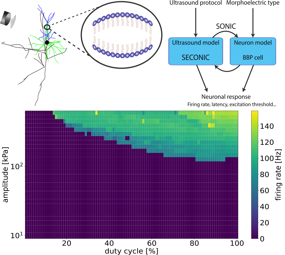 Figure 1. The intramembrane cavitation model in a pyramidal cell (top left), a schematic of the implementation of ultrasound field – neuron coupling (top right) and the firing rate of a L2/3 pyramidal cell as a function of amplitude and duty cycle.
References[1] Plaksin, M., Kimmel, E., & Shoham, S. (2016). Cell-Type-Selective Effects of Intramembrane Cavitation as a Unifying Theoretical Framework for Ultrasonic Neuromodulation. eNeuro, 3(3), ENEURO.0136-15.2016. https://doi.org/10.1523/ENEURO.0136-15.2016
[2] Lemaire, T., Neufeld, E., Kuster, N., & Micera, S. (2019). Understanding ultrasound neuromodulation using a computationally efficient and interpretable model of intramembrane cavitation. Journal of neural engineering, 16(4), 046007. https://doi.org/10.1088/1741-2552/ab1685
[3] Tarnaud, T., Joseph, W., Schoeters, R., Martens, L., & Tanghe, E. (2020). SECONIC: Towards multi-compartmental models for ultrasonic brain stimulation by intramembrane cavitation. Journal of neural engineering, 17(5), 056010. https://doi.org/10.1088/1741-2552/abb73d
Acknowledgement/