IntroductionWhile vagus nerve stimulation (VNS) coupled with motor rehabilitation significantly improves post-stroke recovery [1], its mechanism of action remains poorly understood. Preclinical studies indicate that VNS enhances motor learning when stimulation is precisely paired with task success [2,3], with cholinergic neuromodulation necessary for these effects [4]. These findings collectively suggest that rapid cholinergic signaling may be pivotal in stroke rehabilitation, although this response to VNS has not been well characterized experimentally.
We hypothesize that the immediate cholinergic response to VNS produces rapid changes in cortical oscillatory dynamics through modulation of the muscarinic M-current in cortical neurons.
MethodsWe model the brainstem circuitry connecting the vagus nerve to the cortex with populations of quadratic integrate and fire (Izhikevich) neurons: the nodose ganglion of the vagus nerve, the nucleus of the solitary tract, the locus coeruleus, and cholinergic neurons of the basal forebrain projecting to the cortex (Fig. 1A). Each neuronal population is parameterized using firing rate and adaptation properties fit to published electrophysiological data.
The predicted VNS-triggered cholinergic output derived from basal forebrain neuronal spiking modulates an excitatory-inhibitory (E-I) cortical microcircuit of Hodgkin-Huxley neurons through a conductance-based model of the muscarinic M-current (Fig. 1B).
ResultsPreclinical [2] and preliminary clinical EEG recordings show that VNS immediately reduces cortical gamma power, reflecting a decrease in synchronized spiking activity. Our model generates a dynamic cholinergic output which in turn modulates the M-current within an in silico cortical network [5]. The resulting synaptic activity is used to generate a pseudo-EEG signal that can be compared directly to the spectral changes observed in clinical recordings (Fig. 1B). Expanding upon recent in silico work showing that dynamically increasing acetylcholine concentrations desynchronize cortical microcircuits dependent upon the rate of modulation [6], we determine whether the specific cholinergic response to VNS can account for VNS’s effect on EEG.
DiscussionThe combination of an experimentally constrained brainstem model and a cortical microcircuit subject to cholinergic neuromodulation allows us to test whether rapid cholinergic signaling can account for the immediate decrease in cortical gamma power observed following VNS. This approach allows for explorations of how stimulation parameters—such as inter-stimulation interval, pulse train duration, and stimulation intensity—influence cholinergic output and cortical dynamics beyond what is currently done clinically. In doing so, the model may help identify stimulation paradigms that maximize beneficial neuromodulatory effects, providing mechanistic insight into how VNS protocols can be optimized to improve stroke rehabilitation.
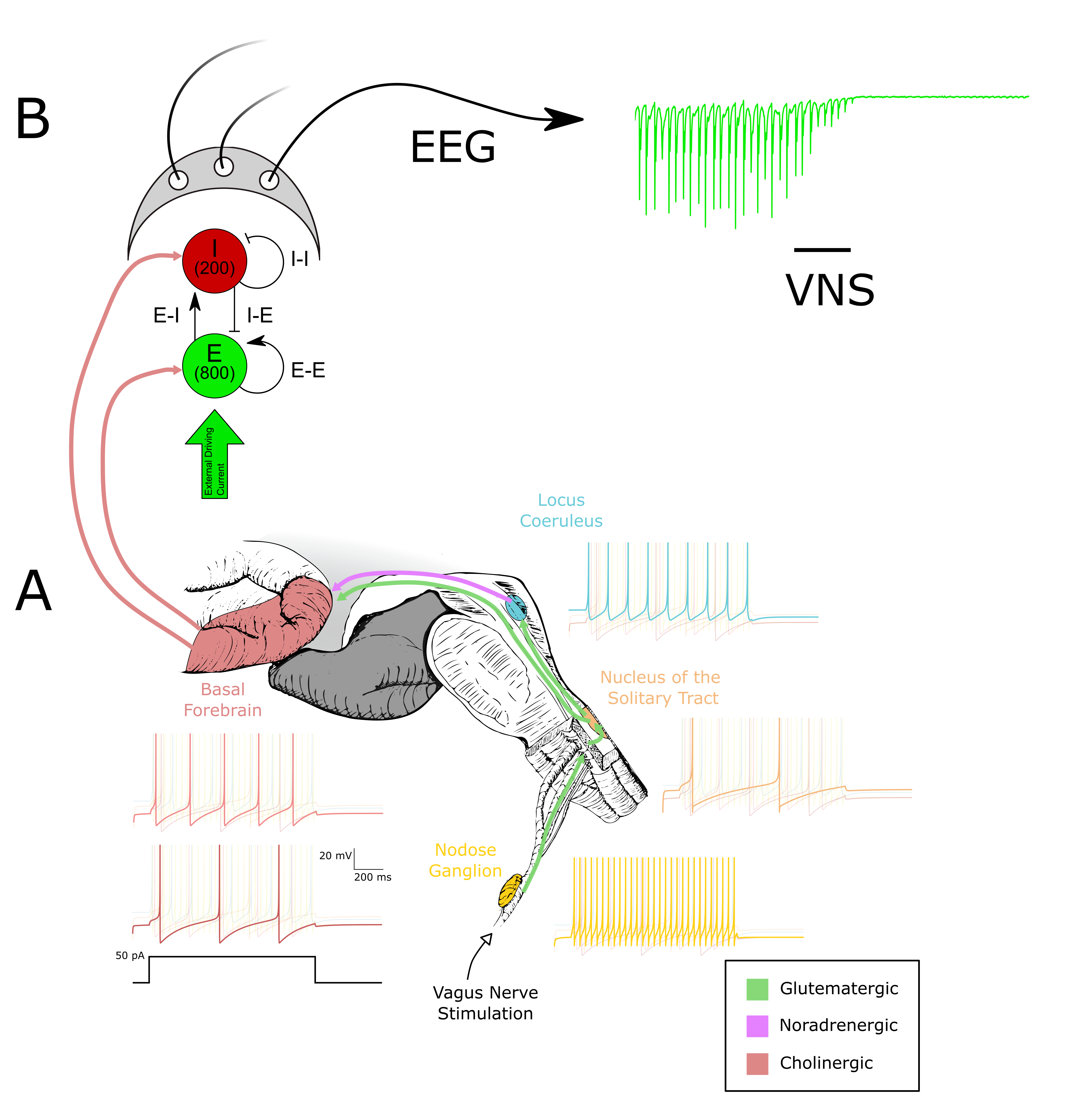 Figure 1. (A) The circuitry between the vagus nerve and the basal forebrain is modeled using quadratic integrate-and-fire (Izhikevich) neurons fitted to experimental data. Representative voltage responses to a 50 pA injection are shown. (B) The output of the basal forebrain in (A) is used to modulate ACh-sensitive K+ channels in an E–I network. Synaptic activity within this network is used to compute an EEG
References1. Dawson, J., et al. (2021). Vagus nerve stimulation paired with rehabilitation for upper limb motor function... The Lancet, 397(10284), 1545–1553.
2. Engineer, N. D., et al. (2011). Reversing pathological neural activity... Nature, 470(7332), 101–104. https://doi.org/10.1038/nature09656
3. Hays, S. A., et al. (2014). The timing and amount of vagus nerve stimulation... Neuroreport, 25(9), 676–682.
4. Bowles, S., et al. (2022). Vagus nerve stimulation drives selective... Neuron, 110(17), 2867–2885.e7.
5. Stiefel, K. M., et al. (2009). The effects of cholinergic neuromodulation... Journal of Computational Neuroscience, 26(2), 289–301.
6. Pandian, S., & Rich, S. (2025). Dynamic cholinergic signaling differentially... (p. 2025.06.20.660675). bioRxiv